. 2024 Jan 15;209(2):153-163.
doi: 10.1164/rccm.202303-0503OC.

Loss of p73 Expression Contributes to Chronic Obstructive Pulmonary Disease

Affiliations

Loss of p73 Expression Contributes to Chronic Obstructive Pulmonary Disease

Bradley W Richmond et al. Am J Respir Crit Care Med. .

Abstract

Rationale: Multiciliated cell (MCC) loss and/or dysfunction is common in the small airways of patients with chronic obstructive pulmonary disease (COPD), but it is unclear if this contributes to COPD lung pathology. Objectives: To determine if loss of p73 causes a COPD-like phenotype in mice and explore whether smoking or COPD impact p73 expression. Methods: p73floxE7-E9 mice were crossed with Shh-Cre mice to generate mice lacking MCCs in the airway epithelium. The resulting p73Δairway mice were analyzed using electron microscopy, flow cytometry, morphometry, forced oscillation technique, and single-cell RNA sequencing. Furthermore, the effects of cigarette smoke on p73 transcript and protein expression were examined using in vitro and in vivo models and in studies including airway epithelium from smokers and patients with COPD. Measurements and Main Results: Loss of functional p73 in the respiratory epithelium resulted in a near-complete absence of MCCs in p73Δairway mice. In adulthood, these mice spontaneously developed neutrophilic inflammation and emphysema-like lung remodeling and had progressive loss of secretory cells. Exposure of normal airway epithelium cells to cigarette smoke rapidly and durably suppressed p73 expression in vitro and in vivo. Furthermore, tumor protein 73 mRNA expression was reduced in the airways of current smokers (n = 82) compared with former smokers (n = 69), and p73-expressing MCCs were reduced in the small airways of patients with COPD (n = 11) compared with control subjects without COPD (n = 12). Conclusions: Loss of functional p73 in murine airway epithelium results in the absence of MCCs and promotes COPD-like lung pathology. In smokers and patients with COPD, loss of p73 may contribute to MCC loss or dysfunction.

Keywords: COPD; cigarette smoke; ciliated cell; p73.


Figures

Figure 1.
Figure 1.
Targeted p73 deletion in the airways results in multiciliated cell (MCC) loss, emphysema-like lung remodeling, and neutrophilic inflammation. All experiments in this figure were performed on adult (4–7 months old) p73Δairway mice and littermate control mice. (A) Low-magnification view of lungs of p73Δairway mice shows emphysema-like lung remodeling but no overt pneumonitis. Scale bars, 100 μm. (B) The airways of p73Δairway mice contain flattened, dysmorphic epithelial cells and areas without overlying epithelial cells (black arrows). Scale bars, 100 μm. (C) Representative images of immunostaining for acetylated α-tubulin (*α-tubulin) showing a near-complete absence of MCCs in the airways of p73Δairway mice. Scale bars, 100 μm. (D) TEM micrographs showing absent MCCs, loss of cell–cell junctions (yellow arrow), and apically oriented nuclei (white arrow) in airway epithelial cells from p73Δairway mice. Scale bars, 2 μm. (E) MLI in p73Δairway and littermate control mice. n = 4–5 mice/group. *P < 0.05; Mann-Whitney U test. (F) Tissue elastance (H) in p73Δairway mice and littermate control mice. n = 8 mice/group. *P < 0.05; Mann-Whitney U test. (G and H) Neutrophil numbers (G) and percentage among CD45+ cells (H) in the left lungs of adult (4–7 months old) p73Δairway mice and littermate control mice. ***P < 0.001; two-way ANOVA with Šidák’s multiple comparison correction. AM = alveolar macrophages; Eos = eosinophils; H&E = hematoxylin and eosin; IM = interstitial macrophages; MLI = mean linear intercept; Mono = monocytes; Neu = neutrophils; TEM = transmission electron microscopy.
Figure 2.
Figure 2.
Deletion of p73 in the airway epithelium results in progressive loss of SCGB1A1+ secretory cells. (A) Quantification of DAPI+ cells per 1 mm basement membrane in 2- and 6-month-old p73Δairway mice and littermate control mice. n = 4–5 mice/group and 5–13 airways/mouse. ****P < 0.0001; one-way ANOVA with Tukey’s multiple comparisons test. (B) Representative images of SCGB1A1 immunostaining in 2- and 6-month-old p73Δairway mice and littermate control mice. Scale bars, 50 μm. Inset, 2× magnification. (C) Percentage of SCGB1A1+ cells among total DAPI+ cells in 2- and 6-month-old p73Δairway mice and littermate control mice. n = 4–5 mice/group and 6–19 airways/mouse. ***P < 0.001 and ****P < 0.0001; one-way ANOVA with Tukey’s multiple comparisons test.
Figure 3.
Figure 3.
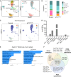
Loss of p73 in the airway epithelium increases cell–cell and cell–basement membrane gene expression in Scgb1a1+ cells. (A) UMAP colored by annotated cell type (left panel) or genotype (right panel). (B) Proportion of each cell type originating from p73Δairway or control mice. (C) Feature plot showing Trp73 expression in MCCs from p73Δairway and control mice. (D) Number of significant differentially expressed genes (DEGs) between p73Δairway and control mice for each cell type. Significance was defined as >0.25-fold expression and adjusted P value < 0.05 (Wilcoxon rank sum test) in genes expressed in at least 25% of the cells in the cluster. (E) Pathways enriched among DEGs in Scgb1a1+ cells from p73Δairway and control mice (KEGG 2019 mouse). (F) Venn diagram showing genes that contributed to the focal adhesion, ECM–receptor interaction, and actin cytoskeleton pathways in E. AT1 = alveolar type I; AT2 = alveolar type II; BASC = bronchioalveolar stem cells; ECM = extracellular matrix; MCCs = multiciliated cells; PNEC = pulmonary neuroendocrine cells; UMAP = Uniform Manifold Approximation and Projection.
Figure 4.
Figure 4.
Acute cigarette smoke suppresses p73 expression in vitro. (A) Murine tracheal epithelial cells (MTECs) were differentiated in air–liquid interface (ALI) culture and then treated with 2.5% cigarette smoke exposure (CSE) or 2.5% phosphate-buffered saline (PBS) (control). Representative immunostaining for p73 (red) and acetylated α-tubulin (*α-tubulin) (green) in both groups at the indicated time points. (B) Quantification of p73+ cells in 12 fields of view for each time point shown in A. ****P < 0.00001 compared with CSE-treated cells at the same time point; Student’s t test. (C) Representative immunostaining for p63 (red) in MTECs treated with 2.5% CSE or 2.5% PBS (control) at the indicated time points. (D) Quantification of p63+ cells in 12 fields of view for each time point shown in C. ****P < 0.00001 compared with CSE-treated cells at the same time point; Student’s t test. (E) Adaptation of the SIU24 in vivo cigarette smoke machine for in vitro use. The apical side of human small airway epithelial cells (HSAECs) in ALI culture was exposed to mainstream cigarette smoke by lifting the top of the cell culture plate. Medium was changed immediately after each exposure to minimize smoke exposure to the basolateral side of each insert. (F) TP73 or (G) TP63 expression was measured by qRT-PCR in HSAECs treated with mainstream CS for 1 or 5 days. *P < 0.05; Kruskal-Wallis test with Dunn’s multiple comparison correction. ns = not significant; WCS = whole cigarette smoke.
Figure 5.
Figure 5.
Chronic cigarette smoke (CS) exposure suppresses p73 expression in vivo. (A) Representative immunostaining for p73 (red) and acetylated α-tubulin (*α-tubulin) (green, left panel) and FoxJ1 (green, right panel) in mice exposed to mainstream CS for 1 or 3 months and unexposed control mice. All immunostaining and micrographs were taken at the same time with the same exposure settings. Data represent quantification of at least three fields of view from three or four inserts performed in triplicate (e.g., a minimum of 27 fields of view per time point). (B and C) qRT-PCR analysis using primers for Trp73 (B) and Foxj1 (C) in whole-lung lysates from mice exposed to mainstream cigarette smoke for 1 or 3 months and unexposed control mice. *P < 0.05, **P < 0.01, and ***P < 0.001; two-way ANOVA with Tukey’s multiple corrections test. ns = not significant.
Figure 6.
Figure 6.
p73+ cells are reduced and p63+ cells are increased in the airways of patients with chronic obstructive pulmonary disease (COPD). (A) Representative images of immunostaining for p73 (red), p63 (green), and DAPI (blue) in small (<2 mm) airways from patients with COPD and deceased organ donors without COPD (control subjects). (B) Quantification of p73+ and p63+ as a percentage of total epithelial cells indicated by DAPI staining. At least three images were analyzed for 11 patients with COPD and 12 control subjects. **P < 0.01, ***P < 0.001, and ****P < 0.0001; one-way ANOVA with Šidák’s multiple comparison correction.

Comment in

References

    1. Agustí A, Celli BR, Criner GJ, Halpin D, Anzueto A, Barnes P, et al. Global Initiative for Chronic Obstructive Lung Disease 2023 report: GOLD executive summary. Am J Respir Crit Care Med . 2023;207:819–837. - PMC - PubMed
    1. Dunican EM, Elicker BM, Henry T, Gierada DS, Schiebler ML, Anderson W, et al. Mucus plugs and emphysema in the pathophysiology of airflow obstruction and hypoxemia in smokers. Am J Respir Crit Care Med . 2021;203:957–968. - PMC - PubMed
    1. Polosukhin VV, Gutor SS, Du RH, Richmond BW, Massion PP, Wu P, et al. Small airway determinants of airflow limitation in chronic obstructive pulmonary disease. Thorax . 2021;76:1079–1088. - PMC - PubMed
    1. Boucher RC. Muco-obstructive lung diseases. N Engl J Med . 2019;380:1941–1953. - PubMed
    1. Ballenger JJ. Experimental effect of cigarette smoke on human respiratory cilia. N Engl J Med . 1960;263:832–835. - PubMed

Publication types