Review
. 2022 Apr 25;7(4):264-273.
doi: 10.1530/EOR-21-0110.

Distal femoral fractures in children

Affiliations
Review

Distal femoral fractures in children

Matías Sepúlveda et al. EFORT Open Rev. .

Abstract

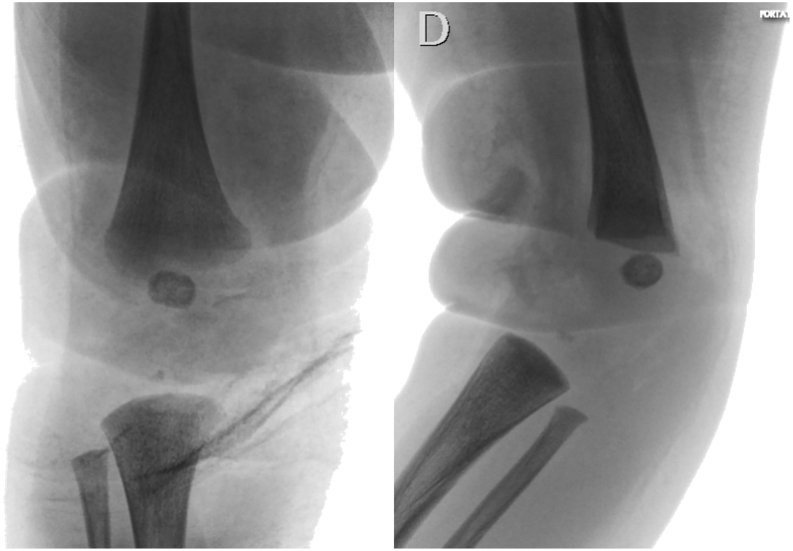
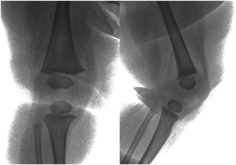
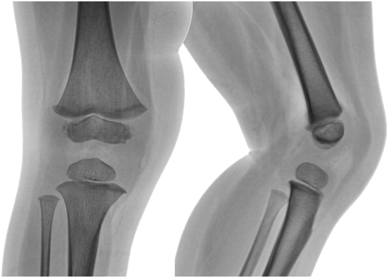
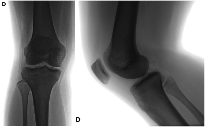
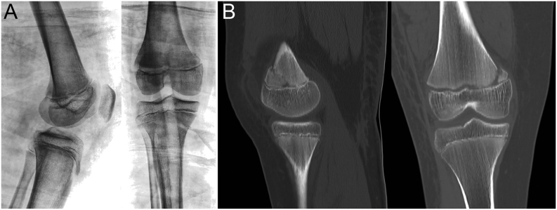
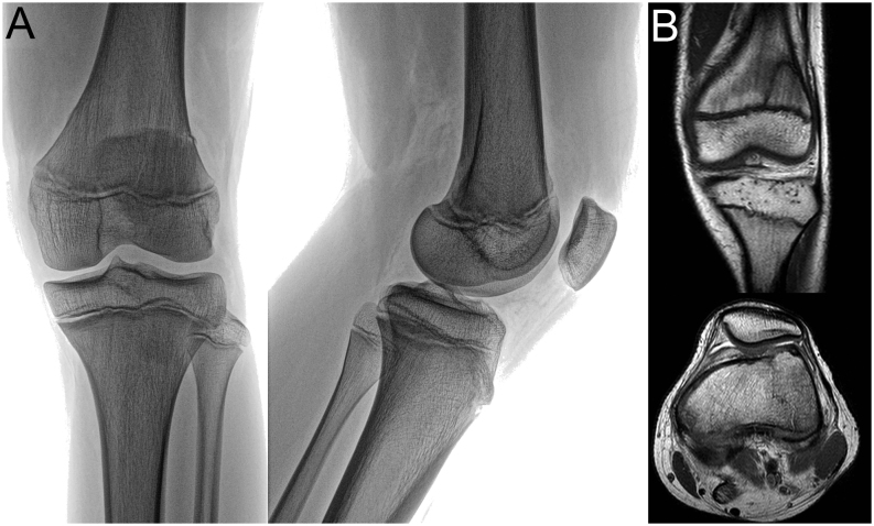
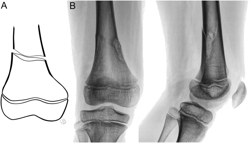
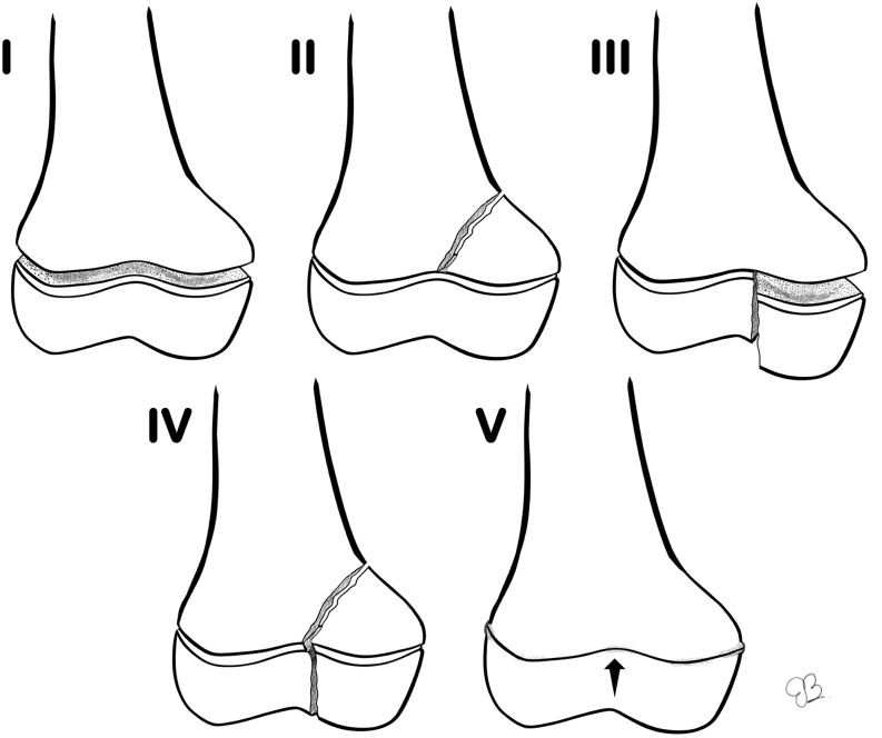
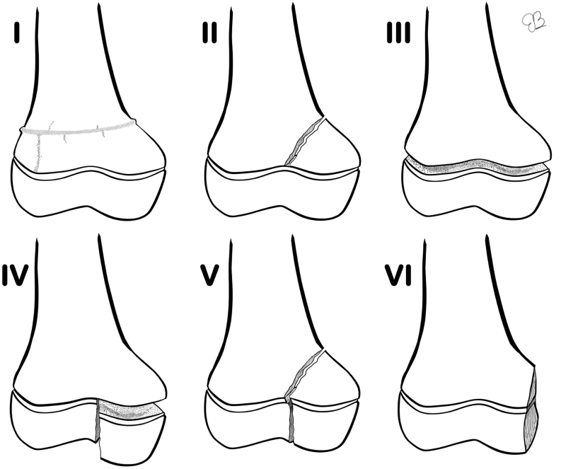
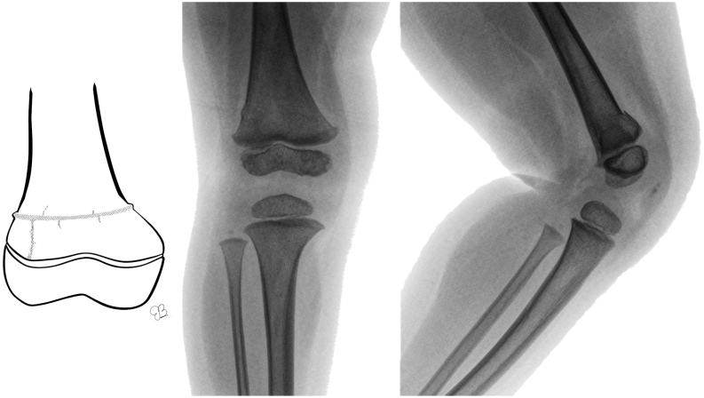
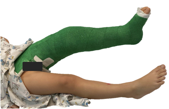
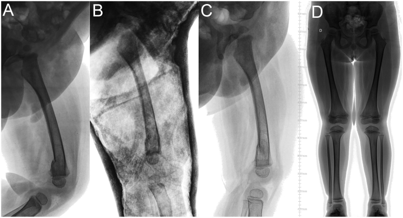
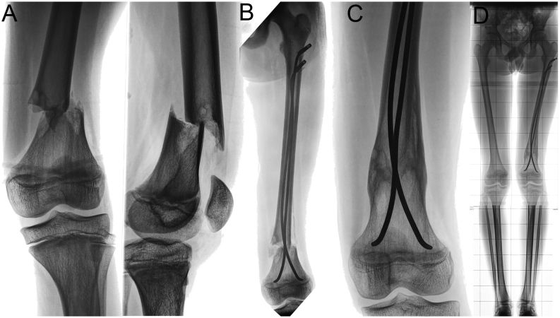
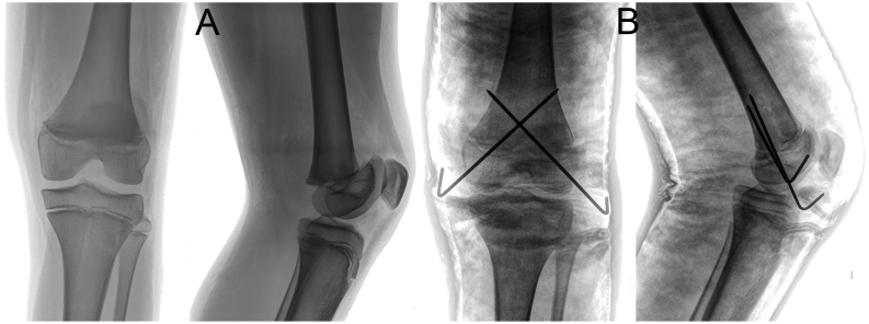
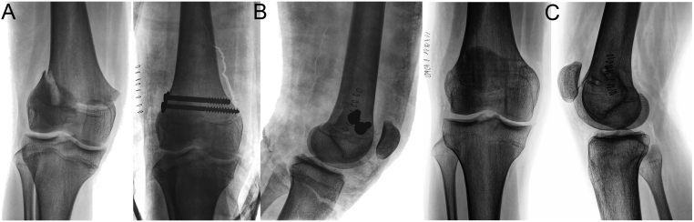
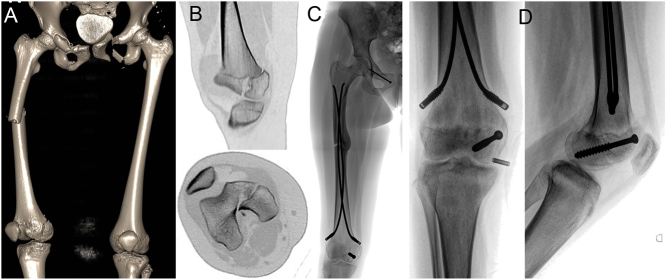
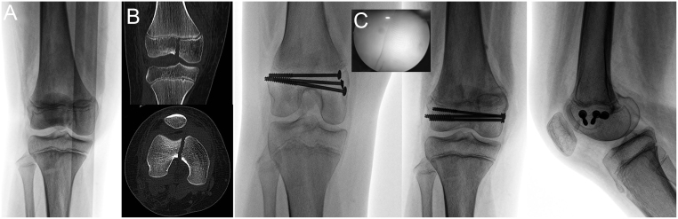
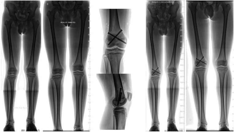
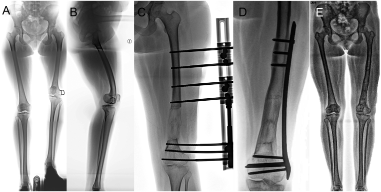
The physis of the distal femur contributes to 70% of femoral growth and 37% of the total limb growth; therefore, physeal injury can lead to important alterations of axes and length. Distal metaphyseal corner-type fracture prior to walking is classically associated with child abuse. In children aged >10 years, sports-related fractures and car accidents are significant contributors. Imaging includes a two-plane radiographic study of the knee. It is recommended to obtain radiographs that include the entire femur to rule out concomitant injuries. In cases of high suspicion of distal metaphyseal fractures and no radiographic evidence, CT or MRI can show the existence of hidden fractures. Fractures with physeal involvement are conventionally classified according to the Salter-Harris classification, but the Peterson classification is also recommended as it includes special subgroups. Conservative and surgical management are valid alternatives for the treatment of these fractures. Choosing between both alternatives depends on factors related to the fracture type. As there is a high risk of permanent physeal damage, long-term follow-up is essential until skeletal maturity is complete.

Keywords: femur; paediatrics; surgery; trauma.


Figures

Figure 1
Figure 1
Radiographic images of the right knee of a neonate. The spherical epiphyseal ossification centre of the distal femur is seen.
Figure 2
Figure 2
Radiographic images of the right knee of a 6-month-old girl. The epiphyseal ossification centre of the distal femur develops curves corresponding to the two condyles.
Figure 3
Figure 3
Radiographic images of right knee of a 2-year-old boy. The distal femoral physis with undulating shape and usual irregularities of the distal margins of the medial and lateral condyle are noted.
Figure 4
Figure 4
Radiographic images of the right knee of a 15-year-old girl. Physeal closure ending.
Figure 5
Figure 5
Scheme of the physis surface with the three main undulations.
Figure 6
Figure 6
(A) Radiographic images of the right knee of a 10-year-old girl, with a distal physeal femoral fracture. (B) CT: sagittal and coronal views show a Salter–Harris type II physeal injury.
Figure 7
Figure 7
(A) Radiographic images of the left knee of a 14-year-old boy, with a distal femoral fracture. (B) MRI: coronal and axial T1 views shows a Salter–Harris II distal femoral physis fracture.
Figure 8
Figure 8
(A) Schematic of distal metaphyseal femoral fracture. (B) Radiographic images of the right knee of a 6-year-old boy with a pathologic bone distal femoral fracture (non-osteogenic fibroma).
Figure 9
Figure 9
Classification according to Salter and Harris for distal femoral fractures in children. Type I, complete separation of the epiphysis from the metaphysis without any bone fracture; type II, line of separation extends along the epiphyseal plate and then out through a portion of the metaphysis; type III, extends from the joint surface to the weak zone of the epiphyseal plate and then extends along the plate to its periphery; type IV, extends from the joint surface through the epiphysis, across the full thickness of the epiphyseal plate, and through a portion of the metaphysis; type V, crushing force applied through the epiphysis to one area of the epiphyseal plate.
Figure 10
Figure 10
Classification according to Peterson for distal femoral fractures in children. Type I, transverse fracture of the metaphysis with extension to the physis; type II, separation of the physis with a portion of metaphysis attached to the physis; type III, separation of the epiphysis from the metaphysis through any layer of the physis disrupting the complete physis; type IV, fracture of the epiphysis extending to and along the physis; type V, fracture that traverses the metaphysis, physis and epiphysis; type VI, Portion of the physis has been removed or is missing.
Figure 11
Figure 11
(A) Schematic of distal Peterson type 1 physeal injury. (B) Radiographic images of the right knee of a 2-year-old girl with this type of fracture.
Figure 12
Figure 12
Photographs of a 3-year-old girl, with a distal femoral fracture, treated with a single-leg hip spica cast, with 15˚ knee flexion.
Figure 13
Figure 13
(A) Lateral radiographic image of the right femur of a 2-year-old boy, with a distal femoral fracture. (B) Radiographic control at 1 week. (C) Radiographic image after cast removal at 4 weeks. (D) Radiographic images (anteroposterior view) of both legs when standing, after 1 year, with no limb-length discrepancy or axis deviation.
Figure 14
Figure 14
(A) Radiographic images of the knee of a 10-year-old boy, with a pathologic bone distal femoral fracture (non-osteogenic fibroma). (B) One-week postoperative radiographic image after closed reduction and anterograde elastic stable endomedular nailing with use of endcaps, maintaining tension for the distal fragment stabilization. (C) Two-month postoperative radiographic images with advance bone healing. (D) The 1-year postoperative radiographic image with no limb length differences or axis deviation.
Figure 15
Figure 15
(A) Radiographic images of the knee of a 13-year-old boy with a distal physeal femoral fracture Salter–Harris type III. (B) Postoperative radiographic images after closed reduction and percutaneous pinning, with two crossed 2.0-mm K-wires and cast protection.
Figure 16
Figure 16
(A) Radiographic images of the knee of a 15-year-old boy with a distal physeal femoral fracture type Salter–Harris II. (B) Postoperative radiographic images after open reduction with periosteal sleeve, screw fixation, and cast protection. (C) Radiographic images after 6 months following screw removal, showing no axis deviation, with physeal closure.
Figure 17
Figure 17
(A) CT scan reconstruction of lower extremities of a 14-year-old girl with polytrauma. Pelvic unstable injuries, a left femur diaphyseal fracture, and distal femoral fracture are diagnosed. (B) CT scan sagittal and axial views show a displaced physeal femoral fracture type Salter–Harris III. (C) The distal femoral fracture was treated by closed reduction and percutaneous stabilization with a cannulated screw. (D) X-ray of the knee after 6 months, with advanced healing of the fracture.
Figure 18
Figure 18
(A) Radiographic images of the knee of a 11-year-old girl, with a distal femoral fracture. (B) Coronal and axial CT views show a displaced Salter–Harris type III fracture. (C) Intraoperative images of arthroscopic-assisted closed reduction. (D) Postoperative radiographic images after fixation with cannulated percutaneous screws.
Figure 19
Figure 19
(A) One year after fracture. Limb-length discrepancy of 1 cm, with no axis deviation. (B) Two years after the fracture. Limb-length discrepancy of 2 cm, with no axis deviation. (C) Epiphysiodesis with percutaneous cannulated screws was performed at age 15 years. (D) Control at 1 month after surgery. (E) Control after 1 year. Final limb-length discrepancy is <1 cm.
Figure 20
Figure 20
(A) Radiographic images of both legs (when standing) of a 20-year-old girl with distal femoral fracture sequelae. A 9-cm left femur shortening and 15˚ distal femur varus deformity on the frontal plane were observed. (B) Lateral one-leg standing radiography shows a 20˚ distal femur procurvatum deformity. (C) Acute distal axis correction and gradual lengthening with a monolateral external fixator was performed. (D) After achieving length correction, percutaneous plating. (E) Radiographic images of both legs standing after 6 months of hardware removal.

References

    1. Salter RB, Harris WR. Injuries involving the epiphyseal plate. Journal of Bone and Joint Surgery 196345587–622. (10.2106/00004623-196345030-00019) - DOI
    1. Alhammoud A, Younis MH, Ahmed AF, Ibrahim T. Surgical versus nonsurgical treatment of distal femur physeal fractures: a systematic review and meta-analysis. Journal of Musculoskeletal Surgery and Research 20193179–183. (10.4103/jmsr.jmsr_53_18) - DOI
    1. Peterson HA.Part 2. Physeal fractures: anatomic sites. Chapter 18. Distal femur. In Epiphyseal Growth Plate Fractures, pp. 595–640. Berlin, Heidelberg: Springer-Verlag, 2007. (10.1007/978-3-540-33802-4) - DOI
    1. Ogden J.Chapter 21: Femur. In Skeletal Injury in the Child, 3rded., pp.857–928. New York: Springer-Verlag, 2000. (10.1007/b97655) - DOI
    1. Suzuki Y, Matsubayashi J, Ji X, Yamada S, Yoneyama A, Imai H, Matsuda T, Aoyama T, Takakuwa T. Morphogenesis of the femur at different stages of normal human development. PLoS ONE 201914 e0221569. (10.1371/journal.pone.0221569) - DOI - PMC - PubMed