. 2023 Nov 6;7(1):e202302332.
doi: 10.26508/lsa.202302332. Print 2024 Jan.

Immunosuppressive low-density neutrophils in the blood of cancer patients display a mature phenotype

Affiliations

Immunosuppressive low-density neutrophils in the blood of cancer patients display a mature phenotype

Christophe Vanhaver et al. Life Sci Alliance. .

Abstract

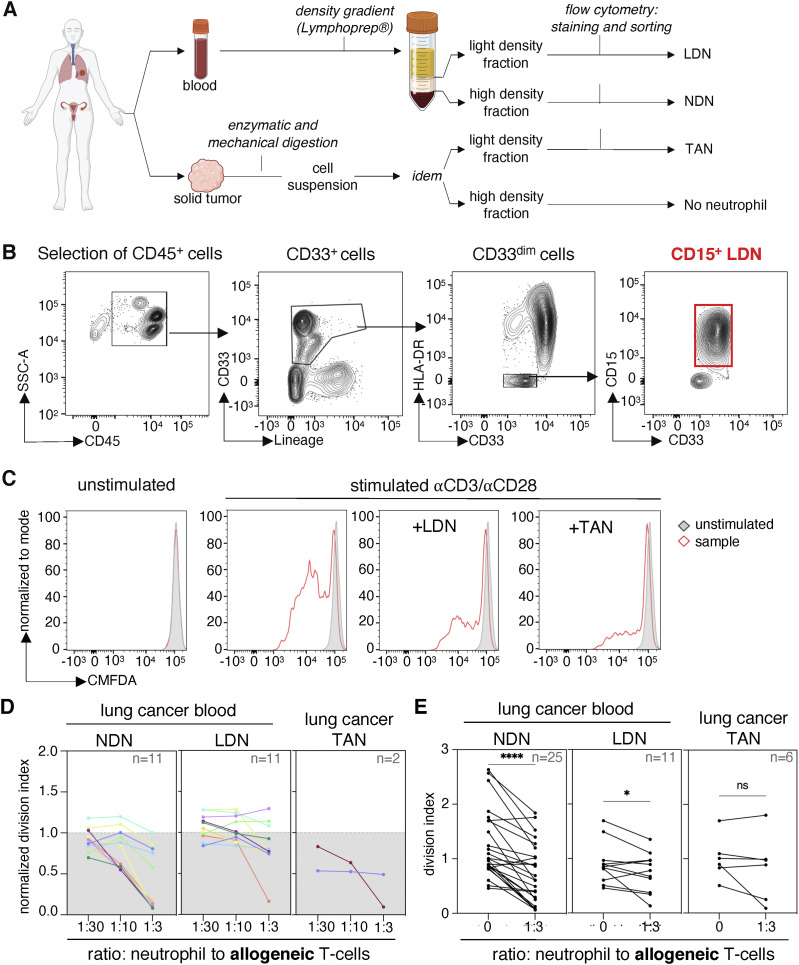
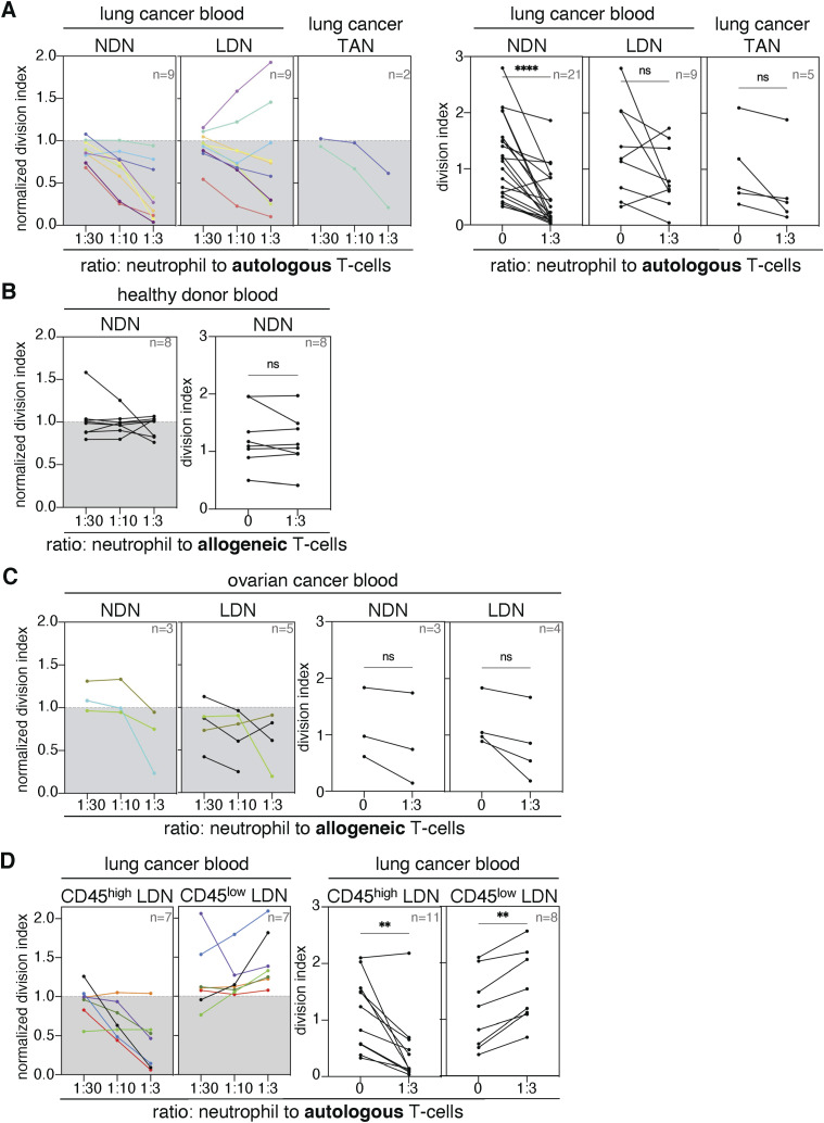
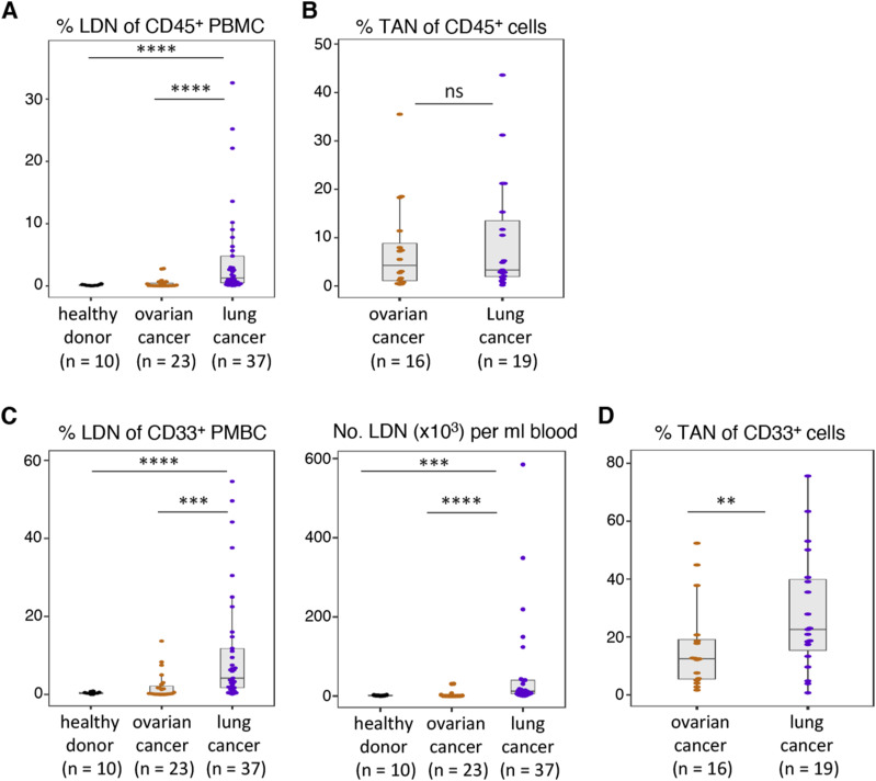
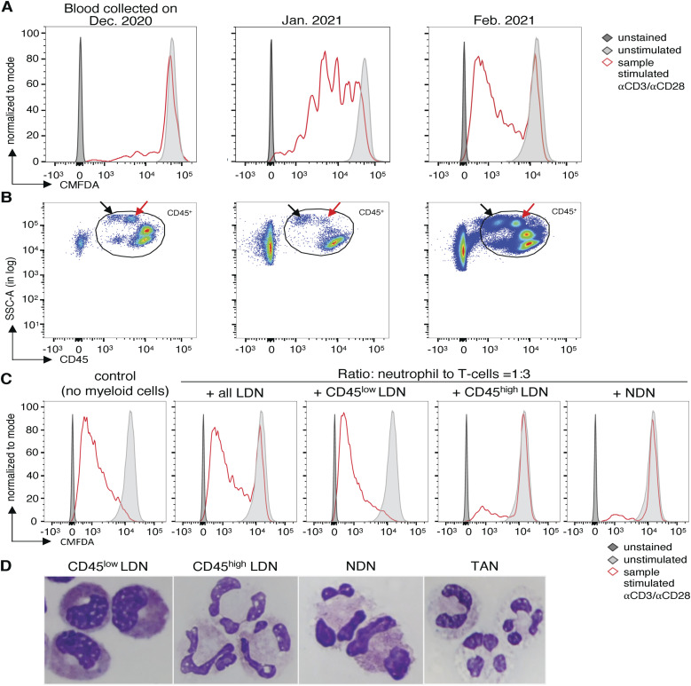
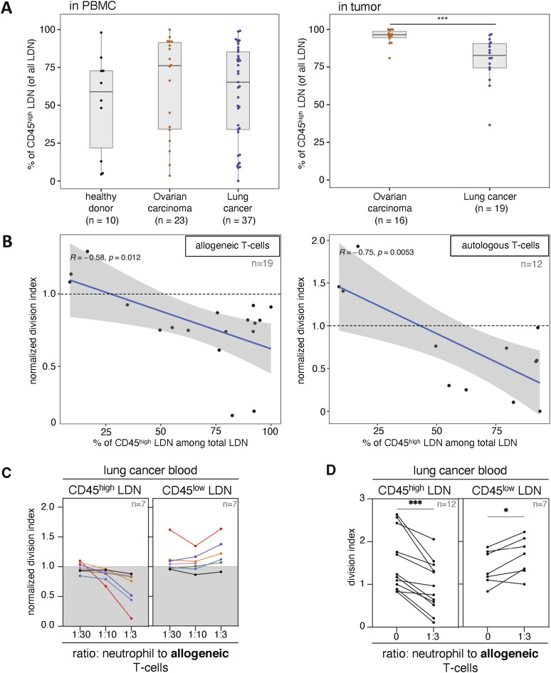
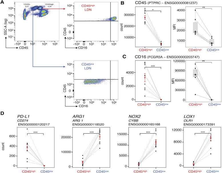
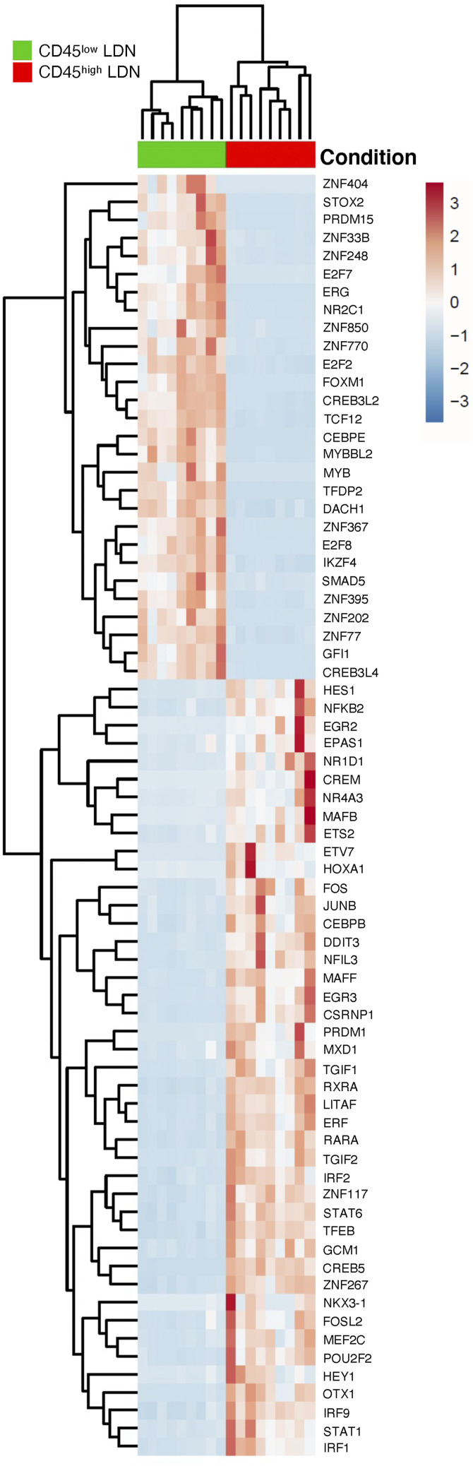
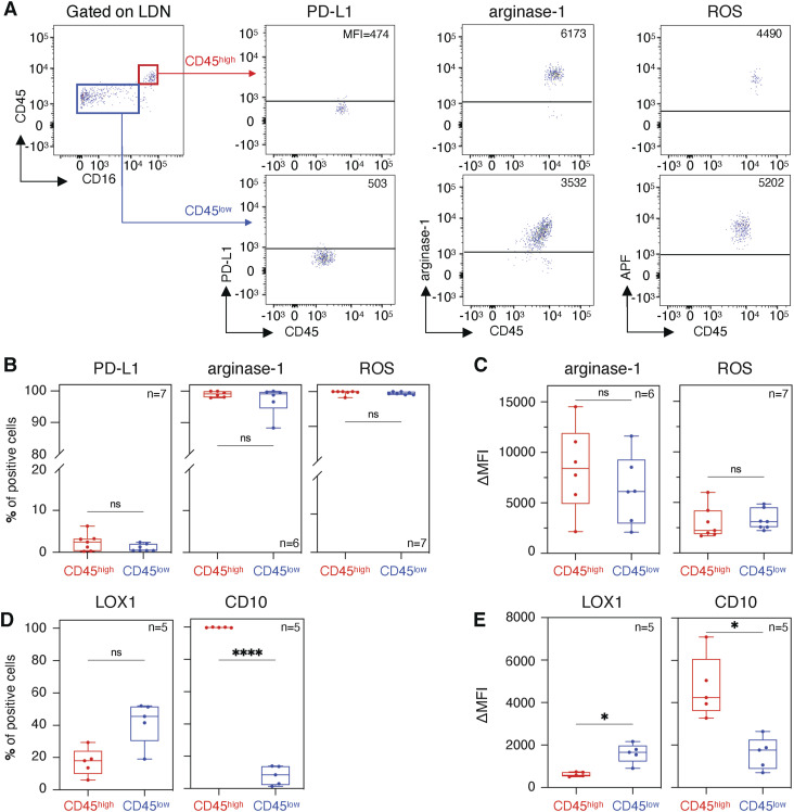
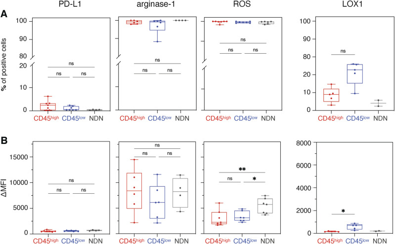
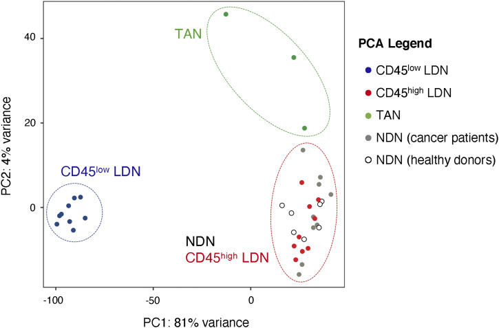
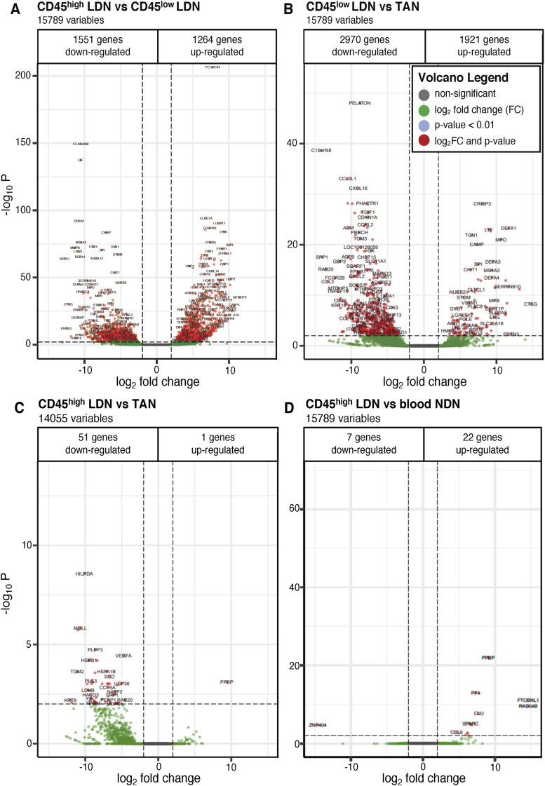
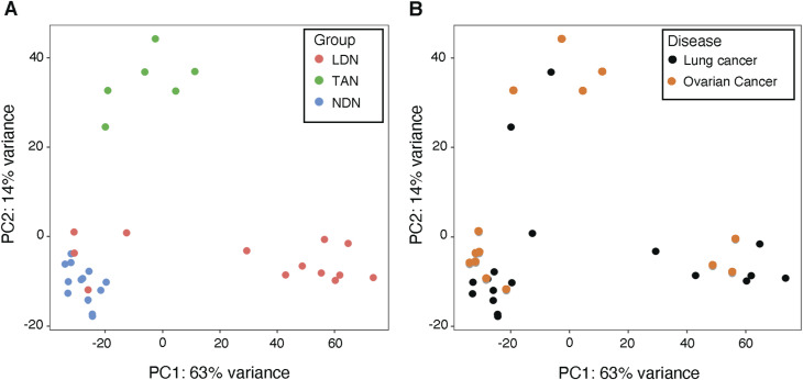
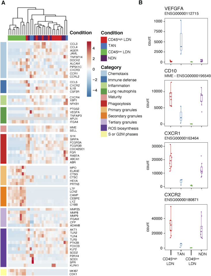
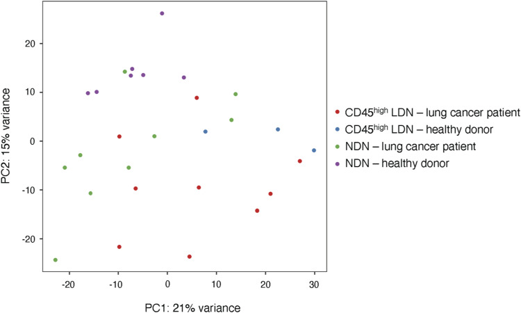
The presence of human neutrophils in the tumor microenvironment is strongly correlated to poor overall survival. Most previous studies have focused on the immunosuppressive capacities of low-density neutrophils (LDN), also referred to as granulocytic myeloid-derived suppressor cells, which are elevated in number in the blood of many cancer patients. We observed two types of LDN in the blood of lung cancer and ovarian carcinoma patients: CD45high LDN, which suppressed T-cell proliferation and displayed mature morphology, and CD45low LDN, which were immature and non-suppressive. We simultaneously evaluated the classical normal-density neutrophils (NDN) and, when available, tumor-associated neutrophils. We observed that NDN from cancer patients suppressed T-cell proliferation, and NDN from healthy donors did not, despite few transcriptomic differences. Hence, the immunosuppression mediated by neutrophils in the blood of cancer patients is not dependent on the cells' density but rather on their maturity.


Conflict of interest statement

The authors declare that they have no conflict of interest.

Figures

Figure 1.
Figure 1.. Normal-density neutrophils (NDN) of NSCLC patients suppress T-cell proliferation more consistently than low-density neutrophils (LDN) and tumor-associated neutrophils (TAN).
(A) Scheme of sample processing and preparation. (B) Gating strategy for the isolation of LDN. (C) Blood LDN and TAN isolated from patient A069 suppress proliferation of allogeneic T cells. 10,000 neutrophils (LDN or TAN) were incubated with 30,000 chloromethylfluorescein diacetate-labeled allogeneic T cells, with a ratio of 1 neutrophil to 3 T cells. The T cells were stimulated with coated anti-CD3 and soluble anti-CD28 antibodies for 4 d. (D) Suppression of T-cell proliferation by NDN, LDN, and TAN from NSCLC patients. The different patients are indicated by colors. T-cell proliferation was calculated using the division index and normalized against stimulated control samples without myeloid cells (control = 1). In (D), data from patients from whom we obtained both NDN and LDN subpopulations. (E) Statistics for all proliferation assays performed using the division index without normalization. The statistical analysis pertains to a ratio of 1 neutrophil to 3 T cells compared with T cells stimulated without myeloid cells. Statistical analyses: Wilcoxon’s matched-pairs signed-rank test. P-values: ns (non-significant), ≥0.05; *, <0.05; and ****, <0.0001.
Figure S1.
Figure S1.. T-cell proliferation assays with low-density neutrophils (LDN), normal-density neutrophils (NDN), and tumor-associated neutrophils from cancer patients and blood donors.
(A, C) Suppression of T-cell proliferation by neutrophils (A) from NSCLC patients and (C) ovarian cancer patients. (B) Suppression of T-cell proliferation by NDN from blood donors. (D) Effect of CD45high or CD45low LDN on the proliferation of autologous T cells. The left panels present data from patients from whom we obtained both subpopulations of neutrophils (LDN and NDN or CD45high and CD45low LDN) from the same blood samples. The different patients are indicated by colors. Proliferation was calculated using the division index and normalized against stimulated control samples without myeloid cells (control = 1). The right panels show statistics for all proliferation assays performed using the division index without normalization. The statistical analysis pertains to a ratio of 1 neutrophil to 3 T cells compared with T cells stimulated alone. Statistical analyses: Wilcoxon’s matched-pairs signed-rank test. P-values: ns (non-significant), ≥ 0.05; **, <0.01; and ****, <0.0001.
Figure S2.
Figure S2.. Frequency and numbers of low-density neutrophils (LDN) and tumor-associated neutrophils (TAN) in healthy donors and cancer patients.
(A) Frequency of LDN among CD45+ cells in the blood of healthy donors, ovarian cancer patients, and NSCLC patients. (B) Frequency of TAN among CD45+ cells within the tumor single-cell suspensions. (C) Frequency of LDN among myeloid (CD33+) cells and numbers of LDN in the blood. (D) Frequency of TAN among myeloid (CD33+) cells within the tumor single-cell suspensions. Statistical analyses: Wilcoxon–Mann–Whitney’s test. P-values: ns (non-significant), ≥ 0.05; **, <0.01; ***, <0.001; and ****, <0.0001. Boxes show the median, and whiskers, the 1,5x interquartile range.
Figure 2.
Figure 2.. Low-density neutrophils (LDN) of cancer patients consist of two subpopulations distinguishable by maturity and the expression of CD45.
(A) Effect of all LDN of NSCLC patient A017 on the proliferation of allogeneic T cells. 30,000 chloromethylfluorescein diacetate-labeled allogeneic T cells were incubated with 10,000 LDN. The T cells were stimulated with coated anti-CD3 and soluble anti-CD28 antibodies for 4 d. (B) Flow cytometry plots on PBMC of NSCLC patient A017 on three dates. He received methylprednisolone from December 19, 2020, to February 07, 2021. Arrows indicate the two LDN subtypes distinguishable by the level of CD45 expression. (C) Detailed analysis of the effect of LDN subtypes and normal-density neutrophils isolated from NSCLC patient A017 on allogeneic T-cell proliferation in February 2021 (third sampling). The proliferation assay was conducted as described before. (D) Cytospin analysis of different populations of neutrophils of another patient. The different neutrophils were isolated on the same day from the same patient. Cells were stained with the May–Grünwald–Giemsa stain and analyzed with a 100x magnification using cytospin.
Figure 3.
Figure 3.. Unlike CD45low low-density neutrophils (LDN), CD45high LDN of NSCLC patients suppress T-cell proliferation.
(A) Frequency of CD45high LDN among all LDN in NSCLC blood and tumor samples. Boxes show the median, and whiskers, the 1.5× interquartile range. (B) Correlation of the frequency of CD45high LDN among all blood LDN with suppression of T-cell proliferation in autologous and allogeneic co-culture assays (1 LDN: 3 T cells). (C) Effect of CD45high or CD45low LDN from NSCLC blood on the proliferation of allogeneic T cells. The different patients are indicated by colors. Proliferation was calculated using the division index and normalized against stimulated control samples without myeloid cells (control = 1). In (C), data from patients from whom we obtained both neutrophil subpopulations. (D) Statistics for all proliferation assays performed using the division index without normalization. The statistical analysis pertains to a ratio of 1 neutrophil to 3 T cells compared with T cells stimulated without myeloid cells. Statistical analyses: Wilcoxon–Mann–Whitney’s test (A) and Wilcoxon’s matched-pairs signed-rank test (D). P-values: *, <0.05; and ***, <0.001.
Figure 4.
Figure 4.. Matched CD45high and CD45low low-density neutrophils (LDN) from NSCLC patients differentially express genes involved in neutrophil function and maturity.
(A) Example of flow cytometric analysis of the differential expression of CD45 and CD16 by CD45high and CD45low LDN from the blood of one representative NSCLC patient. (B, C) Comparison between patient-matched CD45high and CD45low LDN from patients of read counts from the transcriptomic data (n = 9) and the mean fluorescence intensity (MFI) (n = 6) recorded by flow cytometry for (B) CD45 and (C) CD16. Boxes show the median, and whiskers, the 1.5× interquartile range. (D) Comparison of read counts from the transcriptomic data for genes associated with LDN-mediated immunosuppression (CD274, NOX2, CYBB) and LOX-1 between patient-matched CD45high and CD45low LDN from the blood of NSCLC patients (n = 9) (DESeq2 test). Adjusted P-values for RNA-sequencing data. P-adj: PTPRC (count) = 0.028; CD16 (count) = 1.93 × 10−60; CD16 (MFI) = 0.031; CD45 (MFI) = 0.031; OLR1 = 1.95 × 10⁻⁶; ARG1 = 2.644 × 10⁻³; CYBB = 5.44 × 10⁻⁷; CD274 = 1.51 × 10⁻²². Statistical analyses: Wilcoxon’s matched-pairs signed-rank test. P-values: *, <0.05; **, <0.01; and ***, <0.001. In graphs (B, C, D), the triangle corresponds to the mean and the line to the SD.
Figure S3.
Figure S3.. Heatmap of differentially expressed transcription factors between patient-matched CD45high and CD45low low-density neutrophils from the blood of NSCLC patients (n = 9).
Figure 5.
Figure 5.. Protein expression of PD-L1, arginase-1, LOX-1, and CD10 and detection of reactive oxygen species in CD45high low-density neutrophils and CD45low low-density neutrophils.
(A) PD-L1 extracellular staining, arginase-1 intracellular staining, and reactive oxygen species detection by an aminophenyl fluorescein fluorescent probe. The horizontal line corresponds to the positivity threshold determined for each stain by its respective Fluorescence Minus One. (B, C, D, E) Percentage of cells above the positivity threshold and (C, E) the increase in mean fluorescence intensity between the stained sample and Fluorescence Minus One. Patient samples used in panels (B, C, D, E) were different. Boxes show the median, and whiskers, the minimal and maximal values. Statistical analyses: paired t test. P-values: ns (non-significant), ≥0.05; *, <0.05; and ****, <0.0001.
Figure S4.
Figure S4.. Protein expression of PD-L1, arginase-1, and LOX-1 and detection of reactive oxygen species (ROS) in CD45high low-density neutrophils, CD45low low-density neutrophils, and normal-density neutrophils.
(A, B) Percentage of cells above the positivity threshold and (B) the increase in mean fluorescence intensity between the stained sample and Fluorescence Minus One are shown for PD-L1, arginase-1, and LOX-1 stainings and detection of ROS. Statistical analyses: Tukey’s multiple comparisons test (PD-L1, arginase-1, and ROS) and paired t test (LOX-1). P-values: ns (non-significant), ≥ 0.05; *, <0.05; **, <0.01; and ***, <0.001. The patient samples used in PD-L1, arginase-1, and ROS graphs were different than in the LOX-1 graph. Boxes show the median, and whiskers, the minimal and maximal values.
Figure 6.
Figure 6.. Tumor-associated neutrophils differ transcriptomically from CD45high low-density neutrophils and normal-density neutrophils from NSCLC patients.
Principal component analysis of differentially expressed genes in neutrophil subtypes in NSCLC and healthy donors. Normalization of data and principal component analysis were performed with DESeq2 (R). Each point represents one sample (bulk population).
Figure S5.
Figure S5.. Volcano plots of differentially expressed genes between two neutrophil subtypes in blood and tumor from NSCLC patients.
The adjusted P-value threshold was set to P < 0.01 with a cutoff for the log2 fold change (LFC) of 2. (A) CD45high versus CD45low low-density neutrophils (LDN) from blood. (B) Blood CD45low LDN versus tumor-associated neutrophils. (C) Blood CD45high LDN versus tumor-associated neutrophils. (D) Blood CD45high LDN versus blood normal-density neutrophils. (A) For each volcano plot, the number of down- and up-regulated genes is indicated for the first neutrophil subtype named in the title (e.g., (A) shows up- and down-regulated genes in CD45high LDN compared with CD45low LDN).
Figure S6.
Figure S6.. Principal component analysis (PCA) of normal-density neutrophils, low-density neutrophils, and tumor-associated neutrophils of cancer patients.
The two graphs illustrate the same data with different labels. (A) PCA by neutrophil subtype. (B) PCA by pathology. Normalization of data and PCA were performed with DESeq2 (R). Each point represents one sample (bulk population).
Figure 7.
Figure 7.. Tumor-associated neutrophils differ in their expression of genes involved in chemotaxis and maturity compared with CD45high low-density neutrophils (LDN) and normal-density neutrophils (NDN) from NSCLC patients.
(A) Heatmap of selected genes that are differentially expressed in LDN and NDN subpopulations in NSCLC patients. (B) Comparison of read counts from the transcriptomic data for vascular endothelial growth factor A, CD10, CXCR1, and CXCR2 in LDN and NDN subpopulations in NSCLC patients. Boxes show the median, and whiskers, the 1,5x interquartile range.
Figure S7.
Figure S7.. Principal component analysis of CD45high low-density neutrophils and normal-density neutrophils from healthy donors and NSCLC patients.
Normalization of data and principal component analysis were performed with DESeq2 (R). Each point represents one sample (bulk population).
Figure S8.
Figure S8.. Count of CD45high low-density neutrophils from NSCLC patients is not correlated to the neutrophil-to-lymphocyte ratio.

References

    1. Aarts CEM, Hiemstra IH, Béguin EP, Hoogendijk AJ, Bouchmal S, Van Houdt M, Tool ATJ, Mul E, Jansen MH, Janssen H, et al. (2019) Activated neutrophils exert myeloid-derived suppressor cell activity damaging T cells beyond repair. Blood Adv 3: 3562–3574. 10.1182/bloodadvances.2019031609 - DOI - PMC - PubMed
    1. Ballesteros I, Rubio-Ponce A, Genua M, Lusito E, Kwok I, Fernández-Calvo G, Khoyratty TE, Van Grinsven E, González-Hernández S, Nicolás-Ávila JÁ, et al. (2020) Co-option of neutrophil fates by tissue environments. Cell 183: 1282–1297 e18. 10.1016/j.cell.2020.10.003 - DOI - PubMed
    1. Blanter M, Gouwy M, Struyf S (2021) Studying neutrophil function in vitro: Cell models and environmental factors. J Inflamm Res 14: 141–162. 10.2147/JIR.S284941 - DOI - PMC - PubMed
    1. Bolger AM, Lohse M, Usadel B (2014) Trimmomatic: A flexible trimmer for Illumina sequence data. Bioinformatics 30: 2114–2120. 10.1093/bioinformatics/btu170 - DOI - PMC - PubMed
    1. Borregaard N (2010) Neutrophils, from marrow to microbes. Immunity 33: 657–670. 10.1016/j.immuni.2010.11.011 - DOI - PubMed

Publication types