. 2023 Nov 6;13(1):19204.
doi: 10.1038/s41598-023-45852-3.

Surgical delay increases the survival of expanded random-pattern flap in pediatric patients

Affiliations

Surgical delay increases the survival of expanded random-pattern flap in pediatric patients

Jeong Hyun Ha et al. Sci Rep. .

Abstract

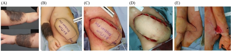
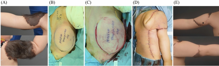
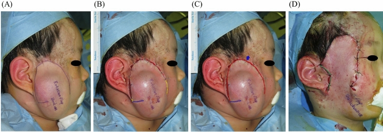
Despite the aid of tissue expansion, the ideal design of random pattern flap is not always available in patients with extensive skin lesions. We investigated the effectiveness of surgical delay on expanded flaps in pediatric patients. Retrospective cohort study was performed on patients who underwent tissue expansion surgery for extensive skin lesions at Seoul National University Children's Hospital. The surgical delay technique was employed for patients with unfavorable flap conditions related to location or transposition angles. The dimensions of skin lesions and flaps were measured based on medical photographs. Fifty patients underwent a total of 66 tissue expansion procedures (49 conventional procedures among 41 patients, 17 surgical delay procedures among 15 patients) from January 2016 to September 2019. Although flaps in the surgical delay group were more narrow-based (p < 0.001), the partial flap loss rate and excised area-to-inflation amount ratio was comparable between the two groups (p = 0.093 and p = 0.194, respectively). Viable flaps, excluding postoperative necrosis, in the surgical delay group were significantly more narrow-based in terms of the length-to-base width ratio and the area-to-base width ratio compared to conventional group (p < 0.01, p < 0.01). Surgical delay can result in outcomes comparable to well-designed random flaps, even in disadvantageous conditions. Patients with large skin lesions but limited areas for expansion may benefit from surgical delay.


Conflict of interest statement

The authors declare no competing interests.

Figures

Figure 1
Figure 1
A 23-months-old female patient underwent tissue expansion on her abdomen for the reconstruction of her Rt. Forearm nevus lesion (A). 8 × 17 cm narrow based distant flap was designed (B). Surgical delay procedures (1st and 2nd) were performed to enhance flap circulation (C, D). The flap was successfully transferred without necrosis and detached from the abdomen (E). (Yellow dotted line is marked along planned flap border; * is marked at distal tip of the flap).
Figure 2
Figure 2
A 17-month-old male patient underwent tissue expansion on his back for the reconstruction of his Rt. Upper extremity (A). An 11 × 15 cm distant flap with a more than 90-degree angle of transposition, carrying a high risk of pedicle kinking, was designed (B). A surgical delay procedure was conducted to improve flap circulation (C). The flap was transferred to his Rt. upper extremity (D), resulting in successful reconstruction without flap necrosis (E). (Yellow dotted line is marked along planned flap border; * is marked at distal tip of the flap).
Figure 3
Figure 3
A 52-month-old male patient underwent tissue expansion on his cheek for facial reconstruction. A flap with an anticipated back-cut was designed (A). A surgical delay procedure was performed to enhance flap circulation, but the back-cut was not incised (B). During the final surgery, the angle and length of the back-cut were determined to transfer the flap most effectively (C, D). (Yellow dotted line is marked along planned flap border; Blu dotted line is marked along the anticipated back-cut; Green dotted line is marked along the actual back-cut made).

References

    1. Adler N, Dorafshar AH, Bauer BS, Hoadley S, Tournell M. Tissue expander infections in pediatric patients: Management and outcomes. Plast. Reconstr. Surg. 2009;124:484–489. doi: 10.1097/PRS.0b013e3181adcf20. - DOI - PubMed
    1. Arneja JS, Gosain AK. Giant congenital melanocytic nevi. Plast. Reconstr. Surg. 2007;120:26e–40e. doi: 10.1097/01.prs.0000267583.63342.0a. - DOI - PubMed
    1. Cherry GW, Austad E, Pasyk K, McClatchey K, Rohrich RJ. Increased survival and vascularity of random-pattern skin flaps elevated in controlled, expanded skin. Plast. Reconstr. Surg. 1983;72:680–687. doi: 10.1097/00006534-198311000-00018. - DOI - PubMed
    1. Saxby PJ. Survival of island flaps after tissue expansion: A pig model. Plast. Reconstr. Surg. 1988;81:30–34. doi: 10.1097/00006534-198801000-00005. - DOI - PubMed
    1. Hudson DA. Maximising the use of tissue expanded flaps. Br. J. Plast. Surg. 2003;56:784–790. doi: 10.1016/S0007-1226(03)00379-5. - DOI - PubMed

Publication types