. 2023 Nov 6;6(1):1124.
doi: 10.1038/s42003-023-05494-7.

The DNA-binding induced (de)AMPylation activity of a Coxiella burnetii Fic enzyme targets Histone H3

Affiliations

The DNA-binding induced (de)AMPylation activity of a Coxiella burnetii Fic enzyme targets Histone H3

Dorothea Höpfner et al. Commun Biol. .

Abstract

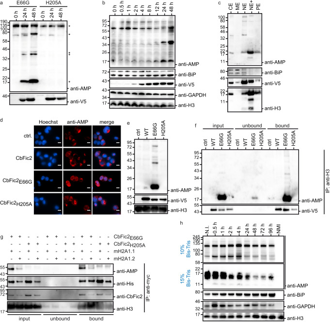
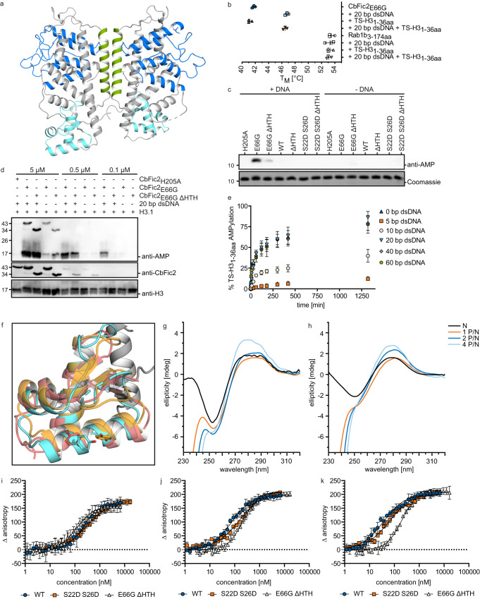
The intracellular bacterial pathogen Coxiella burnetii evades the host response by secreting effector proteins that aid in establishing a replication-friendly niche. Bacterial filamentation induced by cyclic AMP (Fic) enzymes can act as effectors by covalently modifying target proteins with the posttranslational AMPylation by transferring adenosine monophosphate (AMP) from adenosine triphosphate (ATP) to a hydroxyl-containing side chain. Here we identify the gene product of C. burnetii CBU_0822, termed C. burnetii Fic 2 (CbFic2), to AMPylate host cell histone H3 at serine 10 and serine 28. We show that CbFic2 acts as a bifunctional enzyme, both capable of AMPylation as well as deAMPylation, and is regulated by the binding of DNA via a C-terminal helix-turn-helix domain. We propose that CbFic2 performs AMPylation in its monomeric state, switching to a deAMPylating dimer upon DNA binding. This study unveils reversible histone modification by a specific enzyme of a pathogenic bacterium.


Conflict of interest statement

The authors declare no competing interests.

Figures

Fig. 1
Fig. 1. CbFic2 AMPylates Histone H3 in cellulo and in vitro.
a Domain structure prediction and DNA and protein binding prediction of CbFic2 (CBU_0822). CbFic2 is a class II Fic protein consisting of 378 aa. According to SMART analysis, it has a conserved Fic domain (115–223 aa, blue) with the Fic motif HPFDDGNGRIGR (205–216 aa). The inhibitory helix with the sequence TSAIEG (62–67 aa) is located N-terminal to the Fic domain within the DUF4172 domain (4–85 aa, green) of unknown function. The C-terminus contains a helix-turn-helix (HTH) domain (304–362 aa, light blue),. Protein- and DNA-binding regions are predicted with PredictProtein,. Protein binding (RI: 00-33) blue. DNA binding (RI: 00-33) blue, (RI: 34–66) magenta, (RI: 67–100) yellow. RI = reliability index, reliability of positive prediction. The scale of positive prediction ranges from 0 to 100. The higher the score, the more reliable the prediction. b Fluorescent microscopy analysis of protein localization after transient heterologous expression of GFP-CbFic2 full length (CbFic2) or without HTH domain (CbFic2ΔHTH) or the HTH domain alone (CbFic2HTH only) in Cos7 cells. GFP-fusion protein (green) was expressed for 24 h and cell nuclei were stained with Hoechst-33342 (blue). Images were taken by a Leica DMi8 wide field microscope using 100x magnification. Merge of images with GFP and DAPI filter, respectively, reveal co-localization of GFP-CbFic2 to the nucleus. Scale bars: 10 µm. See Supplementary Fig. S1a. c WB analysis of AMPylation patterns of whole cell lysates after transient heterologous expression of CbFic2-GFP or its mutants CbFic2E66G and CbFic2H205A in HEK293 cells. ctrl represents the expression of GFP alone. Fusion protein was expressed for 48 h in HEK293 cells. 20 μg of cleared RIPA lysate per lane were run on Bis-Tris gels and blotted on PVDF. Blots were probed with an anti-AMP antibody, stripped, cut into strips, and treated with antibodies against GFP and histone H3 as expression and loading controls, respectively. See Supplementary Fig. S1b. d WB analysis of AMPylation patterns in acid-soluble nuclear fraction, containing histones, after transient heterologous expression of CbFic2-GFP or its mutants CbFic2E66G and CbFic2H205A in HEK293 cells. ctrl represents the expression of GFP alone. Fusion protein was expressed for 48 h in HEK293 cells. Acid-soluble nuclear proteins were isolated using acid extraction. 10 μg of acid-soluble nuclear fraction per lane were run on Bis-Tris gels and blotted on PVDF. Blots were probed with an anti-AMP antibody, stripped, cut into strips, and treated with antibodies against GFP and histone H3 as expression and loading controls, respectively. See Supplementary Fig. S1c. e WB analysis of AMPylation of recombinant histones by CbFic2E66G in vitro. 0.1 mg ml1 histones were incubated with 0.2 µM CbFic2E66G or CbFic2H205A in the presence of ATP, MgCl2 and DNA at 23 °C for 20 h. 50 ng histones were run on Laemmli gels, blotted on PVDF and probed with an anti-AMP antibody. For loading controls, 1 µg of histones were run on Laemmli gels and stained with Coomassie. f WB analysis of AMPylation after immunoprecipitation against histone H3 on HEK293 lysates after transient heterologous expression of GFP-CbFic2 or its mutants CbFic2E66G and CbFic2H205A. 50 μg of lysate after transient heterologous expression of GFP-CbFic2 were treated in 200 µl with 1 μg anti-H3 antibody and protein A/G magnetic beads. Bound proteins were eluted with 50 μl 1x Laemmli. 10 μl each of the input and unbound sample including 6x Laemmli buffer and 10 μl of the elution (bound) were run on Laemmli gels, blotted on PVDF and probed with an anti-AMP antibody, before being stripped and treated with an antibody against GFP. g WB analysis of AMPylation of the Twinstrep-tagged N-terminal 20 aa of Histone H3 (TS-H31-20aa) and its mutants T3A, T6A, S10A and T11A by CbFic2E66G or CbFic2H205A in vitro. 1 mg ml−1 TS-H31-20aa were incubated with 1 µM CbFic2E66G in the presence of ATP, MgCl2 and DNA at 30 °C for 20 h. 100 ng peptide were run on Tris-Tricine gels, blotted on PVDF and probed with an anti-AMP antibody. For loading controls, 1 µg of peptide was run on Tris-Tricine gels and stained with Coomassie. h WB analysis of AMPylation of the Twinstrep-tagged N-terminal 36 aa of Histone H3 (TS-H31-36aa) and its mutants S10A, S28A and S10A S28A by CbFic2E66G or CbFic2H205A in vitro. 1 mg ml−1 TS-H31-36aa were incubated with 5 µM CbFic2E66G in the presence of ATP, MgCl2 and DNA at 30 °C for 20 h. 100 ng peptide were run on Tris-Tricine gels, blotted on PVDF and probed with an anti-AMP antibody. For loading controls, 1 µg of peptide was run on Tris-Tricine gels and stained with Coomassie. i Representation of modification sites at S10 and S28 (red) within the ARKS motif (red frame) in N-terminal tail of Histone H3.1 by CbFic2 as determined by mutational approaches with WB analysis and MS/MS analysis (Fig. 1g, h; Supplementary Fig. S2a). j WB analysis of AMPylation after anti-myc immunoprecipitation against myc- and his-tagged histone H3.1 and its mutants S10A, S28A or S10A S28A, transiently co-expressed in HEK293 cells with either GFP-CbFic2E66G or GFP-CbFic2H205A. 50 μg of acid-soluble nuclear proteins 48 h post-transfection were treated in 100 µl with 2 μg anti-myc antibody and protein A/G magnetic beads. Bound proteins were eluted with 50 μl 1x Laemmli buffer. 10 μl were run on Bis-Tris gels, blotted on PVDF and probed with an anti-AMP antibody. The blot was stripped, cut into strips, and reprobed with antibodies against CbFic2, and His as expression control of histone H3.1.
Fig. 2
Fig. 2. CbFic2 AMPylates Core Histone Macro-H2A.1.
a WB analysis of AMPylation pattern in THP-1 MDMs before and after induction of V5-CbFic2E66G and V5-CbFic2H205A expression for 24 h and 48 h. The respective stable THP-1 cell lines V5-CbFic2E66G and V5-CbFic2H205A were differentiated into macrophages with PMA for 48 h before inducing the expression of CbFic2 with doxycycline. 20 μg RIPA lysate per lane were run on Bis-Tris gels and blotted on PVDF. The blot was probed with an anti-AMP antibody, stripped, and treated with an antibody against V5 tag. Clearly altered AMPylation bands are marked with an asterisk. b WB analysis of time-resolved AMPylation in THP-1 cells up until 48 h after induction of V5-CbFic2E66G expression by doxycycline. The stable THP-1 cell line V5-CbFic2E66G was induced by doxycycline and samples taken at the indicated time points. 20 μg RIPA lysate per lane was run on Bis-Tris gels and blotted on PVDF. The blot was probed with an anti-AMP antibody, stripped, and treated with antibodies against BiP, GAPDH, and Histone H3 as loading controls, and V5 as expression control of CbFic2, respectively. c WB analysis of AMPylation pattern in fractionated THP-1 MDMs after induction of V5-CbFic2E66G expression for 48 h. The stable THP-1 cell line V5-CbFic2E66G was differentiated into macrophages with PMA for 48 h before inducing the expression of CbFic2 with doxycycline. Cells were fractionated into cytoplasmic (CE), membrane (ME), nuclear soluble (NE), chromatin-bound (NE+) and cytoskeletal protein (PE) extracts using a subcellular protein fractionation kit for cultured cells. 5 μg per fraction were run on Bis-Tris gels and blotted on PVDF. The blot was probed with an anti-AMP antibody, stripped, cut into strips and treated with antibodies against BiP and Histone H3 as loading and fractionation controls, and V5 as expression and fractionation control of CbFic2, respectively. d Immunofluorescence analysis of AMPylation after 48 h of CbFic2 expression in macrophages using anti-AMP antibody. The respective stable THP-1 cell lines CbFic2, CbFic2E66G, CbFic2H205A, and the control cell line (ctrl.) were differentiated into macrophages for 48 h with PMA before inducing the expression of CbFic2 for 48 h using doxycycline. Cells were fixed and permeabilized. Cell nuclei were stained with Hoechst-33342 (blue), and AMPylation was visualized with antibody 17G6 (red). Scale bars: 10 µm. e WB analysis of AMPylation patterns in acid-soluble nuclear fraction, containing histones, after stable expression of V5-CbFic2 or its mutants V5-CbFic2E66G and V5-CbFic2H205A in THP-1 MDMs. ctrl represents the expression of the empty backbone alone. Tagged protein was expressed for 48 h in differentiated THP-1 cells. Acid-soluble nuclear proteins were isolated using acid extraction. 10 μg of acid-soluble nuclear fraction per lane were run on Bis-Tris gels and blotted on PVDF. Blots were probed with an anti-AMP antibody, stripped, cut into strips, and treated with antibodies against V5 and histone H3 as expression and loading controls, respectively. f WB analysis of AMPylation after immunoprecipitation against histone H3 from THP-1 MDMs acid-soluble nuclear fraction after stable expression of V5-CbFic2 or its mutants V5-CbFic2E66G and V5-CbFic2H205A. 50 μg of acid-soluble nuclear fraction after 48 h of stable expression of V5-CbFic2 were treated in 200 µl with 1 μg anti-H3 antibody and protein A/G magnetic beads. Bound proteins were eluted with 50 μl 1x Laemmli. 10 μl each of the input and unbound sample including 6x Laemmli buffer and 10 μl of the elution (bound) were run on Laemmli gels, blotted on PVDF and probed with an anti-AMP antibody, before being stripped and treated with an antibody against V5. g WB analysis of AMPylation after anti-myc immunoprecipitation against myc- and his-tagged Core Histone Macro-H2A.1 isoforms mH2A1.1 or mH2A1.2, both transiently co-expressed in HEK293 cells with either GFP-CbFic2E66G or CbFic2H205A. 50 μg of acid-soluble nuclear proteins 48 h post-transfection were treated in 100 µl with 2 μg anti-myc antibody and protein A/G magnetic beads. Bound proteins were eluted with 50 μl 1x Laemmli buffer. 10 μl each of the input and unbound sample including 6x Laemmli buffer and 10 μl of the elution (bound) were run on Bis-Tris gels, blotted on PVDF and probed with an anti-AMP antibody. The blot was stripped, cut into strips, and reprobed with antibodies against Histone H3 as loading control, V5 as expression control of CbFic2, and His as expression control of mH2A1, respectively. h WB analysis of AMPylation patterns over the time course of infection of murine J774 macrophages by virulent NMI C. burnetii. J774 cells were infected with C. burnetii, and at indicated time points lysed by RIPA. 20 μg of lysate per lane was run on Bis-Tris gels (gel percentages indicated to the left; for a full presentation of blots see Supplementary Fig. S2b) and blotted on PVDF. Blots were probed with an anti-AMP antibody, stripped, cut into strips, and treated with antibodies against BiP, GAPDH, and histone H3 as loading controls. N.I.: not infected, NMI: NMI cells alone.
Fig. 3
Fig. 3. CbFic2 activity is stimulated by DNA binding.
a Crystal structure of CbFic2 in the apo state. Dimer interface (17–41 aa) in green, Fic domain (96–223 aa) in blue, wHTH domain (300–367 aa) in light blue. For protein:protein contacts of the dimer interface, see Supplementary Fig. S3a. b Analysis of the thermostability of CbFic2 in the presence or absence of DNA by TSA. 4 µg (4 µM) TS-CbFic2E66G or 2 µg Rab1b3-174 aa (ctrl) in 20 mM HEPES pH 7.0, 50 mM NaCl, 1 mM MgCl2, 2 mM DTT supplemented with 5x SYPRO® Orange were measured in the presence or absence of 4 µM 20 bp dsDNA or 4 µM TS-H31 36aa as indicated. Samples were heated from 25–95 °C at a rate of 1 °C min−1 and fluorescence (ex. 465 nm, em. 590 nm) measured in a RT-PCR cycler. The melting temperature TM, as the inflection point of fluorescence increase during thermal protein unfolding, was determined at the zero point of the second derivative of each melting curve. Scatter plots represent technical triplicates as mean value with standard deviation as error bars using GraphPad Prism 8.0. c WB analysis of TS-H31-36aa AMPylation by CbFic2H205A, CbFic2E66G, CbFic2E66G ΔHTH, CbFic2, CbFic2ΔHTH, CbFic2S22D S26D and CbFic2S22D S26D ΔHTH in the absence or presence of DNA in vitro. 50 µM TS-H31-36aa were incubated with 1 µM of the indicated CbFic2 versions with or without 4 µM 20 bp dsDNA in the presence of 1 mM ATP and 1 mM MgCl2 at 37 °C for 8 h. 100 ng peptide were run on Tris-Tricine gels, blotted on PVDF and probed with an anti-AMP antibody. For loading controls, 1 µg of peptide was run on Tris-Tricine gels and stained with Coomassie. d WB analysis of full-length H3.1 AMPylation by CbFic2H205A, CbFic2E66G and CbFic2E66G ΔHTH in the absence or presence of DNA in vitro. 1 mg ml−1 H3.1 was incubated with 5 µM CbFic2H205A or 5 µM, 0.5 µM or 0.1 µM CbFic2 CbFic2E66G and CbFic2E66G ΔHTH with or without 5 µM 20 bp dsDNA in the presence of 1 mM ATP and 1 mM MgCl2 at 23 °C oN. 50 ng H3.1 was run on 15% glycine gels, blotted on PVDF and probed with an anti-AMP antibody. For loading controls, the WB was stripped and reprobed with anti-CbFic2 and ani-H3 antibodies. e Intact MS analysis and quantification of time-resolved TS-H31-36aa AMPylation by CbFic2E66G in the absence or presence of 5 bp, 10 bp, 20 bp, 40 bp, 60 bp dsDNA. 50 µM TS-H31-36aa was incubated with 5 µM of CbFic2E66G in the presence of 5 µM DNA as indicated, 2 mM ATP, 4 mM MgCl2 at 37 °C for 22 h. AMPylation was measured by the mass increase of 329 Da, and AMPylated peaks were quantified by intensity after deconvolution. AMPylation was defined as a decrease in unAMPylated peptide over time, to reduce the complexity of multiple AMPylation. Each data point represents the mean of biological triplicates; error bars correspond to standard deviation. See Supplementary Fig. S3d and Supplementary Table 3. f Structural superposition of crystal structures of Z-DNA- (orange) and B-DNA-binding (red) HTH domains with that of the HTH domain of CbFic2 (cyan). B-DNA binding: human transcription factor E2F4 (PDB: 1cf7, chain C, red); Z-DNA binding: domain hZαADAR1 of human double-stranded RNA-specific adenosine deaminase ADAR1 (PDB: 1qbj, chain D, orange). The conserved tyrosine of Z-DNA-binding domains is highlighted as stick. g, h CD measurement of 20 bp dsDNA with g 100% GC or h 40% GC content in the absence or presence of increasing amounts of CbFic2. 1 μM of dsDNA was mixed with CbFic2 to final concentrations of 1 μM ([P]/[N] = 1), 2 μM ([P]/[N] = 2) and 4 μM ([P]/[N] = 4). [P] and [N] stand for protein concentration and DNA concentration, respectively. Before each measurement, samples were incubated for 1 h at 25 °C. CD spectra between 230 and 320 nm were collected using a 0.75 cm quartz cell. See Supplementary Fig. S3e, f. ik Analysis of the binding affinity of CbFic2, CbFic2E66G ΔHTH and CbFic2S22D S26D against i 20 bp, j 40 bp and k 60 bp dsDNA by fluorescence anisotropy. 1 nM 5‘-FITC-labeled DNA was mixed with a dilution series from 20 μM CbFic2 using a pipetting robot in a 384w format. Values were baseline corrected by anisotropy values of free DNA. Fit corresponds to “Specific binding with Hill slope” (3) (GraphPad Prism 8.0). Data shown correspond to the mean of technical triplicates, error bars to the standard deviation. See Supplementary Fig. S3g, h and Supplementary Table 2.
Fig. 4
Fig. 4. CbFic2 is regulated by a monomer/dimer equilibrium.
a WB analysis of TS-H31-36aa-AMP deAMPylation by CbFic2H205A, CbFic2E66G, CbFic2E66G ΔHTH, CbFic2, CbFic2ΔHTH, CbFic2S22D S26D and CbFic2S22D S26D ΔHTH in the absence or presence of DNA in vitro. 50 µM TS-H31-36aa-AMP were incubated with 1 µM of the indicated CbFic2 versions with or without 4 µM 20 bp dsDNA in the presence of 1 mM MgCl2 at 37 °C for 8 h. 100 ng peptide was run on Tris-Tricine gels, blotted on PVDF and probed with an anti-AMP antibody. For loading controls, 1 µg of peptide was run on Tris-Tricine gels and stained with Coomassie. b Intact MS analysis and quantification of time-resolved TS-H31-36aa-AMP deAMPylation by CbFic2, CbFic2S22D S26D or CbFic2ΔHTH in the absence or presence of DNA in vitro. 50 µM TS-H31-36aa-AMP were incubated with 0.5 µM CbFic2 as indicated in the presence of 5 µM DNA, 1 mM MgCl2 at 37 °C for 24 h. DeAMPylation was measured by the mass loss of 329 Da, and peaks were quantified by intensity after deconvolution. deAMPylation was defined as an increase in unAMPylated peptide over time. Each data point represents the mean of biological triplicates; error bars correspond to standard deviation. See Supplementary Fig. S4a and Supplementary Table 3. c WB analysis of auto-AMPylation of CbFic2 in cis/trans. 0.3 µM CbFic2 versions as indicated were incubated alone or in the presence of another CbFic2 version, in the presence or absence of 2.5 µM 20 bp dsDNA, in the presence of 1 mM ATP and 1 mM MgCl2 for 8 h at 37 °C. 50 ng protein was run on Laemmli gels, blotted on PVDF and probed with an anti-AMP antibody. For loading controls, blots were stripped and incubated with an anti-CbFic2 antibody. See Supplementary Fig. S4b. d Intact MS analysis of auto-AMPylation of CbFic2 and CbFic2E66G over the time course of incubation with ATP in the presence or absence of DNA. 0.2 mg ml−1 (4 µM) CbFic2 or CbFic2E66G were incubated in 20 mM HEPES pH 7.5, 150 mM NaCl, 1 mM MgCl2, 1 mM TCEP, 1 mM ATP both in the presence and absence of 5 μM 20 bp dsDNA at 37 °C and analyzed by MS. The degree of automodification was detected by the specific mass gain of AMPylation of 329 Da. AMPylation was quantified by the ratio of the specific signal intensity to the total intensity of all CbFic2 signals. As CbFic2 shows multiple auto-AMPylations (see supplement for detailed depiction), data represent the decrease of unAMPylated CbFic2. Each data point represents the mean of biological triplicates; error bars correspond to standard deviation. See Supplementary Fig. S4c. e Fluorescence anisotropy analysis of the influence of CbFic2 auto-AMPylation and the presence of ATP on DNA binding. 1 nM 5‘-FITC-labeled 20 bp dsDNA was mixed with a dilution series from 20 μM auto-AMPylated CbFic2E66G-AMP or CbFic2E66G in the presence of 1 mM ATP using a pipetting robot in a 384w format. Values were baseline corrected by anisotropy values of free DNA. Fit corresponds to “Specific binding with Hill slope” (3) (GraphPad Prism 8.0). Data shown correspond to the mean of technical triplicates, error bars to the standard deviation. See Supplementary Table 2. f WB analysis of concentration-dependent auto-AMPylation of CbFic2. From a starting concentration of 50 µM CbFic2 versions as indicated, protein was diluted to 15 µM, 5 µM, 1.5 µM, 0.5 µM and 0.15 µM, and incubated in the presence or absence of 50 µM, 15 µM or 5 µM or 4 µM (for protein concentrations of or below 1.5 µM) 20 bp dsDNA, respectively, in the presence of 1 mM ATP and 1 mM MgCl2 for 8 h at 37 °C. 50 ng protein was run on Laemmli gels, blotted on PVDF and probed with an anti-AMP antibody. For loading controls, blots were stripped and incubated with an anti-CbFic2 antibody. g WB analysis of Histone H3.1 AMPylation by CbFic2E66G, CbFic2E66G ΔHTH, CbFic2, CbFic2ΔHTH, CbFic2S22D S26D at low enzyme concentrations in the absence or presence of DNA in vitro. 0.1 mg ml−1 Histone H3.1 was incubated with 0.5 µM of the indicated CbFic2 versions with or without 5 µM 20 bp dsDNA in the presence of 1 mM ATP and 1 mM MgCl2 at 37 °C for 8 h. 100 ng Histone H3.1 were run on Tris-Tricine gels, blotted on PVDF and probed with an anti-AMP antibody. For loading controls, blot was stripped, cut into strips and reprobed with anti-Histone H3 and anti-CbFic2 antibodies.
Fig. 5
Fig. 5. CbFic2 shows DNA-induced dimerization.
ac Analysis of concentration-dependent dimerization of a CbFic2, b CbFic2S22D S26D and c CbFic2ΔHTH by analytical size exclusion chromatography. CbFic2 was injected at indicated concentrations onto a Superdex 75 pg 10/300 (Cytiva), run at 0.5 ml min−1 in 20 mM HEPES pH 7.5, 150 mM NaCl, 1 mM MgCl2, 1 mM TCEP, and protein was detected by absorbance at 280 nm. Data intensity was normalized to the internal control of vitamin B12 (tR = 38.5 min). Arrows indicate the gel filtration standard (BioRad) comprising bovine γ-globulin (MW 158 kDa), chicken ovalbumin (MW 44 kDa), horse myoglobin (MW 17 kDa) and vitamin B12 (MW 1.35 kDa). d Superimposition of CbFic2 crystal structure (lighter shades; light green dimerization interface; middle blue Fic domain; cyan HTH domain) with AlphaFold model (darker shades; dark green dimerization interface; dark blue Fic domain; turquoise HTH domain) shows structural flexibility in the HTH domain by a kink in the long connecting helix between HTH domain (turquoise) and Fic domain (blue). e Proposed model for DNA-binding induced dimerization. Superimposition of HTH domain (turquoise) of CbFic2 AlphaFold structure with human transcription factor E2F4 (PDB: 1cf7, chain C, red) bound to B-DNA suggests that bound 20 bp DNA to CbFic2 might span across both monomers and thereby stimulate dimerization. f 90° turn of (e) around the vertical axis. gi Analysis of DNA-induced dimerization by in-solution FP-fusion FRET time course measurements in 20 mM HEPES pH 7.5, 150 mM NaCl, 1 mM MgCl2, 1 mM TCEP. After 3 min, donor CyPet-CbFic2 or its mutants (D) and after another 10 min, acceptor YPet-CbFic2 or its mutants (A) were added at concentrations of 0.5 µM (resulting in total CbFic2 concentrations of 1 µM). After another 10 min incubation, 4 µM of dsDNA was added three times in succession (DNA), with each incubation lasting 10 min. g represents 0.5 µM CyPet- and YPet-CbFic2 followed by 4 µM 10 bp, 20 bp, 40 bp or 60 bp dsDNA, h 0.5 µM CyPet- and YPet-CbFic2S22D S26D followed by 4 µM 10 bp, 20 bp, 40 bp or 60 bp dsDNA and i 0.5 µM CyPet- and YPet-CbFic2 or -CbFic2S22D S26D or -CbFic2ΔHTH or -CbFic2S22D S26D ΔHTH each followed by 4 µM 20 bp dsDNA. Measurements were performed at 25 °C, with an excitation wavelength of 405 nm and an emission wavelength of 530 nm. Intensities were normalized to the value at 760 s corresponding to the endpoint intensity of donor addition. j WB analysis of hetero-dimer formation after co-IP of recombinant HA- and V5- CbFic2 using an anti-HA antibody. 0.2 µM HA- and 0.2 µM V5-tagged CbFic2, CbFic2S22DS26D or CbFic2ΔHTH, respectively, were incubated with 4 µM 20 bp or 40 bp dsDNA in the presence of 2 µg anti-HA antibody in 20 mM HEPES pH 7.0, 150 mM NaCl, 1 mM MgCl2, 20% (v/v) glycine, 0.1% (v/v) Tween 20 and precipitated by protein A/G magnetic beads. For each CbFic2 version, a control assay without HA-tagged protein was prepared in addition, to control for unspecific binding of CbFic2. HA-tagged CbFic2 and its binding partners were eluted by 0.1 M glycine, pH 2.0. Samples were separated by 12% Tris-glycine gels, blotted, blocked and detection of the potential heterodimer was performed via anti-V5 tag antibody. k CbFic2 regulation by DNA-induced dimerization. Suggested model of CbFic2 regulation on the basis of our data.

Similar articles

Cited by

References

    1. Khater S, Mohanty D. In silico identification of AMPylating enzymes and study of their divergent evolution. Sci. Rep. 2015;5:10804. doi: 10.1038/srep10804. - DOI - PMC - PubMed
    1. Garcia-Pino A, Zenkin N, Loris R. The many faces of Fic: structural and functional aspects of Fic enzymes. Trends Biochem. Sci. 2014;39:121–129. doi: 10.1016/j.tibs.2014.01.001. - DOI - PubMed
    1. Kinch LN, Yarbrough ML, Orth K, Grishin NV. Fido, a novel AMPylation domain common to fic, doc, and AvrB. PLoS ONE. 2009;4:1–9. doi: 10.1371/journal.pone.0005818. - DOI - PMC - PubMed
    1. Luong P, et al. Kinetic and structural insights into the mechanism of AMPylation by VopS Fic domain. J. Biol. Chem. 2010;285:20155–20163. doi: 10.1074/jbc.M110.114884. - DOI - PMC - PubMed
    1. Xiao J, Worby CA, Mattoo S, Sankaran B, Dixon JE. Structural basis of Fic-mediated adenylylation. Nat. Struct. Mol. Biol. 2010;17:1004–1010. doi: 10.1038/nsmb.1867. - DOI - PMC - PubMed

Publication types