

Immune checkpoint analysis in ear cancer
- PMID: 37932810
- PMCID: PMC10626725
- DOI: 10.1186/s13005-023-00395-w
Immune checkpoint analysis in ear cancer
Abstract
Background: Among cutaneous squamous cell carcinomas, the ear (ecSCC) is one of the most common sites. Loco regional lymph node metastasis is found in six to eleven percent of cases, corresponding to increased metastasis compared to other sites. The aim of this study was to test the markers PD-L1, PD-1, CD4, CD8, and FoxP3 for suitability as prognostic predictive markers.
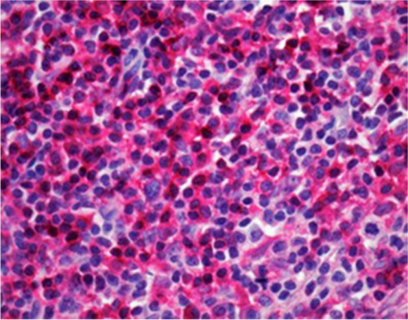
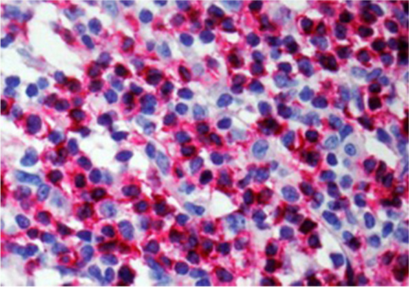
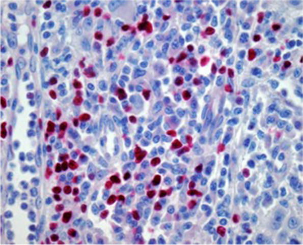
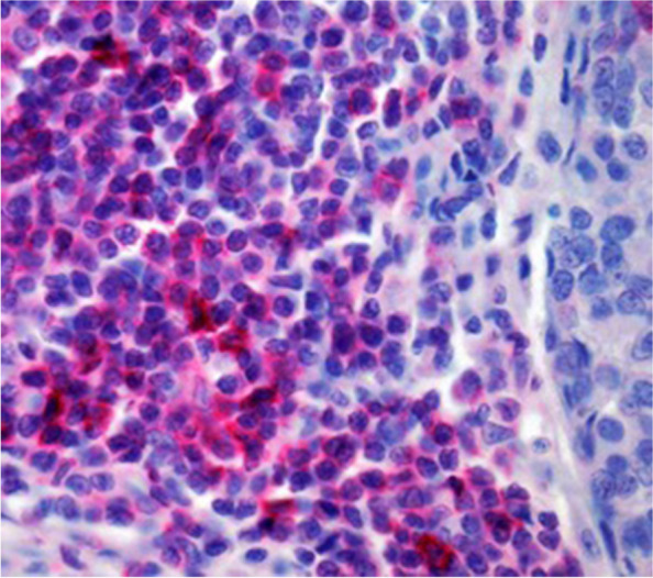
Methods: Sixty-four patients with ecSCC were included in this study. The expression of immunohistochemical markers (PD-L1, PD-1, CD4, CD8, FOXP3) was correlated with retrospective clinic pathological parameters (lymph node metastasis, distant metastasis, lymph node metastasis during follow-up, disease progression, disease-specific death).
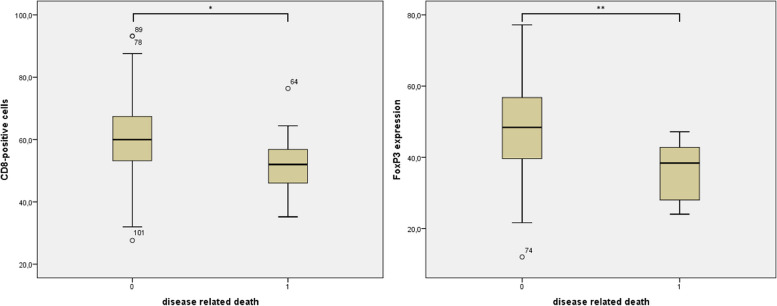
Results: There was a correlation between increased disease specific death and a weak Foxp3 (p = 0.003) or reduced CD8 (p = 0.04). A PD-L1 expression > 1% was found in 39.1% of patients.
Conclusion: The investigated markers (CD4, CD8, FoxP3, PD-1, PD-L1) seem overall rather inappropriate for prognostic evaluation in ecSCC. Only the correlation of disease specific death with CD8 or FoxP3 seems to be worth testing in larger collectives.
© 2023. The Author(s).
Conflict of interest statement
The authors declare no competing interests.
Figures

References
-
- Leiter U, Heppt MV, Steeb T, Amaral T, Bauer A, Becker JC. S3 guideline for actinic keratosis and cutaneous squamous cell carcinoma (cSCC) – short version, part 2: epidemiology, surgical and systemic treatment of cSCC, follow-up, prevention and occupational disease. J Dtsch Dermatol Ges. 2020;18(4):400–13. - PubMed
-
- Salmaninejad A, Khoramshahi V, Azani A, Soltaninejad E, Aslani S, Zamani MR, u. a. PD-1 and cancer: molecular mechanisms and polymorphisms. Immunogenetics. 22. Juni 2017; - PubMed
MeSH terms
Substances
LinkOut - more resources
Full Text Sources
Research Materials
