. 2022:2:55.
doi: 10.1038/s43586-022-00136-4. Epub 2022 Jul 21.

Optogenetics for light control of biological systems

Affiliations

Optogenetics for light control of biological systems

Valentina Emiliani et al. Nat Rev Methods Primers. 2022.

Abstract

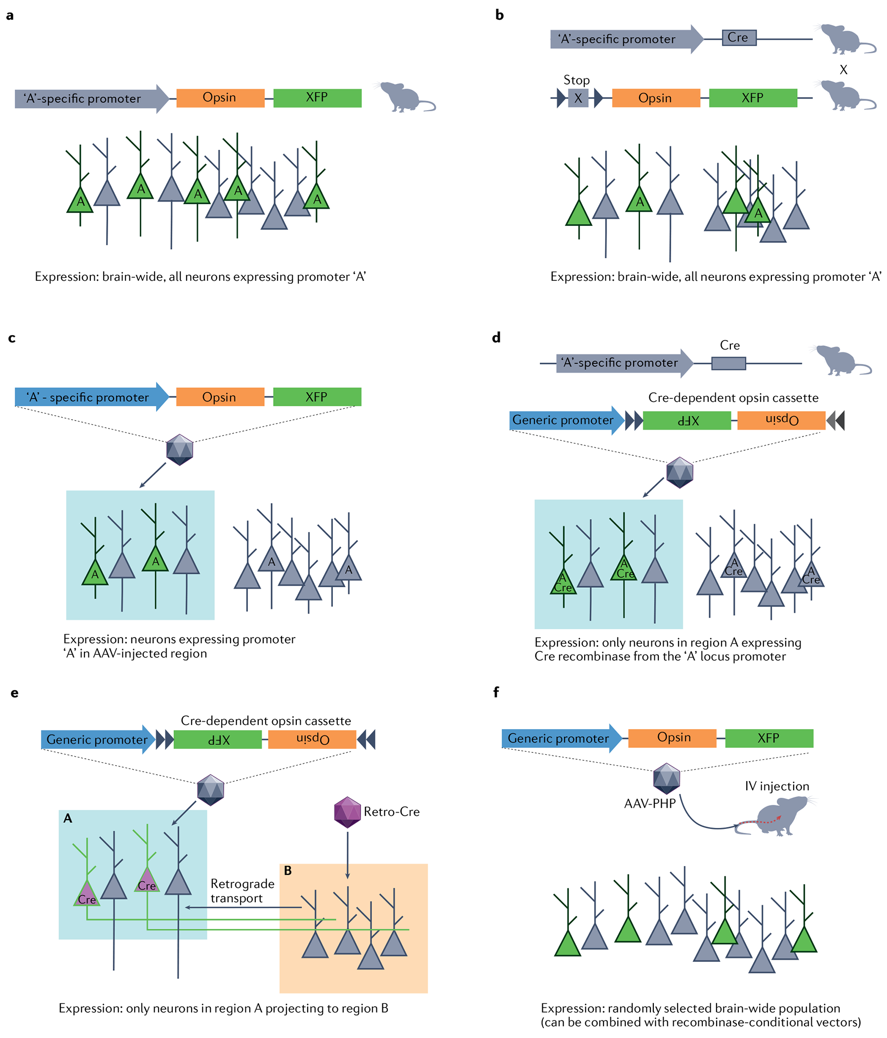
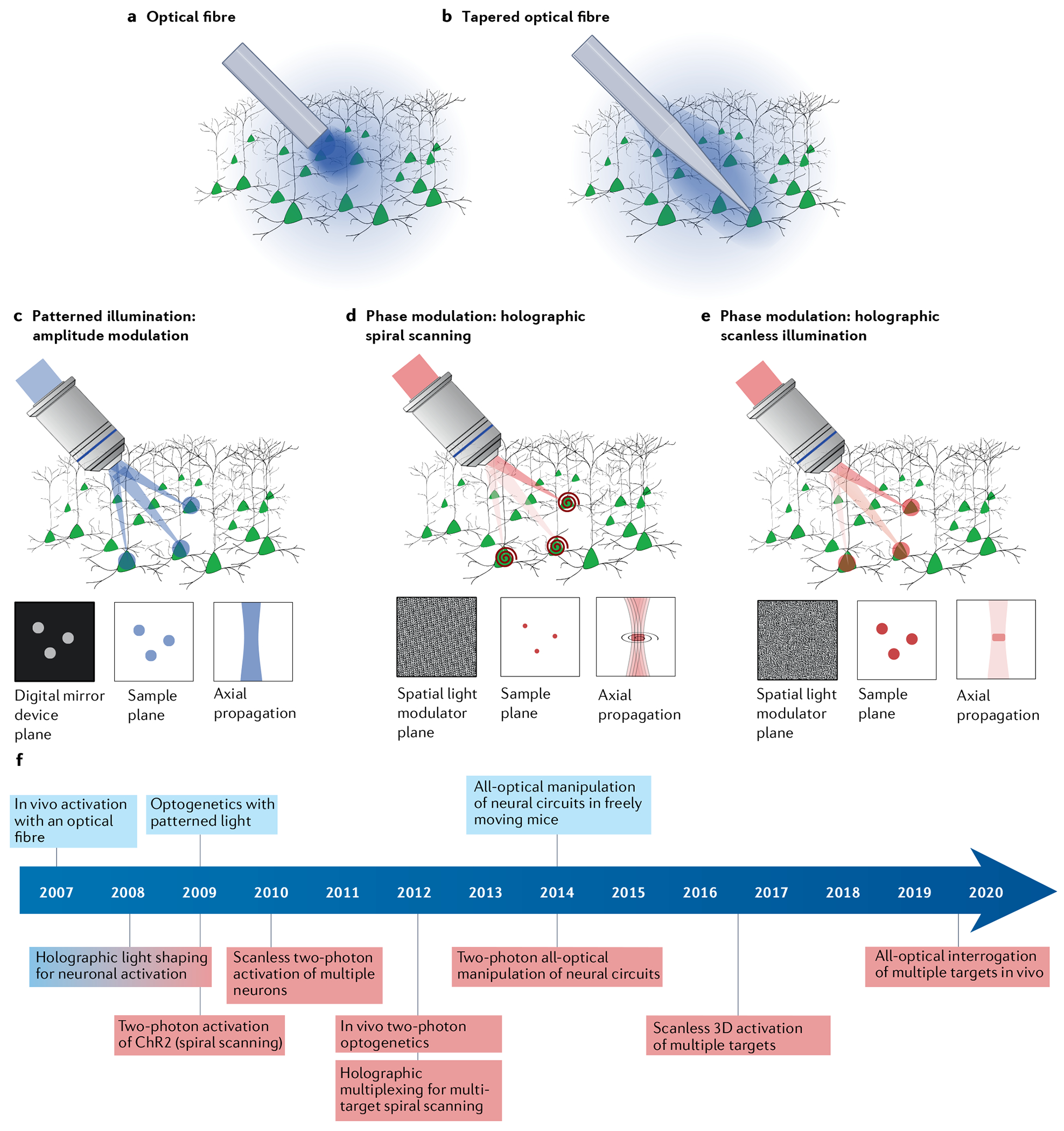
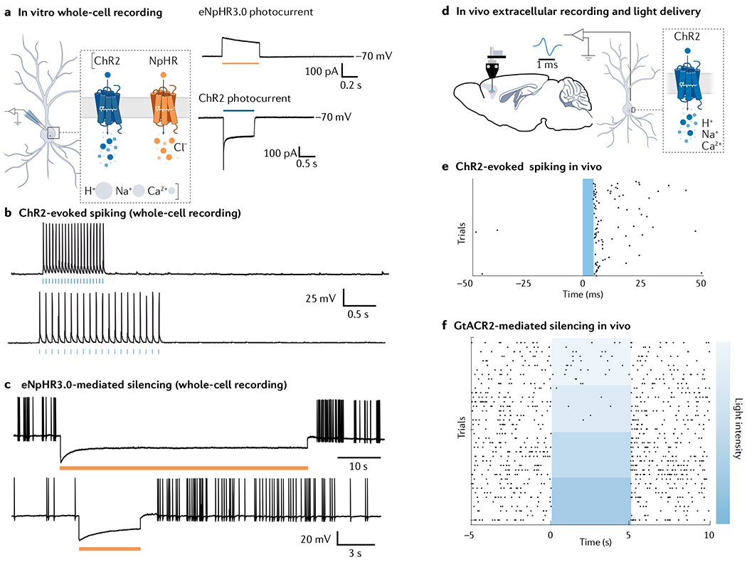
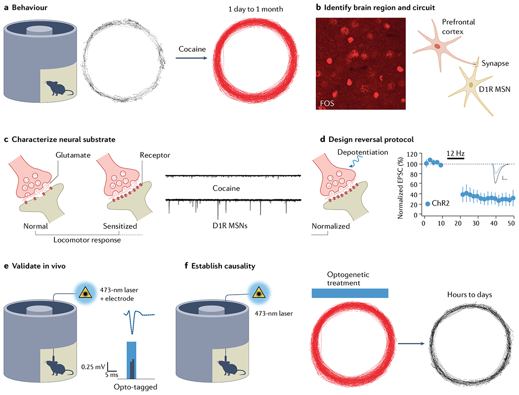
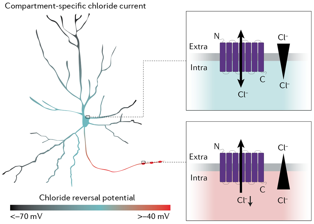
Optogenetic techniques have been developed to allow control over the activity of selected cells within a highly heterogeneous tissue, using a combination of genetic engineering and light. Optogenetics employs natural and engineered photoreceptors, mostly of microbial origin, to be genetically introduced into the cells of interest. As a result, cells that are naturally light-insensitive can be made photosensitive and addressable by illumination and precisely controllable in time and space. The selectivity of expression and subcellular targeting in the host is enabled by applying control elements such as promoters, enhancers and specific targeting sequences to the employed photoreceptor-encoding DNA. This powerful approach allows precise characterization and manipulation of cellular functions and has motivated the development of advanced optical methods for patterned photostimulation. Optogenetics has revolutionized neuroscience during the past 15 years and is primed to have a similar impact in other fields, including cardiology, cell biology and plant sciences. In this Primer, we describe the principles of optogenetics, review the most commonly used optogenetic tools, illumination approaches and scientific applications and discuss the possibilities and limitations associated with optogenetic manipulations across a wide variety of optical techniques, cells, circuits and organisms.


Conflict of interest statement

Competing interests Z.-H.P. is a co-inventor on patents related to optogenetic vision restoration and is also a co-founder and scientific advisor of Ray Therapeutics. The other authors declare no competing interests.

Figures

Fig. 1 |
Fig. 1 |. Principles of optogenetics.
DNA encoding a sensory photoreceptor derived from a microorganism, plant or animal (orange) is cloned under regulation of control elements that allow targeting of specific host cells (blue), packed into a vector such as a viral vector or bacteria and injected into the tissue, organ or organism of interest. Targeted cell (orange) now expresses light-sensitive protein and can be controlled with light in various ways, depending on the specific photoreceptor expressed.
Fig. 2 |
Fig. 2 |. The optogenetic actuator toolbox.
a | Key advances in development of optogenetic tools. Not all available tools are highlighted. Major developments are shown above the arrow, and first applications of channelrhodopsins (ChRs) to model organisms including humans are shown below. b | Tools for optogenetic manipulation of membrane voltage and local ion concentrations (top), second messenger, G-protein signalling and kinase signalling (middle) and the light-controlled interaction of photoreceptors with tethered partner proteins for subcellular application (bottom). Light–oxygen–voltage (LOV) domain-based dimerizers expose an ‘aged’ signalling peptide after light-triggered unfolding of the Jɑ-helix. Cryptochrome 2 (Cry2) and phytochrome B (PhyB) interact with CiBN or PIF domains after blue or red light absorption, respectively,. c | Commonly used optogenetic tools for excitation or inhibition of neuronal activity including cation-conducting ChRs eTsChR, Cheriff, CoChR, CrChR2TC (REF.), ChroME and derivatives, SSFO/Soul,, ChRmine, bReaChES and f-Chrimson, chloride and potassium-conducting ChRs (for example, GtACR1, GtACR2 (REF.) and HcKCR1 (REF.)), inward directed proton pumps (for example, NsXeR) and outward-directed proton, sodium and chloride pumps (for example, Arch3.0 (REF.), eKR2 (REF.), eNpHR3.0 (REF.)), all plotted according to their peak excitation wavelength and temporal kinetics. d | Soluble enzyme bPAC and rhodopsin–guanyl cyclase CaRhGC produce cAMP and cGMP following illumination, whereas non-bleaching opsins mOPN4 (REF.), eOPN3 (REF.), PPO and JellyOP activate different G-protein pathways. e | Genetically encoded sensors with diverse excitation spectra (x axis) can be used to monitor changes in Ca2+ voltage and pH, such as GCaMP and R-CaMP and FRGeco for Ca2+ (REF.), ASAP3 (REF.), Voltron, VARNAM, Quasar and Archon for voltage, and pHluorin and pHmScarlet for pH. In experiments combining sensors and actuators, both need to be chosen carefully to minimize optical crosstalk. ec, extracellular; GPCR, G-protein-coupled receptor; ic, intracellular; optoGPCR, hybrid between structurally related opsin and GPCR; RTK, receptor tyrosine kinase.
Fig. 3 |
Fig. 3 |. Cell type-specific targeting of optogenetic tools.
a | Transgenic mice constitutively expressing an opsin gene from their genome allow simple experiments that only require addition of light delivery apparatus. Promoter ‘A’ activity (indicated by A) will lead to transgene expression (green). b | Transgenic animal expressing a recombinase such as Cre under control of a cell type-specific promoter is crossed with a second line carrying a conditional expression cassette encoding the desired opsin. Dual transgenic offspring will then show organism-wide expression of the opsin in all cells that underwent promoter activation at any stage of development (green). Cre expression (indicated by A) is unnecessary once the conditional expression cassette was activated. c | Where a short minimal promoter sequence is available, targeted viral vector injections can be used to restrict expression spatially as well as by the gene expression profile. A viral vector containing the specific minimal promoter sequence upstream of the opsin gene will lead to expression in specific cells expressing the promoter (indicated by A), only in the region targeted with the injection (blue box). d | Approaches in parts a and b can be combined to achieve both spatial and gene expression specificity in cases where short specific promoters are not available, or where promoter activity is not specific during development. e | Projection neurons can be addressed by injection of an axon terminal-transducing, retrograde travelling viral vector encoding for the opsin or a recombinase into the target region. Recombinase-encoding viral vector is injected in a projection target (area B, red box) and travels retrogradely. A second viral injection of conditional expression cassette encoding the desired opsin into an upstream region (area A, blue box) will then lead to opsin expression only in neurons within area A that project to area B. f | Adeno-associated virus (AAV) capsids engineered for improved blood–brain barrier penetration allow brain-wide (mostly sparse) expression of an opsin through intravenous injection of the viral vector. IV, intravenous.
Fig. 4 |
Fig. 4 |. Optical approaches for optogenetic stimulation.
a–c | Single-photon wide-field illumination (blue) of all genetically targeted opsin-expressing neurons using excitation through optical fibres: illumination using a flat-cleaved optical fibre causes high peak light power density at the fibre–tissue interface (part a); a tapered fibre increases the optical fibre–tissue interface resulting in a reduced peak light power density (part b); and single-photon multi-target patterned illumination by spatially shaping the intensity of the excitation beam by means of a digital micromirror device, placed in a plane conjugated to the sample plane (part c). Light distribution at the digital mirror device plane and at the sample plane only differ by a spatial scaling factor corresponding to the magnification of the optical system. Axial resolution is proportional to the square of the lateral spot dimensions. d,e | Two-photon multi-target illumination by holographic light shaping: a spatial light modulator placed at a plane conjugated with the objective back aperture generates a 3D distribution of holographic spots, which are scanned with a spiral trajectory to cover the cell surface — axial extension of the generated spot is optimized to illuminate upper and lower cell membranes (part d); and a spatial light modulator is used to generate multiple extended spots with a size large enough to cover the whole cell soma — temporal focusing is used to maintain micrometre axial resolution independently of lateral spot size (part e). f | Timeline indicating critical optical developments that have enabled new optogenetic experiments throughout the past 15 years. Single-photon and two-photon milestones coloured blue and red, respectively. Holographic light shaping for neuronal activation was developed simultaneously for single-photon and two-photon activation, indicated by red–blue gradient for the milestone in part f. ChR2, channelrhodopsin 2.
Fig. 5 |
Fig. 5 |. Expected results in optogenetic experiments.
a | Expression of optogenetic actuators such as channelrhodopsin 2 (ChR2) or NpHR in neurons leads to emergence of light-driven photocurrents, which can be recorded using the whole-cell patch clamp technique (left). Cells expressing chloride-conducting NpHR will show an outward current (top right, voltage clamp recording with cell resting at −70 mV) whereas cells expressing cation-conducting ChR2 will show an inward photocurrent (bottom right, voltage clamp recording with cell resting at −70 mV). b | Whole-cell current-clamp recordings in a neuron expressing excitatory ChR2, showing action potentials evoked by brief light pulses (blue bars). c | Hyperpolarization and silencing of spontaneously occurring action potentials in a neuron expressing eNpHR3.0. d | Extracellular recordings, coupled with local light delivery, used to reveal activity of neurons in vivo, using the awake behaving optrode configuration. e | Raster plot showing action potentials (black dots) occurring rapidly after a 5-ms blue light pulse delivered into the target brain region. f | Raster plot showing activity of neurons expressing inhibitory anion-conducting GtACR2, showing increased inhibition of action potential firing with increasing light intensity. Part f is reprinted from REF., CC BY 4.0 (https://creativecommons.org/licenses/by/4.0/).
Fig. 6 |
Fig. 6 |. Establishing links of causality with optogenetics.
Experimental road map based on identifying the neural correlate of behavioural sensitization to cocaine. a | When injected, cocaine elicits a locomotor response quantified in a cyclotron. Response is enhanced upon second injection of same dose. b | Fos is an immediate early gene highlighting the neurons particularly active, which provided the entry point to identifying the medial prefrontal cortex to nucleus accumbens projection as the behavioural relevant circuit. c | Slice electrophysiology enables observation of selective potentiation of glutamate transmission onto dopamine type 1 receptor medium spiny neurons (D1R MSNs). d | Depotentiation protocol (long-term depression (LTD) at 12 Hz) validated in slices restores standard transmission. e | In vivo validation involves opto-tagging, where spontaneously occurring spikes (grey, dashed trace) are compared with optogenetically evoked spikes (blue trace). Waveform and latency are important parameters. f | LTD protocol is eventually applied in vivo to reverse sensitization. ChR2, channelrhodopsin 2; EPSC, excitatory postsynaptic current. Part d adapted with permission from REF., AAAS.
Fig. 7 |
Fig. 7 |. Optogenetic application for vision restoration, cardiac research and plant modification.
A | Strategies for optogenetic restoration of vision following photoreceptor degeneration. Visual processing pathways in normal retina, illustrating the rod/cone, ON/OFF pathways and antagonistic centre–surround receptive fields of retinal ganglion cells (ON cells, including rod bipolar cells and AII amacrine cells (AII), shown in grey tones; OFF cells shown in black; ON and OFF regions receptive field of retinal ganglion cells indicated + and −, respectively) (part Aa). Ubiquitous expression of a depolarizing optogenetic tool (green) in all retinal ganglion cells to convert them into ON cells (part Ab). Targeting a depolarizing optogenetic tool in ON bipolar cells to produce ON and OFF response in retinal ganglion cells and, possibly, the centre–surround receptive fields (part Ac). B | Optogenetics in cardiac research. Cell-specific targeting used for sympathetic (red) and parasympathetic (blue) nervous control of the heart using tyrosine hydroxylase (TH) and choline acetyltransferase (ChAT) promoters; cardiomyocytes (CM) from upper or lower chambers of the heart (atria (A) or ventricles (V)) can be selectively light-sensitized; and specific targeting of the fast conduction system (CS), cardiac fibroblasts (FB), vascular cells (VC) or macrophages (M) is also of interest (part Ba). Rhythm control can include optical pacemaking by short pulses (top trace), heart rate modulation by low-level constant (middle trace) or pulsed light by activating sympathetic nervous system (increase) or parasympathetic nervous system (decrease), and arrhythmias can be terminated to restore normal rhythm through a single long pulse (bottom trace), series of pulses and/or spatially patterned light (part Bb). Cardiotoxicity testing, a required component in drug development, enabled by high-throughput screening (HTS) optogenetic platforms, which can integrate patient-derived induced pluripotent stem cell-derived CM (iPSC-CM) for personalized therapy (part Bc). C | Optogenetic approaches in plants. Carbon dioxide entering through stomata with loss of water and oxygen (part Ca); and (Cb–Cg) expression of rhodopsins to control plant cell behaviour (scale bars: 15 μm): absorbance spectra of anion channelrhodopsins GtACR1 (black) in relation to endogenous relevant plant photoreceptors (part Cb); optical fibre illumination of a leaf from an Arabidopsis plant mounted in a microscope set-up (part Cc) for simultaneous optical stimulation and electric recordings of guard cells embedded in the leaf epidermis (part Cd); representative membrane voltage recording from wild-type tobacco (red) and tobacco with stable GtACR1-expressing guard cell (black) in response to a 525 nm light pulse (10 s) of 0.57 mW mm−2 in presence of background red light (630 nm, 0.018 mW mm−2) to elicit stomatal opening (part Ce); and closure of stomatal aperture only induced in GtACR1-expressing cells in presence of green light (green bar in part Cf; green light spot in part Cg). BC, bipolar cells; GC, guanyl cyclase; RBC, rod bipolar cells. Part A adapted with permission from REF., Annual Reviews. Part Cc, image courtesy of S. Scherzer and A. Reyer. Parts Cd, Cf and Cg adapted with permission from REF., AAAS.
None

References

    1. Famintzin A Die Wirkung der Lichtes auf die Bewegung der Chlamidomonas pulvisculus Ehr., Euglena viridis Ehr. und Orcillatoria insignis Tw. in Melanges Biologiques tires du Bulletin de I’Ácademie Imperial des Sciences De St. Petersbourg 73–93 (1866).
    1. Kuhne WF Zur Photochemie der Netzhaut [German] (Carl Winter’s Universitatsbuchhandlung, 1877).
    1. Darwin C The Power of Movements in Plants (Appleton, 1881).
    1. Zemelman BV, Lee GA, Ng M & Miesenböck G Selective photostimulation of genetically chARGed neurons. Neuron 33, 15–22 (2002). - PubMed
    1. Boyden ES, Zhang F, Bamberg E, Nagel G & Deisseroth K Millisecond-timescale, genetically targeted optical control of neural activity. Nat. Neurosci 8, 1263–1268 (2005). - PubMed

LinkOut - more resources