

The potential role of synovial cells in the progression and treatment of osteoarthritis
- PMID: 37933282
- PMCID: PMC10582617
- DOI: 10.1002/EXP.20220132
The potential role of synovial cells in the progression and treatment of osteoarthritis
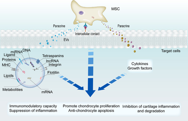
Abstract
Osteoarthritis (OA), the commonest arthritis, is characterized by the progressive destruction of cartilage, leading to disability. The Current early clinical treatment strategy for OA often centers on anti-inflammatory or analgesia medication, weight loss, improved muscular function and articular cartilage repair. Although these treatments can relieve symptoms, OA tends to be progressive, and most patients require arthroplasty at the terminal stages of OA. Recent studies have shown a close correlation between joint pain, inflammation, cartilage destruction and synovial cells. Consequently, understanding the potential mechanisms associated with the action of synovial cells in OA could be beneficial for the clinical management of OA. Therefore, this review comprehensively describes the biological functions of synovial cells, the synovium, together with the pathological changes of synovial cells in OA, and the interaction between the cartilage and synovium, which is lacking in the present literature. Additionally, therapeutic approaches based on synovial cells for OA treatment are further discussed from a clinical perspective, highlighting a new direction in the treatment of OA.
Keywords: cartilage; inflammation; osteoarthritis; synovial cells; synovium; treatment.
© 2023 The Authors. Exploration published by Henan University and John Wiley & Sons Australia, Ltd.
Conflict of interest statement
The authors declare no conflicts of interest.
Figures


References
-
- Hunter D. J., March L., Chew M., Lancet 2020, 396, 1711. - PubMed
Publication types
LinkOut - more resources
Full Text Sources