Review
. 2023 Mar 30;3(3):20220002.
doi: 10.1002/EXP.20220002. eCollection 2023 Jun.

Recent development of pH-responsive theranostic nanoplatforms for magnetic resonance imaging-guided cancer therapy

Affiliations
Review

Recent development of pH-responsive theranostic nanoplatforms for magnetic resonance imaging-guided cancer therapy

Xu Li et al. Exploration (Beijing). .

Abstract

The acidic characteristic of the tumor site is one of the most well-known features and provides a series of opportunities for cancer-specific theranostic strategies. In this regard, pH-responsive theranostic nanoplatforms that integrate diagnostic and therapeutic capabilities are highly developed. The fluidity of the tumor microenvironment (TME), with its temporal and spatial heterogeneities, makes noninvasive molecular magnetic resonance imaging (MRI) technology very desirable for imaging TME constituents and developing MRI-guided theranostic nanoplatforms for tumor-specific treatments. Therefore, various MRI-based theranostic strategies which employ assorted therapeutic modes have been drawn up for more efficient cancer therapy through the raised local concentration of therapeutic agents in pathological tissues. In this review, we summarize the pH-responsive mechanisms of organic components (including polymers, biological molecules, and organosilicas) as well as inorganic components (including metal coordination compounds, metal oxides, and metal salts) of theranostic nanoplatforms. Furthermore, we review the designs and applications of pH-responsive theranostic nanoplatforms for the diagnosis and treatment of cancer. In addition, the challenges and prospects in developing theranostic nanoplatforms with pH-responsiveness for cancer diagnosis and therapy are discussed.

Keywords: MRI‐guided; cancer therapy; nanoplatform; pH‐responsive.


Conflict of interest statement

The authors declare no conflict of interest.

Figures
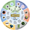
SCHEME 1
SCHEME 1
Schematic illustration of pH‐responsive theranostic nanoplatforms. Various therapeutic modes for cancer under magnetic resonance imaging guidance (inner ring). pH‐responsive nanoplatforms based on various nanomaterials (outer ring). Reproduced with permission.[ 23 ] Copyright 2013, Elsevier Ltd.; Reproduced with permission.[ 24 ] Copyright 2016, American Chemical Society; Reproduced with permission.[ 58 ] Copyright 2017, Elsevier Ltd.; Reproduced with permission.[ 27 ] Copyright 2013, Elsevier B.V.; Reproduced with permission.[ 65 ] Copyright 2016, American Chemical Society; Reproduced with permission.[ 67 ] Copyright 2015, Elsevier Ltd.; Reproduced with permission.[ 71 ] Copyright 2019, Wiley‐VCH Verlag GmbH & Co. KGaA; Reproduced with permission.[ 83 ] Copyright 2017, WILEY‐VCH Verlag GmbH & Co. KGaA; Reproduced with permission.[ 97 ] Copyright 2019, Wiley‐VCH Verlag GmbH & Co. KGaA; Reproduced with permission.[ 91 ] Copyright 2014, WILEY‐VCH Verlag GmbH & Co. KGaA; Reproduced with permission.[ 103 ] Copyright 2021, American Chemical Society; Reproduced with permission.[ 102 ] Copyright 2019, WILEY‐VCH Verlag GmbH & Co. KGaA.
FIGURE 1
FIGURE 1
pH‐responsive nanoplatforms based on polymer, biological molecule, and organosilica respectively. (A) Polymer‐based nanoplatform. Schematic representation of the preparation of the cancer‐recognizable magnetic resonance imaging contrast agents (CR‐CAs) and pH‐dependent structural transformation in CR‐CAs. Inset: Chemical structural representation of the protonation of imidazole groups in poly(ethylene glycol)‐b‐poly(l‐histidine) (PEG‐p(l‐His)) at acidic pH. Reproduced with permission.[ 23 ] Copyright 2013, Elsevier Ltd. (B) Biological molecule‐based nanoplatform. Schematic representation of the preparation of pH low insertion peptide (pHLIP)‐modified Fe3O4 NPs and pH‐dependent structural transformation of pHLIP. Reproduced with permission.[ 59 ] Copyright 2021, American Chemical Society. (C) Organosilica‐based nanoplatform. Schematic representation of the preparation of gold NPs‐conjugated mesoporous silica NPs and pH‐controlled release. Reproduced with permission.[ 65 ] Copyright 2016, American Chemical Society.
FIGURE 2
FIGURE 2
pH‐responsive nanoplatforms based on metal coordination compound, metal oxide, and metal salt respectively. (A) Metal coordination compound nanoparticles‐based nanoplatform. Schematic representation of pH‐dependent disintegration of the coordinatively unsaturated shell (Fe‐GA) and pH‐dependent release of Fe3+ at different pH values. Reproduced with permission.[ 71 ] Copyright 2019, Wiley‐VCH Verlag GmbH & Co. KGaA. (B) Metal oxide nanoparticles‐based nanoplatform. Schematic illustration of the composition of ultrathin Gd2O3 nanoscrolls and their pH‐responsive biodegradation as well as DOX release. Reproduced with permission.[ 97 ] Copyright 2019, Wiley‐VCH Verlag GmbH & Co. KGaA. (C) Metal salt nanoparticles‐based nanoplatform. Schematic illustration of the hybrid structure of PEGMnCaP and release profiles of Mn ions under physiological conditions at different pH. Reproduced with permission.[ 106 ] Copyright 2016, Macmillan Publishers Limited.
FIGURE 3
FIGURE 3
pH‐responsive nanoplatforms for T1 MR imaging. (A) Gd‐based T1 magnetic resonance imaging (MRI) CAs. The nanoplatform (MSNPs) is based on cell membrane‐coated NaGdF4‐CaCO3 nanocomposites. The spin interaction between crystal lattices and surrounding protons is structurally blocked by multilayers outside the T1 source (blue line). The energy transfer from protons to crystal lattices via spin‐lattice interaction is then activated due to the pH‐responsive capability of MSNPs under acidic microenvironments (orange line). The in vitro and in vivo MRI performance of MSNPs. Reproduced with permission.[ 102 ] Copyright 2019, WILEY‐VCH Verlag GmbH & Co. KGaA. (B) Mn‐based T1 MRI CAs. The nanoplatform (Mn‐LDH) is based on Mn–doped layered double hydroxides. The in vitro and in vivo MRI performance of Mn‐LDH and schematic illustration of structure related multifunctional properties of Mn‐LDH. Reproduced with permission.[ 83 ] Copyright 2017, WILEY‐VCH Verlag GmbH & Co. KGaA.
FIGURE 4
FIGURE 4
pH‐responsive nanoplatforms for T2 MR imaging. (A) FeOx‐based T2 magnetic resonance imaging (MRI) CAs. The nanoplatform (ATF‐IONP‐Gem) is based on amino‐terminal fragment (ATF) peptide‐conjugated iron oxide nanoparticles (IONPs). The in vivo MRI performance of nontargeted IONP‐Gem and ATF‐IONP‐Gem. Reproduced with permission.[ 140 ] Copyright 2013, American Chemical Society. (B) MnO x ‐based T2 MRI CAs. The nanoplatform (MnO x ‐SPNs) is based on ultrathin MnO x nanosheets and semiconducting polymer nanoparticles (SPNs). Schematic illustration of MR signal amplification activated by introducing SPNs or BSA. The in vitro and in vivo MRI performance of MnO x ‐SPNs. Reproduced with permission.[ 151 ] Copyright 2020, Elsevier Inc.
FIGURE 5
FIGURE 5
pH‐responsive nanoplatforms for T1‐T2 dual modal MR imaging. (A) Gd/Fe‐based magnetic resonance imaging (MRI) CAs. The nanoplatform (FA‐PYFGN‐CDDP) is based on folic acid (FA) functionalized SPIONs‐Gd2O3 core–shell nanocomposites. The in vitro MRI performance of FA‐PYFGN‐CDDP and Fe3O4. And the in vivo MRI performance of FA‐PYFGN‐CDDP, nontargeted PYFGN‐CDDP, and Fe3O4. Reproduced with permission.[ 156 ] Copyright 2017, American Chemical Society. (B) Mn/Fe‐based MRI CAs. The nanoplatform (Fe3O4/MnO x ‐GO) is based on Fe3O4 and MnO x co‐integrated graphene oxide (GO) nanosheets. The in vitro (black line: pH 7.4 and red line: pH 6.0) and in vivo MRI performance of Fe3O4/MnO x ‐GO. Reproduced with permission.[ 29 ] Copyright 2014, WILEY‐VCH Verlag GmbH & Co. KGaA.
FIGURE 6
FIGURE 6
pH‐responsive nanoplatforms for (A) chemotherapy. The nanoplatform (USIO NCs/DOX@CM) is based on cell membrane (CM)‐coated and DOX‐loaded ultrasmall iron oxide nanoclusters (USIO NCs). Schematic illustration for UTMD‐promoted delivery of USIO NCs/DOX@CM to achieve pH‐responsive tumor theranostics. The therapeutic outcomes of USIO NCs/DOX@CM‐UTMD and other groups. Reproduced with permission.[ 161 ] Copyright 2020, Elsevier Ltd. (B) Photodynamic therapy. The nanoplatform (hMUC) is based on honeycomb MnO x NPs and Ce6‐sensitized up‐conversion nanoparticles (UCNPs). Schematic illustration of the systemic delivery of hMUC in vivo, and corresponding diagnosis and therapy for tumor. The therapeutic outcomes of hMUC+NIR and other groups. Reproduced with permission.[ 96 ] Copyright 2018, American Chemical Society.
FIGURE 7
FIGURE 7
pH‐responsive nanoplatforms for (A) photothermal therapy. The nanoplatform (MnO x /Ti3C2−SP) is based on soybean phospholipid (SP)‐modified MnO x /Ti3C2 nanosheets. Schematic illustration of MnO x /Ti3C2−SP nanosheets as the photothermal agents for cancer cell ablation. The magnetic resonance imaging (MRI) and PTT performance of MnO x /Ti3C2−SP. Reproduced with permission.[ 135 ] Copyright 2017, American Chemical Society. (B) Chemodynamic therapy. The nanoplatform (FePt@FeO x @TAM‐PEG) is based on core–shell structured FePt@FeO x and pH‐sensitive drug tamoxifen (TAM). Schematic illustration of acidity‐unlocked FePt@FeO x @TAM‐PEG with positive feedback loop promoting Fenton‐like reactions for self‐boosting tumor specific chemodynamic therapy. The MRI and CDT performance of FePt@FeO x @TAM‐PEG. Reproduced with permission.[ 181 ] Copyright 2021, Wiley‐VCH GmbH. (C) Immunotherapy. The nanoplatform (NC‐aP) is based on PD‐1 antibody‐conjugated Fe3O4 nanoclusters. Schematic illustration of magnetic nanoclusters armed with PD‐1 antibody improved adoptive T cell therapy for solid tumors. The therapeutic outcomes of NC‐aP under magnetic field and other groups. Reproduced with permission.[ 184 ] Copyright 2019, American Chemical Society.
FIGURE 8
FIGURE 8
pH‐responsive nanoplatforms for combined therapy. (A) Chemotherapy and photothermal therapy. The nanoplatform (Mn‐APPMSF) is based on Fe(III)‐AQ4N prodrugs and Mn(II)‐loaded semiconducting polymer dots‐hybridized mesoporous silica. Schematic illustration of the functional principle of Mn‐APPMSF in tumor microenvironments. The PTT performance and therapeutic outcomes of Mn‐APPMSF/laser and other groups. Reproduced with permission.[ 190 ] Copyright 2018, The Royal Society of Chemistry. (B) Chemotherapy and magnetic hyperthermia. The nanoplatform (γ‐SD/PLL) is based on covalent organic frameworks‐coated γ‐Fe2O3 NPs. Schematic representation of magnetically induced heat‐ and acid‐triggered Dox release from γ‐SD/PLL in conditions that mimic cancer cell physiology. The magnetic resonance imaging performance, DOX release profiles, and therapeutic outcomes of γ‐SD/PLL. Reproduced with permission.[ 192 ] Copyright 2020, American Chemical Society.
FIGURE 9
FIGURE 9
pH‐responsive nanoplatforms for combined therapy. (A) Chemotherapy and photodynamic therapy (PDT). The nanoplatform ((UCNP@PFNS/AQ4N)@MnCaP) is based on a core of PFNS‐modified up‐conversion nanoparticles (UCNPs) and a shell of Mn–doped Ca3(PO4)2. Schematic representation of (UCNP@PFNS/AQ4N)@MnCaP for synergetic PDT and chemotherapy under magnetic resonance imaging (MRI) guidance. The MRI and therapeutic outcomes of (UCNP@ PFNS/AQ4N)@MnCaP. Reproduced with permission.[ 111 ] Copyright 2019, Elsevier Ltd. (B) Chemotherapy and chemodynamic therapy. The nanoplatform (Dox‐CACN) is based on DOX‐loaded iron oxide nanoclusters via DNA‐programmed self‐assembly. Schematic representation of dual key co‐activated nanoplatform for switchable MRI monitoring ferroptosis‐based synergistic therapy. The therapeutic outcomes of Dox‐CACN and other groups. Reproduced with permission.[ 128 ] Copyright 2022, Elsevier Inc.
FIGURE 10
FIGURE 10
pH‐responsive nanoplatforms for combined therapy. (A) Chemodynamic therapy and limotherapy. The nanoplatform (MCDION‐Se) is based on nanoselenium (nano‐Se)‐coated MnCO3‐deposited nanoparticles. Schematic illustration of the cascade reaction of MCDION‐Se in the intracellular environment. The magnetic resonance imaging (MRI) and therapeutic outcomes of MCDION‐Se and other groups. Reproduced with permission.[ 195 ] Copyright 2019, Elsevier Ltd. (B) Chemotherapy, radiotherapy, and photothermal therapy. The nanoplatform (Gd2Hf2O7@PDA@PEG‐Pt‐RGD) is based on polydopamine (PDA)‐modified and cisplatin‐loaded Gd2Hf2O7 NPs. Schematic illustration of Gd2Hf2O7@PDA@PEG‐Pt‐RGD for MRI‐guided synergistic therapy. The MRI and therapeutic outcomes of Gd2Hf2O7@PDA@PEG‐Pt‐RGD and other groups. Reproduced with permission.[ 129 ] Copyright 2020, American Chemical Society.

References

    1. Cheng L., Wang C., Feng L., Yang K., Liu Z., Chem. Rev. 2014, 114, 10869. - PubMed
    1. Zhu L., Wang D., Wei X., Zhu X., Li J., Tu C., Su Y., Wu J., Zhu B., Yan D., J. Controlled Release 2013, 169, 228. - PubMed
    1. Zhang S., Qian X., Zhang L., Peng W., Chen Y., Nanoscale 2015, 7, 7632. - PubMed
    1. Zhang L., Yang Z., Zhu W., Ye Z., Yu Y., Xu Z., Ren J., Li P., ACS Biomater. Sci. Eng. 2017, 3, 1690. - PubMed
    1. Estrella V., Chen T., Lloyd M., Wojtkowiak J., Cornnell H. H., Ibrahim‐Hashim A., Bailey K., Balagurunathan Y., Rothberg J. M., Sloane B. F., Johnson J., Gatenby R. A., Gillies R. J., Cancer Res. 2013, 73, 1524. - PMC - PubMed

LinkOut - more resources