. 2023 Dec 11;51(22):12261-12274.
doi: 10.1093/nar/gkad974.

Differing structures and dynamics of two photolesions portray verification differences by the human XPD helicase

Affiliations

Differing structures and dynamics of two photolesions portray verification differences by the human XPD helicase

Iwen Fu et al. Nucleic Acids Res. .

Abstract

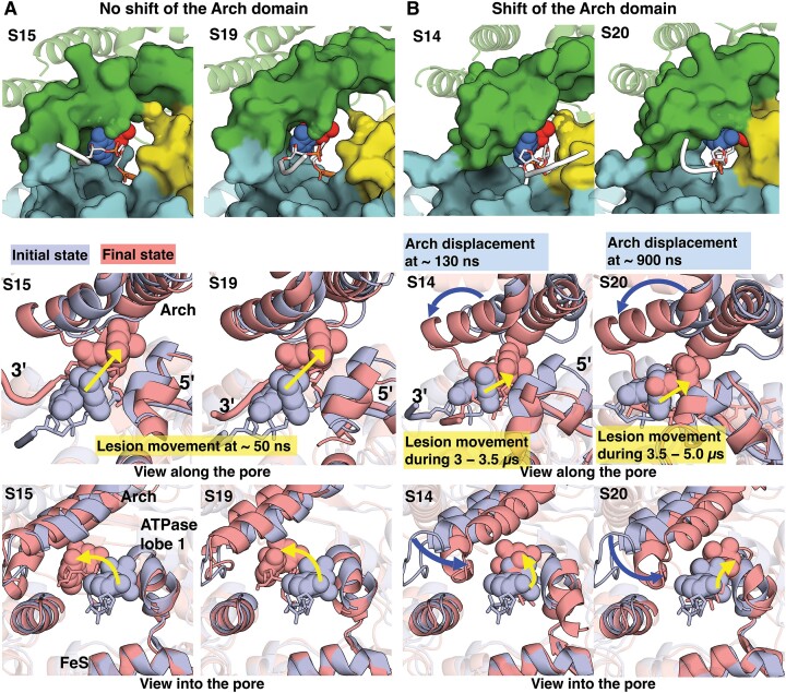
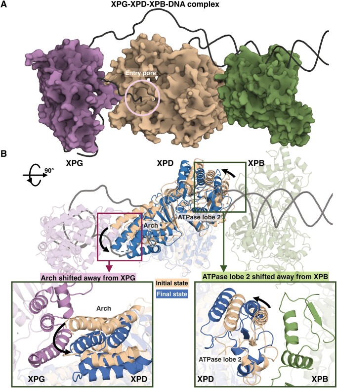
Ultraviolet light generates cyclobutane pyrimidine dimer (CPD) and pyrimidine 6-4 pyrimidone (6-4PP) photoproducts that cause skin malignancies if not repaired by nucleotide excision repair (NER). While the faster repair of the more distorting 6-4PPs is attributed mainly to more efficient recognition by XPC, the XPD lesion verification helicase may play a role, as it directly scans the damaged DNA strand. With extensive molecular dynamics simulations of XPD-bound single-strand DNA containing each lesion outside the entry pore of XPD, we elucidate strikingly different verification processes for these two lesions that have very different topologies. The open book-like CPD thymines are sterically blocked from pore entry and preferably entrapped by sensors that are outside the pore; however, the near-perpendicular 6-4PP thymines can enter, accompanied by a displacement of the Arch domain toward the lesion, which is thereby tightly accommodated within the pore. This trapped 6-4PP may inhibit XPD helicase activity to foster lesion verification by locking the Arch to other domains. Furthermore, the movement of the Arch domain, only in the case of 6-4PP, may trigger signaling to the XPG nuclease for subsequent lesion incision by fostering direct contact between the Arch domain and XPG, and thereby facilitating repair of 6-4PP.


Figures

Graphical Abstract
Graphical Abstract
Figure 1.
Figure 1.
Initial structures of XPD complexed with ssDNA containing a lesion near the DNA entry pore and the positioning and orientation of the lesion. (A) The chemical and crystal structures of the pyrimidine (6−4) pyrimidone photoproduct (6−4PP) and the cyclobutane pyrimidine dimer (CPD). Left: The 6−4PP lesion, taken from a Rad4–Rad23 structure bound to 24-bp DNA containing this lesion (PDB ID 6CFI (68)), has two roughly perpendicular thymine rings, which are linked by a single C6−C4 covalent bond. Right: The CPD lesion with cis-syn stereochemistry, taken from the crystal structure of a nucleosome containing a CPD lesion (PDB ID 5B24 (69)), has two open book-like thymine bases that are covalently cross-linked and contains a cyclobutane ring that is formed by bonds between the C5’s and C6’s of the adjacent thymine bases. (B) Initial structure of XPD in complex with ssDNA (grey) containing the 6−4PP lesion (red sticks and spheres). The XPD protein contains four domains: ATPase lobe 1 (yellow), ATPase lobe 2 (pink), Arch (green) and FeS (cyan) containing an FeS cluster (indicated as orange for Fe atoms and yellow spheres for S atoms). XPD has a central tunnel that accommodates ssDNA with its 3′- and 5′-end extended toward the DNA entry and exit pores, respectively. The box highlights the DNA entry pore, formed by the Arch, FeS and ATPase lobe 1 domains. (C) A zoom-in view of the DNA entry pore depicts the initial structures of the XPD complex with lesion-containing ssDNA. Note that the initial structures for each model are the first snapshot after minimization in the MD simulation. Full details concerning the preparation of the initial models for the MD simulations of the XPD-ssDNA complex are given in Materials and methods.
Figure 2.
Figure 2.
The CPD backbone is translocated in the 3′ to 5′ direction into the unoccupied space within the pore, but its modified bases are blocked from entering into the pore. (A) Backbone translocation of CPD occurs during 0–1.9 μs via two main DNA bending conformational transitions that take place over hundreds of nanoseconds (Supplementary MovieS1); the DNA bending is monitored by the nucleotide phosphate (crosslinked P atom of CPD) to phosphate (dA) distance. A DNA bend between the CPD and its 5′-dA occurs early in the simulation (∼200 ns). The DNA becomes more bent after 1.9 μs during the translocation and this bent DNA is then retained throughout the simulation. At the initial state (the snapshot after minimization in the MD simulations), CPD is placed outside the pore with its base pointing away from the ATPase lobe 1 domain and oriented toward the region near the Arch and the FeS domains. H384 forms hydrogen bonds with the bases of CPD. R112 and R196 anchor the backbones of CPD and dA. Y192 and Y211 interact with the backbone of dA. The nucleotide phosphate to phosphate distance is ∼ 11.6 Å. Stage 1 (during 0.2−1.9 μs): R380 and K216, both positioned on the 5′-side of CPD, together pull the 5′-phosphate group of CPD to outcompete its backbone interactions with R196 and R112. As a result, the backbone of CPD is translocated toward its 5′ direction and is lifted away from FeS and pulled toward the region near the Arch domain and ATPase lobe 1 domains. A bend is formed between dA and CPD because the phosphate group of dA is still anchored stably by Y192 after the backbone of CPD is translocated in the 3′−5′ direction. Now, R196 interacts with the crosslinked phosphate of CPD. The nucleotide phosphate to phosphate distance is ∼8 Å. Stage 2 (1.9–8 μs): K128 and R380 together pull the phosphate group of CPD to outcompete its backbone interactions with K216; as a result, the backbone of CPD is pointed away from the ATPase lobe 1 domain. The nucleotide phosphate to phosphate distance is ∼ 6.3 Å, which is retained throughout the simulation. The best representative frames from each stage are displayed, revealing the pathway of the CPD translocation in the 3′ to 5′ direction along the entry pore (Supplementary MovieS1). The top panel shows the XPD-ssDNA interactions near the entry pore; the hydrogen bonds are denoted by the yellow dashed lines. The bottom panel is a view into the entry pore (surface rendering), showing the positioning of the nucleotides near the pore during the MD simulation; the 3′-end d(TG) nucleotides are not shown for clarity. The structures are color-coded as in Figure 1. H135, R380 and H384 are labeled and displayed as sphere rendering, highlighting their relative positions with respect to CPD. (B) Superimposed structures with CPD prior to translocation (light-blue) and after translocation (red). A view along the entry pore showing that during the CPD transition, dA is pointed away from the position between the ATPase lobe 1 helix and the FeS helix within the pore; this creates the space for the DNA bend/loop between CPD and dA. We have superimposed the structures from stage 2 where there is a DNA bend between CPD and dA within the pore and the initial state (prior to the process of CPD translocation). The Arch domain does not lift away from the DNA; thus, it does not contribute to the extra space for the DNA bend between the CPD and dA.
Figure 3.
Figure 3.
The 6−4PP lesion initially positioned outside the entry undergoes a 3′→5′ translocation via its modified bases flipping into the unoccupied space within the pore. Top panel shows the best representative structure of each simulation. Middle and bottom panels reveal the superimposed structures with 6−4PP prior to translocation (initial structure, light-blue) and after translocation (best structure, red) with a view along and into the entry pore, respectively. The movements of the Arch domain and the lesion are highlighted as dark-blue and yellow arrows, respectively. The lengths of the arrows are approximate indicators of the extents of the movements. (A) In simulations S15 and S19, the pathway of base-translocation of 6−4PP into the entry pore is driven by the movement of the modified bases, which takes place early in the simulation (at ∼50 ns). The bases move toward the Arch domain (bottom panel) and in a 3′ to 5′ direction (middle panel) into the pore. The Arch domain retains its initial position and shows no notable displacement during the base-translocation of the 6−4PP lesion into the pore. (B) In simulations S14 and S20, the pathway of base-translocation of 6−4PP involves the movements of the Arch domain (dark-blue arrow) and the lesion (yellow arrow), which take place at different times. The Arch domain is shifted toward the lesion as well as the ATPase lobe 1 and the FeS domains. The lesion is then moved in a 3′ to 5′ direction into the pore. Details concerning simulation S20 that shows the base-translocation of 6−4PP via multiple conformational transitions that take place over 5 μs is given in Figure 4. See Supplementary Figure S7 for simulation S14.
Figure 4.
Figure 4.
The 6−4PP is translocated in the 3′ to 5′ direction as its bases flip into the unoccupied space within the pore. (A) The base-translocation of 6−4PP occurs during 0–5 μs via multiple conformational transitions that take place over microseconds. Superimposed structures with 6−4PP prior to translocation (light-blue) and after translocation (red) with a view along the entry pore show that two main movements contribute to the base-translocation of 6−4PP. One is the displacement of the Arch domain (dark-blue arrow) and the other is the movement of the lesion in a 3′ to 5′ direction (yellow arrow). The plot shows the relative motion of the Arch and the FeS domains near the entry pore; this is obtained by monitoring the Cα RMSD of the Arch domain after fitting the stable region of the FeS Cα atoms to the initial structure; the Arch domain deviates significantly from its initial structure by greater than ∼8 Å at ∼0.9 μs in going from Stage 1 to Stage 2. For the movement of the lesion, we measured the distance between the base 5T O2 atom of 6−4PP and the N atom of A218 of the ATPase lobe 1 domain, d= O2(5T)−N(A218). The 3′→5′ movement of 6−4PP occurs at ∼3.5 μs during the transition between Stage 2 and Stage 3, which is reflected in the distance d decrease from ∼9 to ∼4.5 Å. (B) Stages 1−4 illustrate the pathway of 6−4PP’s base-translocation in the 3′ to 5′ direction along the entry pore (Supplementary MovieS2). The best representative frames from each stage are displayed. The top panel shows the XPD-ssDNA interactions near the entry pore with a view along the entry pore; the hydrogen bonds are denoted by the yellow dashed lines. The bottom panel is a view into the entry pore (surface rendering), showing the positioning and orientation of the 6−4PP lesion near the pore during the MD simulation; the 3′-end d(TG) nucleotides are not shown for clarity. The structures are color-coded as in Figure 1. R380, D219 and L220 are labeled and shown as sphere rendering, highlighting their relative positions with respect to the lesion. Stage 1 (during 0−0.9 μs): 6−4PP is placed outside the pore with its bases pointing toward the ATPase lobe 1 domain. R196 anchors the backbones of 6−4PP, and Y192 and Y211 interact with the phosphate group of 5′-dA. R380 and D219 are positioned on the 5′-side of 6−4PP. The distance of O2(5T)−N(A218) is ∼10 Å. Stage 2 (0.9–3.5 μs): after the Arch domain is shifted toward the lesion and the ATPase lobe 1 domain (Supplementary Figures S8A), R380 is hydrogen-bonded with the O4 atom of the 5T Also, R196 interacts with the 5′-P and the crosslinked P atoms of 6−4PP. Together, these interactions reorient the bases of 6−4PP away from the ATPase lobe 1 domain. The distance (d) of O2(5T)−N(A218) is ∼9 Å. R380 and D219 are still positioned slightly on the 5′-side of 6−4PP. Stage 3 (4.5–5 μs): 6−4PP is moved in a 3′→5′ direction closer to A218 during the transition between 3.5 and 4.5 μs, reflected in the decreased distance (d) of O2(5T)−N(A218) from ∼9 to ∼4.5 Å. D219 is hydrogen-bonded with N3 and O2 of 5T base. Now, both R380 and D219 are positioned on the 3′-side of 6−4PP. R196 and Y192 retain their interactions with the backbone of 6−4PP. Stage 4 (5–8 μs): the bases of 6−4PP are re-oriented toward A218, further shortening the distance d by ∼1 Å. Thus, the N3 and O2 atoms of the 5T base are engulfed by a small pocket formed by the ATPase lobe 1 helix with residues 215−221 (PKIADLV). R196 remains anchored to the backbone of the lesion and Y192 anchors to the backbone of 5′-dA. The extensive interactions of the XPD pore and the bases of the 6−4PP within the pore are detailed in Supplementary Figure S8B.
Figure 5.
Figure 5.
The displacement of the Arch domain changes the overall conformation of XPD within the complex of XPG-XPD-XPB bound to the open bubble substrate, altering direct XPG-XPD and XPD-XPB interactions, which could facilitate signaling for the subsequent incision step. (A) A model of XPG (purple), XPD (olive) and XPB (green) bound to the open bubble DNA (black), derived from the structural model provided by (45). In this model, we replaced the XPB and XPD helicases with those in the existing cryo-EM structure of TFIIH-XPA-DNA (44); XPG was replaced with the existing XPG crystal structure (PDB 3Q8K) (85). In this complex, XPD has direct contacts with XPG mainly via the Arch domain and direct contacts with XPB via the ATPase lobe 2 domain. (B) Superimposed structures of XPD prior to the shift of the Arch domain (olive) and after the shift of the Arch domain (blue) within the XPG-XPD-XPB-DNA complex highlight the changes in overall conformation of XPD, including the shift of the Arch domain away from XPG (left inset box) and the shift of the ATPase lobe 2 away from XPB (right inset box). Thus, it is likely that the excision process is helped by the change of overall XPD conformation in the pre-incision complex due to the lesion-imposed shift of the Arch domain that can be rapidly sensed by the XPG nuclease for the subsequent incision step.

References

    1. Ganesan A., Hanawalt P.. Photobiological origins of the field of genomic maintenance. Photochem. Photobiol. 2016; 92:52–60. - PMC - PubMed
    1. Mao P., Wyrick J.J., Roberts S.A., Smerdon M.J.. UV-induced DNA damage and mutagenesis in chromatin. Photochem. Photobiol. 2017; 93:216–228. - PMC - PubMed
    1. Cadet J., Douki T.. Formation of UV-induced DNA damage contributing to skin cancer development. Photochem. Photobiol. Sci. 2018; 17:1816–1841. - PubMed
    1. Sancar A. Mechanisms of DNA repair by photolyase and excision nuclease (Nobel Lecture). Angew. Chem. Int. Ed Engl. 2016; 55:8502–8527. - PubMed
    1. Hoeijmakers J.H. DNA damage, aging, and cancer. N. Engl. J. Med. 2009; 361:1475–1485. - PubMed

Publication types