

Deep learning performance for detection and classification of microcalcifications on mammography
- PMID: 37934382
- PMCID: PMC10630180
- DOI: 10.1186/s41747-023-00384-3
Deep learning performance for detection and classification of microcalcifications on mammography
Abstract
Background: Breast cancer screening through mammography is crucial for early detection, yet the demand for mammography services surpasses the capacity of radiologists. Artificial intelligence (AI) can assist in evaluating microcalcifications on mammography. We developed and tested an AI model for localizing and characterizing microcalcifications.
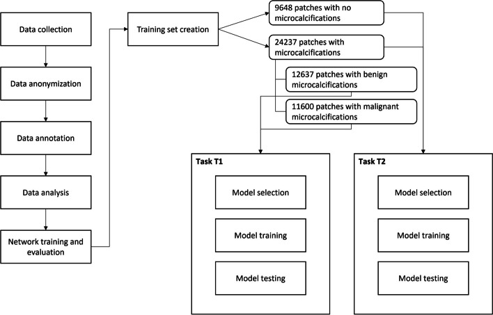
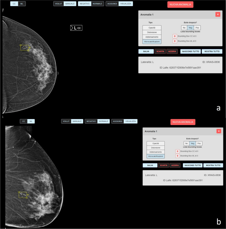
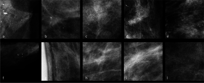
Methods: Three expert radiologists annotated a dataset of mammograms using histology-based ground truth. The dataset was partitioned for training, validation, and testing. Three neural networks (AlexNet, ResNet18, and ResNet34) were trained and evaluated using specific metrics including receiver operating characteristics area under the curve (AUC), sensitivity, and specificity. The reported metrics were computed on the test set (10% of the whole dataset).
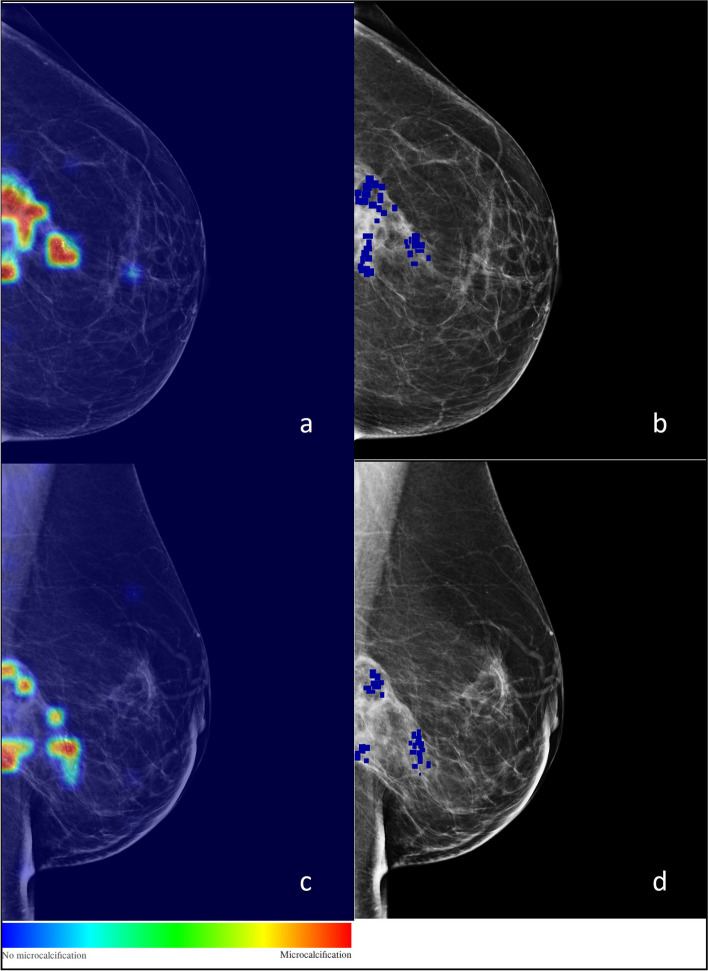
Results: The dataset included 1,000 patients aged 21-73 years and 1,986 mammograms (180 density A, 220 density B, 380 density C, and 220 density D), with 389 malignant and 611 benign groups of microcalcifications. AlexNet achieved the best performance with 0.98 sensitivity, 0.89 specificity of, and 0.98 AUC for microcalcifications detection and 0.85 sensitivity, 0.89 specificity, and 0.94 AUC of for microcalcifications classification. For microcalcifications detection, ResNet18 and ResNet34 achieved 0.96 and 0.97 sensitivity, 0.91 and 0.90 specificity and 0.98 and 0.98 AUC, retrospectively. For microcalcifications classification, ResNet18 and ResNet34 exhibited 0.75 and 0.84 sensitivity, 0.85 and 0.84 specificity, and 0.88 and 0.92 AUC, respectively.
Conclusions: The developed AI models accurately detect and characterize microcalcifications on mammography.
Relevance statement: AI-based systems have the potential to assist radiologists in interpreting microcalcifications on mammograms. The study highlights the importance of developing reliable deep learning models possibly applied to breast cancer screening.
Key points: • A novel AI tool was developed and tested to aid radiologists in the interpretation of mammography by accurately detecting and characterizing microcalcifications. • Three neural networks (AlexNet, ResNet18, and ResNet34) were trained, validated, and tested using an annotated dataset of 1,000 patients and 1,986 mammograms. • The AI tool demonstrated high accuracy in detecting/localizing and characterizing microcalcifications on mammography, highlighting the potential of AI-based systems to assist radiologists in the interpretation of mammograms.
Keywords: Artificial intelligence; Machine learning; Mammography; Microcalcifications; Neural networks (computer).
© 2023. The Author(s).
Conflict of interest statement
FP and DO are members of the
Figures
References
-
- Bevers TB, Helvie M, Bonaccio E et al (2018) Breast Cancer Screening and Diagnosis, Version 3.2018, NCCN Clinical Practice Guidelines in Oncology. J Natl Compr Canc Netw 16(11):1362–1389. 10.6004/jnccn.2018.0083 - PubMed
Publication types
MeSH terms
LinkOut - more resources
Full Text Sources
Medical