. 2023 Dec 22;8(24):e171140.
doi: 10.1172/jci.insight.171140.

Dabrafenib protects from cisplatin-induced hearing loss in a clinically relevant mouse model

Affiliations

Dabrafenib protects from cisplatin-induced hearing loss in a clinically relevant mouse model

Matthew A Ingersoll et al. JCI Insight. .

Abstract

The widely used chemotherapy cisplatin causes permanent hearing loss in 40%-60% of patients with cancer. One drug, sodium thiosulfate, is approved by the FDA for use in pediatric patients with localized solid tumors for preventing cisplatin-induced hearing loss, but more drugs are desperately needed. Here, we tested dabrafenib, an FDA-approved BRAF kinase inhibitor and anticancer drug, in a clinically relevant multidose cisplatin mouse model. The protective effects of dabrafenib, given orally twice daily with cisplatin, were determined by functional hearing tests and cochlear outer hair cell counts. Toxicity of the drug cotreatment was evaluated, and levels of phosphorylated ERK were measured. A dabrafenib dose of 3 mg/kg BW, twice daily, in mice, was determined to be the minimum effective dose, and it is equivalent to one-tenth of the daily FDA-approved dose for human cancer treatment. The levels of hearing protection acquired, 20-25 dB at the 3 frequencies tested, in both female and male mice, persisted for 4 months after completion of treatments. Moreover, dabrafenib exhibited a good in vivo therapeutic index (> 25), protected hearing in 2 mouse strains, and diminished cisplatin-induced weight loss. This study demonstrates that dabrafenib is a promising candidate drug for protection from cisplatin-induced hearing loss.

Keywords: Cancer; Drug therapy; Otology; Protein kinases; Therapeutics.


Figures

Figure 1
Figure 1. Dabrafenib protects against cisplatin-induced hearing loss following a single high dose of cisplatin.
(A) Schedule of administration of dabrafenib and cisplatin in FVB mice: 30 mg/kg cisplatin was administered once on day 1 while 12 mg/kg dabrafenib was administered for 3 days, twice a day. Auditory testing was performed before treatment began and 21 days after cisplatin administration. (B) ABR threshold shifts following protocol in A. (C) Weight change over 21 days following protocol in A. (D) Kaplan-Meier survival curves of mouse cohorts following protocol in A. Carrier alone (black), cisplatin alone (yellow), dabrafenib alone (blue), and dabrafenib plus cisplatin (purple). Data shown as means ± SEM; *P < 0.05, **P < 0.01, ***P < 0.001 compared with cisplatin alone by 2-way ANOVA with Bonferroni’s post hoc test.
Figure 2
Figure 2. Dabrafenib-treated mice have significantly lower ABR threshold shifts compared with cisplatin alone–treated mice.
(A) Schedule of administration of dabrafenib and cisplatin in a translational, multicycle treatment protocol using CBA/CaJ mice. Each cycle consisted of 4 days of treatment with 3 mg/kg cisplatin in the morning and 15, 3, or 0.6 mg/kg dabrafenib in the morning and evening. A 10-day recovery period followed the 4 days of treatment. This cycle was repeated for a total of 3 times. Auditory testing occurred before treatment began, immediately after cycle 3 (day 42), and 4 months after cycle 3 (day 165). (B) ABR threshold shifts recorded immediately after the completion of cycle 3 (day 42) in protocol shown in A. (C) Amplitudes of ABR wave 1 at 16 kHz from B. (D) ABR threshold shifts of female and (E) male mice recorded immediately after the completion of cycle 3. (F) ABR threshold shifts recorded 4 months after the completion of cycle 3 (day 165). Carrier (black), cisplatin alone (yellow), 15 mg/kg dabrafenib alone (purple), 3 mg/kg dabrafenib alone (orange), 15 mg/kg dabrafenib plus cisplatin (blue), 3 mg/kg dabrafenib plus cisplatin (red), and 0.6 mg/kg dabrafenib plus cisplatin (green). Data shown as means ± SEM; *P < 0.05, **P < 0.01, ***P < 0.001 compared with cisplatin alone by 2-way ANOVA with Bonferroni’s post hoc test.
Figure 3
Figure 3. Dabrafenib-treated mice have significantly lower DPOAE threshold shifts compared with cisplatin alone–treated mice.
(A) DPOAE threshold shifts recorded immediately after the completion of cycle 3 (day 42) in protocol shown in Figure 2A. (B) DPOAE threshold shifts of female and (C) male mice recorded immediately after the completion of cycle 3. (D) DPOAE threshold shifts recorded 4 months after the completion of cycle 3 (day 165). Carrier (black), cisplatin alone (yellow), 15 mg/kg dabrafenib alone (purple), 3 mg/kg dabrafenib alone (orange), 15 mg/kg dabrafenib plus cisplatin (blue), 3 mg/kg dabrafenib plus cisplatin (red), and 0.6 mg/kg dabrafenib plus cisplatin (green). Data shown as means ± SEM; *P < 0.05, **P < 0.01, ***P < 0.001 compared with cisplatin alone by 2-way ANOVA with Bonferroni’s post hoc test.
Figure 4
Figure 4. EP remains unchanged after cisplatin treatment.
(A) Representative EP measured from a CBA/CaJ mouse. The times of insertion into the endolymph and withdrawal are shown below the trace. (B) Average EP measurements from mice before the treatment protocol in Figure 2A began. Additionally, males and females are graphed individually. (C) Average EP measurements of mice treated with carrier or cisplatin at different time points throughout protocol. Groups from left to right are as follows: untreated mice before protocol began, carrier-treated mice measured immediately after cycle 3 (day 42), cisplatin-treated mice measured immediately after cycle 3, and cisplatin-treated mice measured 4 months after cycle 3 (day 165). Data shown as means ± SEM; all groups compared with one another by 2-way ANOVA with Bonferroni’s post hoc test.
Figure 5
Figure 5. Dabrafenib protects from cisplatin-induced OHC death.
(A) Representative myosin VI–stained confocal images of the 8, 16, and 32 kHz regions of the cochlea collected immediately after the completion of cycle 3 (day 42) of protocol shown in Figure 2A. (B) Number of OHCs per 160 μm at the 8, 16, and 32 kHz regions of cochlea collected immediately after the completion of cycle 3. (C) Representative myosin VI–stained confocal images of the 8, 16, and 32 kHz regions of the cochlea collected 4 months after the completion of cycle 3 (day 165). (D) Number of OHCs per 160 μm at the 8, 16, and 32 kHz regions of cochlea collected 4 months after the completion of cycle 3. Carrier (black), cisplatin alone (yellow), 15 mg/kg dabrafenib alone (purple), 3 mg/kg dabrafenib alone (orange), 15 mg/kg dabrafenib plus cisplatin (blue), 3 mg/kg dabrafenib plus cisplatin (red), and 0.6 mg/kg dabrafenib plus cisplatin (green). Data shown as means ± SEM; *P < 0.05, **P < 0.01, ***P < 0.001 compared with cisplatin alone by 2-way ANOVA with Bonferroni’s post hoc test. n = 4–5.
Figure 6
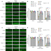
Figure 6. Dabrafenib attenuates ERK phosphorylation in the cochlear organ of Corti during the multicycle cisplatin treatment protocol.
(A) Representative images of cochlear cryosections stained with DAPI (blue) and phosphorylated ERK (green) on day 4 of the protocol in Figure 2A. Mice were sacrificed 45 minutes following the last cisplatin injection of cycle 1. Total n = 3 mice from each experimental group were tested. (B) Representative images of cochlear cryosections on day 32. Mice were sacrificed 45 minutes following the last cisplatin injection of cycle 3. Experimental groups from left to right are as follows: carrier alone, cisplatin alone, 3.0 mg/kg dabrafenib alone, and 3.0 mg/kg dabrafenib + cisplatin. Total n = 3 mice from each experimental group were tested. Scale bars: 60 μm.
Figure 7
Figure 7. Dabrafenib-treated mice have less weight loss during the multicycle cisplatin protocol.
(A) Weight loss over the 42-day treatment protocol shown in Figure 2A. Carrier (black), cisplatin alone (yellow), 15 mg/kg dabrafenib alone (purple), 3 mg/kg dabrafenib alone (orange), 15 mg/kg dabrafenib plus cisplatin (blue), 3 mg/kg dabrafenib plus cisplatin (red), and 0.6 mg/kg dabrafenib plus cisplatin (green). (B) Kaplan-Meier survival curves of mouse cohorts going to day 42 following protocol in Figure 2A. Data shown as means ± SEM; *P < 0.05, **P < 0.01, ***P < 0.001 compared with cisplatin alone by 2-way ANOVA with Bonferroni’s post hoc test.
Figure 8
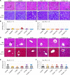
Figure 8. Dabrafenib and cisplatin do not cause significant damage to the kidneys or liver.
(A) Representative H&E and PAS images of the kidney at 20× original magnification. Treatment groups from left to right are as follows: carrier alone, cisplatin alone, 3 mg/kg dabrafenib alone, 3 mg/kg dabrafenib plus cisplatin, 15 mg/kg dabrafenib alone, and 15 mg/kg dabrafenib plus cisplatin. (B) Kidneys collected immediately after cycle 3 and (C) 4 months after cycle 3 were stained with H&E and PAS and scored in a blinded manner by an experienced pathologist. Score of 0 indicates no visible damage while a score of 4 indicates very severe damage. (D) Representative H&E- and Masson’s trichrome– stained images of the liver at 20× original magnification. (E) Histology scores of liver samples collected immediately after cycle 3 and (F) 4 months after cycle 3 (165 days) blindly scored by experienced pathologist. 0 = normal, 1 = mild damage, 2 = moderate damage, 3 = severe damage, and 4 = very severe (fulminant) damage. Data shown as means ± SEM; all groups compared with one another by 2-way ANOVA with Bonferroni’s post hoc test.

References

    1. Xu H, et al. Common variants in ACYP2 influence susceptibility to cisplatin-induced hearing loss. Nat Genet. 2015;47(3):263–266. doi: 10.1038/ng.3217. - DOI - PMC - PubMed
    1. Dasari S, Tchounwou PB. Cisplatin in cancer therapy: molecular mechanisms of action. Eur J Pharmacol. 2014;740:364–378. doi: 10.1016/j.ejphar.2014.07.025. - DOI - PMC - PubMed
    1. Ding D, et al. Review: ototoxic characteristics of platinum antitumor drugs. Anat Rec (Hoboken) 2012;295(11):1851–1867. doi: 10.1002/ar.22577. - DOI - PubMed
    1. Steyger PS. Mechanisms of aminoglycoside- and cisplatin-induced ototoxicity. Am J Audiol. 2021;30(3s):887–900. doi: 10.1044/2021_AJA-21-00006. - DOI - PMC - PubMed
    1. Breglio AM, et al. Cisplatin is retained in the cochlea indefinitely following chemotherapy. Nat Commun. 2017;8(1):1654–1659. doi: 10.1038/s41467-017-01837-1. - DOI - PMC - PubMed

Publication types