. 2023 Dec 22;8(24):e172977.
doi: 10.1172/jci.insight.172977.

PGF2α signaling drives fibrotic remodeling and fibroblast population dynamics in mice

Affiliations

PGF2α signaling drives fibrotic remodeling and fibroblast population dynamics in mice

Luis R Rodriguez et al. JCI Insight. .

Abstract

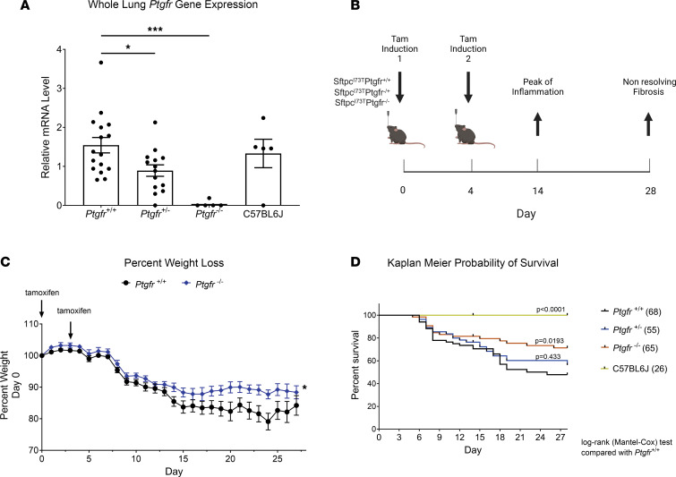
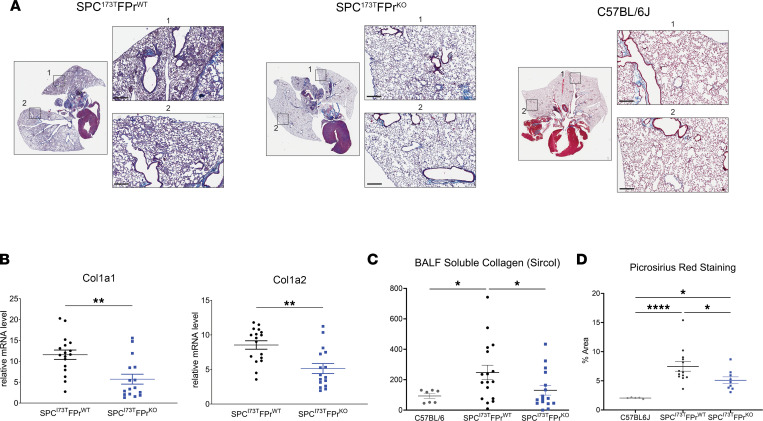
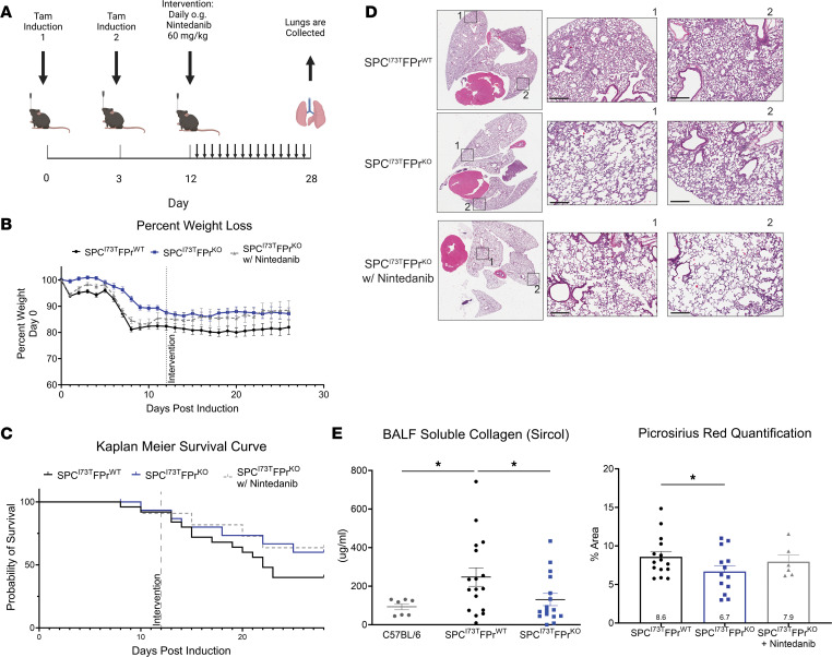
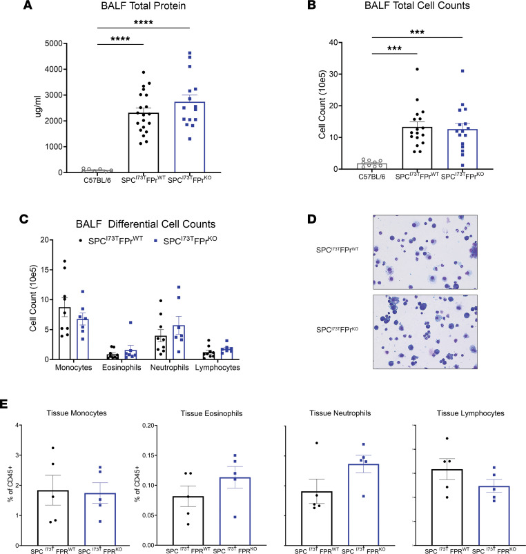
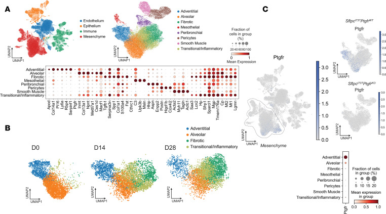
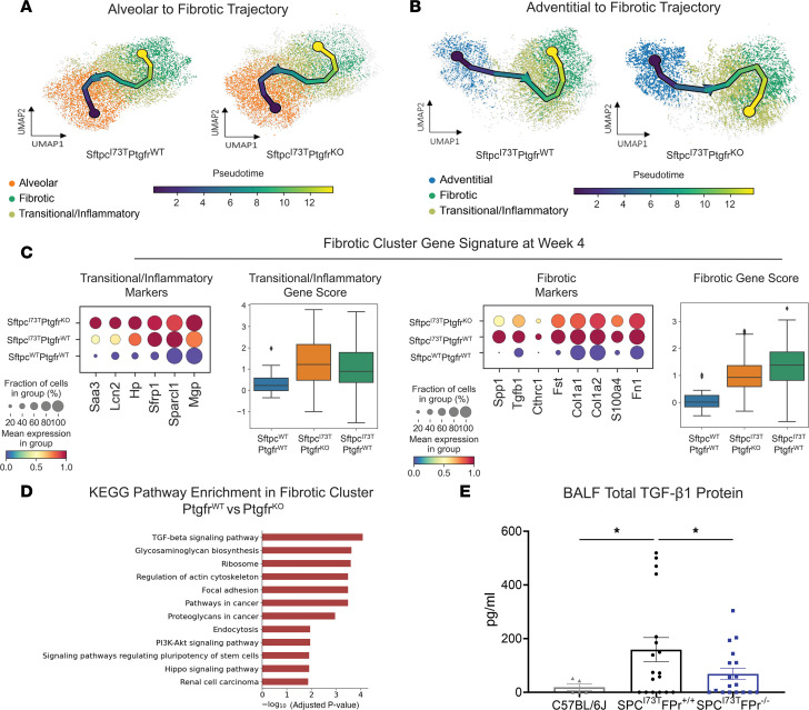
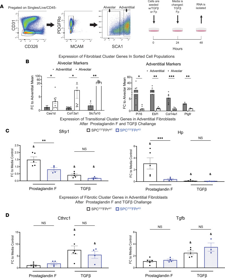
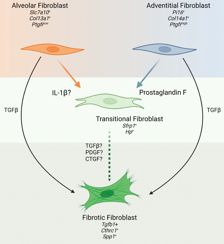
Idiopathic pulmonary fibrosis (IPF) is a chronic parenchymal lung disease characterized by repetitive alveolar cell injury, myofibroblast proliferation, and excessive extracellular matrix deposition for which unmet need persists for effective therapeutics. The bioactive eicosanoid, prostaglandin F2α, and its cognate receptor FPr (Ptgfr) are implicated as a TGF-β1-independent signaling hub for IPF. To assess this, we leveraged our published murine PF model (IER-SftpcI73T) expressing a disease-associated missense mutation in the surfactant protein C (Sftpc) gene. Tamoxifen-treated IER-SftpcI73T mice developed an early multiphasic alveolitis and transition to spontaneous fibrotic remodeling by 28 days. IER-SftpcI73T mice crossed to a Ptgfr-null (FPr-/-) line showed attenuated weight loss and gene dosage-dependent rescue of mortality compared with FPr+/+ cohorts. IER-SftpcI73T/FPr-/- mice also showed reductions in multiple fibrotic endpoints for which administration of nintedanib was not additive. Single-cell RNA-Seq, pseudotime analysis, and in vitro assays demonstrated Ptgfr expression predominantly within adventitial fibroblasts, which were reprogrammed to an "inflammatory/transitional" cell state in a PGF2α /FPr-dependent manner. Collectively, the findings provide evidence for a role for PGF2α signaling in IPF, mechanistically identify a susceptible fibroblast subpopulation, and establish a benchmark effect size for disruption of this pathway in mitigating fibrotic lung remodeling.

Keywords: Eicosanoids; Fibrosis; Molecular biology; Pulmonology.


Figures

Figure 1
Figure 1. Deletion of Ptgfr reduces morbidity and mortality in IER-SftpcI73T mice.
(A) Ptgfr mRNA content of whole lung mRNA isolated from generated lines of IER-SftpcI73T/Ptgfr mice deficient in 0, 1, 2 Ptgfr alleles assayed by qPCR. Ordinary 1-way ANOVA testing was performed, with statistical significance between groups denoted by *P < 0.05 and **P < 0.005. (B) Schematic of single, split i.p. or split OG dosing strategy employed for TAM induction of IER-SftpcI73T/Ptgfr–/– and IER-SftpcI73T/FPr+/+ cohorts. (C) Representative weight loss curve from a single cohort containing IER-SftpcI73T/Ptgfr–/– (n = 20) and IER-SftpcI73T/Ptgfr++–(n = 13) controls; mixed-effects modeling was performed with time x genotype. *P < 0.05. (D) Aggregate Kaplan-Meier curve for IER-SftpcI73T/Ptgfr mice from 3 cohorts separately induced with either single i.p. or split i.p. doses of TAM in corn oil, with total numbers of each Ptgfr genotype shown. Negative controls consisted of SftpcWT C57BL/6 mice given TAM or uninduced IER-SftpcI73T/Ptgfr–/– animals. P values versus IER-SftpcI73T/Ptgfr–/– obtained by log-rank testing are shown.
Figure 2
Figure 2. Ptgfr deficiency mitigates collagen expression and deposition after induction of SftpcI73T.
(A) Representative histology from IER-SftpcI73T/Ptgfr+/+ and IER-SftpcI73T/Ptgfr–/– mice 28 days after TAM and development of fibrosis. Images are derived from Masson’s trichrome–stained sections. Scale bars: 300 μM. (B) Relative fold mRNA levels between IER-SftpcI73T/Ptgfr+/+ and IER-SftpcI73T/Ptgfr–/– measured via qPCR demonstrates decreased Col1a1 and Col1a2 in IER-SftpcI73T/Ptgfr–/– mice 28 days after TAM induction. Statistical significance testing was performed using 2 tailed Welch’s t test. **P < 0.005. (C) Quantification of soluble collagen in BALF from mice during fibrotic remodeling reveals a lower concentration in IER-SftpcI73T/Ptgfr–/– mice. (D) PSR staining for collagen fibrils indicates mitigation of collagen deposition in IER-SftpcI73T/Ptgfr–/– mice. Quantification was performed using ImageJ; data represent percentage of total section area. All quantified data in this figure are derived from IER-SftpcI73T/Ptgfr+/+ (n = 17) and IER-SftpcI73T/Ptgfr–/– (n = 16). In C and D, ordinary 1-way ANOVA testing was performed. *P < 0.05, ****P < 0.00005.
Figure 3
Figure 3. Nintedanib intervention is not additive to Ptgfr deficiency in IER-SftpcI73T mice.
(A) Daily nintedanib intervention (60 mg/kg) was initiated at D12 after TAM induction. Following 16 days of intervention, surviving mice were euthanized and processed to evaluate fibrotic endpoints. (B) Weight loss as a percent of starting weight was tracked throughout the study; nintedanib intervention in IER-SftpcI73T/Ptgfr–/– mice did not reduce mean weight loss. (C) Kaplan-Meier survival analysis by log-rank testing demonstrates a nonsignificant improved probability of survival in IER-SftpcI73T/Ptgfr–/– that was not improved through nintedanib intervention. (D) Representative histology from IER-SftpcI73T/Ptgfr+/+, IER-SftpcI73T/Ptgfr–/–, and nintedanib-treated IER-SftpcI73T/Ptgfr–/– mice 28 days after TAM and development of fibrosis. Images are derived from H&E-stained sections. Scale bars: 300 μM. (E) Soluble collagen in BALF as measured by Sircol assay and fibrillar collagen in histological sections measured by PSR staining demonstrated a significant decrease in IER-SftpcI73T/Ptgfr–/– mice; again, nintedanib treatment did not improve these outcomes. Quantification of PSR was performed using ImageJ, and data represent percentage of total section area. Survival and weight loss data are derived from IER-SftpcI73T/Ptgfr+/+ (n = 26), IER-SftpcI73T/Ptgfr–/– without nintedanib (n = 12), and IER-SftpcI73T/Ptgfr–/– with nintedanib (n = 12). Soluble collagen and PSR analysis included IER-SftpcI73T/Ptgfr+/+ (n = 14), IER-SftpcI73T/Ptgfr–/– without nintedanib (n = 11), and IER-SftpcI73T/Ptgfr–/– with nintedanib (n = 7). Ordinary 1-way ANOVA testing was performed. *P < 0.05.
Figure 4
Figure 4. Ptgfr deficiency has no effect on early lung injury and inflammation in IER-SftpcI73T.
(A and B) Quantification of total protein and total cell counts in BALF did not result in a significant difference between IER-SftpcI73T/Ptgfr+/+ (n = 17) and IER-SftpcI73T/Ptgfr–/– (n = 16) mice 14 days after TAM induction. Ordinary 1-way ANOVA testing was performed. ***P < 0.005, ****P < 0.0005. (C) BALF cell differential determined by quantification of modified Giemsa-stained cytospins yielded no significant difference between IER-SftpcI73T/Ptgfr+/+ (n = 9) and IER-SftpcI73T/Ptgfr–/– (n = 7) mice 14 days after TAM induction. (D) Representative Giemsa-stained images from IER-SftpcI73T/Ptgfr+/+ and IER-SftpcI73T/Ptgfr–/– mice 14 days after TAM induction. Total original magnification, ×100. (E) Flow cytometry quantification of whole-lung single-cell suspensions confirms that there is no differential immune cell infiltration between IER-SftpcI73T/Ptgfr+/+ (n = 5) and IER-SftpcI73T/Ptgfr–/–(n = 5) mice 14 days after TAM induction. Ordinary 1-way ANOVA testing was performed for this analysis.
Figure 5
Figure 5. Ptgfr expression is limited to adventitial and alveolar fibroblasts.
(A) UMAP clustering 94,258 cells identifies 4 primary cell compartments in IER-SftpcI73T/Ptgfr+/+, IER-SftpcI73T/Ptgfr–/–, and uninduced controls. Subclustering of the mesenchymal compartment identifies 8 mesenchymal clusters defined by marker genes depicted as a gradient dot plot. (B) UMAP projections of Pdgfra+ mesenchymal populations across time identifies 2 injury-specific clusters (fibrotic and transitional/inflammatory). (C) UMAP projection of all cells identifies the restriction of Ptgfr expression to the mesenchymal compartment and lack of Ptgfr expression in IER-SftpcI73T/Ptgfr–/– mice. Gradient dot plot of Ptgfr expression within the mesenchyme demonstrates increased expression and increased percent expression of Ptgfr in adventitial fibroblasts as compared with alveolar fibroblasts.
Figure 6
Figure 6. Ptgfr deficiency alters fibroblast lineage trajectory through fibrotic remodeling.
(A) UMAP analysis of reclustered alveolar, transitional/inflammatory, and fibrotic fibroblasts with superimposed vector from pseudotime trajectory analysis reveals no Ptgfr-dependent effect on terminal node. (B) UMAP analysis of reclustered adventitial, transitional/inflammatory, and fibrotic fibroblasts with superimposed vector from pseudotime trajectory analysis demonstrates a Ptgfr-dependent effect on terminal node. In IER-SftpcI73T/Ptgfr–/–, the terminal node is found in the transitional/inflammatory cluster, while SftpcI73T/Ptgfr+/+ samples have a vector terminating in the fibrotic cluster. (C) Comparative analysis of gene expression within the fibrotic cluster of IER-SftpcI73T/Ptgfr+/+ and IER-SftpcI73T/Ptgfr–/– mice is presented by gradient gene expression dot plots. Genes seen in dot plots were combined to generate a score and plotted in a box plot. Marker genes associated with the transitional/inflammatory cluster are comparatively elevated in IER-SftpcI73T/Ptgfr–/– mice, while fibrotic marker genes are elevated in the IER-SftpcI73T/Ptgfr+/+ mice. All comparisons achieved statistical significance. (D) KEGG pathway enrichment analysis comparing the fibrotic clusters identifies multiple pathways associated with cytoskeletal rearrangement, mesenchymal activation, and TGF-β signaling that are upregulated in IER-SftpcI73T/Ptgfr+/+ mice. (E) Measurement of BALF TGF-β1 via ELISA demonstrates a significant decrease in IER-SftpcI73T/Ptgfr–/– mice (n = 18) as compared with IER-SftpcI73T/Ptgfr+/+ mice (n = 18). Ordinary 1-way ANOVA testing was performed. *P < 0.05.
Figure 7
Figure 7. In vitro prostaglandin F2α (PGF) challenge promotes adventitial fibroblast entry into the transitional/inflammatory state.
(A) Sorting strategy for the isolation of adventitial and alveolar fibroblast used in IER-SftpcI73T/Ptgfr+/+ and IER-SftpcI73T/Ptgfr–/– prior to induction by TAM. Initial gating was performed on the CD45CD31CD326McamPdgfra+ population. Adventitial fibroblasts are Sca1+, and the Sca1 population is made up of the alveolar fibroblasts. After sorting, cells were seeded for 48 hours on tissue culture plastic with either 10 ng/mL TGF-β1, 500 nM PGF, or media control. (B) Gene expression analysis via qPCR in untreated adventitial and alveolar fibroblasts quantifying the expression of population-specific marker genes confirms the identity of target fibroblasts. (C) Quantification of transitional cluster maker genes after 48-hour challenge demonstrates the potential for FPr to induce the transitional state in adventitial fibroblasts. This is not observed in adventitial fibroblasts lacking the FPr. (D) Quantification of fibrotic cluster marker genes after 48-hour challenge confirms that TGF-β promotes entry adventitial fibroblasts into the fibrotic state independent of FPr status. Ordinary 1-way ANOVA testing was performed. *P < 0.05, **P < 0.005, ***P < 0.0005. Statistical significance between treatment and media control denoted by &P < 0.05.
Figure 8
Figure 8. Role of PGF as a driver of fibroblast heterogeneity in pulmonary fibrosis.
Application of single-cell discovery and in vitro validation identifies PGF as a driver of adventitial fibroblast activation into the transitional fibroblast state. Paired with work by Tsukui et al. demonstrating potential of alveolar (lipo) fibroblasts to enter the same transitional state through IL-1B stimulation (23), there is now evidence for differential drivers of transitional fibroblast differentiation within fibroblast population subsets. Concurrently, classical profibrotic TGF-β signaling drives entry of adventitial and alveolar fibroblasts as an “override” into a terminally activated fibroblast state expressing Cthrc1. Contextualizing these findings with previous work by Oga et al. (32) suggests that PGF-stimulated transitional fibroblast may be more sensitive to a second hit (e.g., TGF-β, PDGF, or CTGF), accelerating the expansion of the terminal fibrotic fibroblast population.

Update of

References

    1. Raghu G, et al. Diagnosis of idiopathic pulmonary fibrosis. An official ATS/ERS/JRS/ALAT clinical practice guideline. Am J Respir Crit Care Med. 2018;198(5):e44–e68. doi: 10.1164/rccm.201807-1255ST. - DOI - PubMed
    1. Raghu G, et al. Incidence and prevalence of idiopathic pulmonary fibrosis. Am J Respir Crit Care Med. 2006;174(7):810–816. doi: 10.1164/rccm.200602-163OC. - DOI - PubMed
    1. Katzenstein ALA, Myers JL. Idiopathic pulmonary fibrosis - clinical relevance of pathologic classification. Am J Respir Crit Care Med. 1998;157(4 pt 1):1301–1315. doi: 10.1164/ajrccm.157.4.9707039. - DOI - PubMed
    1. Lederer DJ, Martinez FJ. Idiopathic pulmonary fibrosis. N Engl J Med. 2018;379(8):797–798. - PubMed
    1. Raghu G, et al. Idiopathic pulmonary fibrosis (an update) and progressive pulmonary fibrosis in adults: an official ATS/ERS/JRS/ALAT clinical practice guideline. Am J Respir Crit Care Med. 2022;205(9):e18–e47. doi: 10.1164/rccm.202202-0399ST. - DOI - PMC - PubMed

Publication types