. 2023 Dec 6;13(12):jkad240.
doi: 10.1093/g3journal/jkad240.

A genome-wide overexpression screen reveals Mycobacterium smegmatis growth inhibitors encoded by mycobacteriophage Hammy

Affiliations

A genome-wide overexpression screen reveals Mycobacterium smegmatis growth inhibitors encoded by mycobacteriophage Hammy

Isabel Amaya et al. G3 (Bethesda). .

Abstract

During infection, bacteriophages produce diverse gene products to overcome bacterial antiphage defenses, to outcompete other phages, and to take over cellular processes. Even in the best-studied model phages, the roles of most phage-encoded gene products are unknown, and the phage population represents a largely untapped reservoir of novel gene functions. Considering the sheer size of this population, experimental screening methods are needed to sort through the enormous collection of available sequences and identify gene products that can modulate bacterial behavior for downstream functional characterization. Here, we describe the construction of a plasmid-based overexpression library of 94 genes encoded by Hammy, a Cluster K mycobacteriophage closely related to those infecting clinically important mycobacteria. The arrayed library was systematically screened in a plate-based cytotoxicity assay, identifying a diverse set of 24 gene products (representing ∼25% of the Hammy genome) capable of inhibiting growth of the host bacterium Mycobacterium smegmatis. Half of these are related to growth inhibitors previously identified in related phage Waterfoul, supporting their functional conservation; the other genes represent novel additions to the list of known antimycobacterial growth inhibitors. This work, conducted as part of the HHMI-supported Science Education Alliance Gene-function Exploration by a Network of Emerging Scientists (SEA-GENES) project, highlights the value of parallel, comprehensive overexpression screens in exploring genome-wide patterns of phage gene function and novel interactions between phages and their hosts.

Keywords: Mycobacterium smegmatis; cytotoxicity; mycobacteriophage.


Conflict of interest statement

Conflicts of interest statement The authors declare no conflict of interest.

Figures

Fig. 1.
Fig. 1.
The genome of phage Hammy. The Hammy genome is shown as a line with kbp markers and genes represented by boxes—those above the line are transcribed rightwards and those below are transcribed leftwards. Numbers inside the box correspond to gene numbers and predicted functions are indicated above or below each gene. Box shading corresponds to cytotoxicity scoring, with white boxes designating genes found to have no effect on M. smegmatis growth (cytotoxicity score 0), hatched box indicating omitted gene 19 not tested in this study, and blue representing observed toxicity in our assay. The saturation of blue boxes corresponds to the severity of growth inhibition using the following scores: light blue (score 1; reduction in colony size; genes 12, 29, 36, 53, 60, 61, 63, 67, and 84), medium blue (score 2; 1–3 log reduction in viability; genes 32, 90, and 92), and dark blue (score 3; >3-log reduction in viability; genes 9, 20, 34, 50, 51, 54, 56, 58, 68, 69, 77, and 78).
Fig. 2.
Fig. 2.
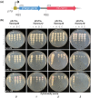
Expression of phage genes from the pExTra plasmid. a) Recombinant pExTra plasmids constructed in this study encode Hammy gene sequences downstream of the pTet promoter and upstream of mcherry. The two genes in this pExTra operon are transcriptionally linked, each bearing their own translational signals. b) Results of representative cytotoxicity assays are shown to demonstrate the range of observed growth defects. In each assay, colonies of M. smegmatis mc2155 transformed with the specified pExTra plasmid were resuspended, serially diluted, and spotted on 7H11 Kan media containing 0, 10, or 100 ng/ml aTc. Triplicate colonies (a, b, c) were tested for each gene alongside a positive control strain (+) transformed with pExTra02 (expressing wildtype Fruitloop 52) and a negative control strain (−) transformed with pExTra03 (expressing Fruitloop 52 I70S).
Fig. 3.
Fig. 3.
Comparison of Hammy and Waterfoul patterns of cytotoxicity. a) The proportion of Waterfoul (top) and Hammy (bottom) genes tested in this study or Heller et al. (2022) that were assigned each score (0–3) is represented as a stacked bar chart. Heller et al. scored two cytotoxic genes (Waterfoul 8 and 86) as 3* as recovery of pExTra trasformants was inhibited by presence of these gene inserts even in the absence of aTc. b) The proportion of Waterfoul (top) and Hammy (bottom) cytotoxic genes (score 13) that have NKF or that fall into various functional classes is represented as a stacked bar chart with different colors indicating functional class as described in the key. c) Shown is a chart listing the 45 out of 46 gene phamilies shared by Hammy and Waterfoul that were tested in both studies. Phamily number designations and functions are listed (NKF, no known function; TMD, transmembrane domain) next to representative homologous genes from Hammy and Waterfoul, with boxes shaded by cytotoxicity score. A binary indicator of whether homologous genes were both classified as toxic or nontoxic is illustrated by black or red shading, and % protein identity is indicated by the purple gradient boxes. d) For both Hammy (top) and Waterfoul (bottom), cytotoxicity scores (0–3) for each gene are plotted in genomic order along the horizontal axis, showing clustering of cytotoxic genes in the early and late lytic regions. e) Aligned maps of Hammy and Waterfoul provide a zoomed-in view of two genomic regions harboring clusters of cytotoxic genes, with boxes numbered by gene and shaded corresponding to the cytotoxicity scores (Heller et al. 2022) as in panels a and c. Gene phamilies found in both genomes are labeled by phamily numbers (taken from phagesDB.org as of February 27, 2023) with asterisks indicating those gene phamilies that were found to be cytotoxic (scores 1–3) or nontoxic consistently across both genomes.
Fig. 4.
Fig. 4.
Overproduction of multidomain mycobacteriophage lysin a proteins. a) lysA genes from various mycobacteriophages were expressed from pExTra and tested in the plate-based cytotoxicity assay alongside pExTra02 (+) and pExTra03 (−) controls. b) The same strains were evaluated in an endpoint liquid growth assay. Two colonies of each transformed M. smegmatis strain were grown until saturation and subcultured in duplicate 24-well plates. Upon reaching an OD600 of ∼0.2, one set of cultures was induced by addition of 100 ng/ml aTc and the other left as an uninduced control. Growth was monitored over 24 h of induction at 37 °C with shaking. Plate images from a representative experiment showing lysis and culture appearance after 24 h with (right image) or without induction (left image) are shown. c) The tested Lysin A proteins harbor various enzymatic domains as predicted by HHPRED and as previously described (Payne and Hatfull 2012), including two putative peptidase domains, P1 (1CV8_A; probability 99.2%) and P2 (3NPF_B; probability >88%), a G19-like glycosyl hydrolase domain (5H7T_A; probability >92%), and a G25-like glycosyl hydrolase domain (cd06419; probability >99%). Relationships between these Lysin A proteins are represented by the phylogenetic tree (left; Clustal Omega and Splitstree (Huson and Bryant 2006)) and % amino acid identity values on the right.

References

    1. Anders KR, Barekzi N, Best AA, Frederick GD, Mavrodi DV, Vazquez E; SEA-PHAGES; Amoh NYA, Baliraine FN, Buchser WJ, et al. . 2017. Genome sequences of mycobacteriophages amgine, amohnition, Bella96, cain, DarthP, Hammy, krueger, LastHope, peanam, PhelpsODU, phrank, SirPhilip, slimphazie, and unicorn. Genome Announc. 5(49):e01202–17. doi:10.1128/genomeA.01202-17. - DOI - PMC - PubMed
    1. Catalão MJ, Milho C, Gil F, Moniz-Pereira J, Pimentel M. 2011. A second endolysin gene is fully embedded in-frame with the lysA gene of mycobacteriophage Ms6. Plos One. 6(6):e20515. doi:10.1371/journal.pone.0020515. - DOI - PMC - PubMed
    1. Catalão MJ, Pimentel M. 2018. Mycobacteriophage lysis enzymes: targeting the mycobacterial cell envelope. Viruses. 10(8):428. doi:10.3390/v10080428. - DOI - PMC - PubMed
    1. Cresawn SG, Bogel M, Day N, Jacobs-Sera D, Hendrix RW, Hatfull GF. 2011. Phamerator: a bioinformatic tool for comparative bacteriophage genomics. BMC Bioinformatics. 12(1):395. 10.1186/1471-2105-12-395. - DOI - PMC - PubMed
    1. Dedrick RM, Guerrero Bustamante CA, Garlena RA, Pinches RS, Cornely K, Hatfull GF. 2019. Mycobacteriophage ZoeJ: a broad host-range close relative of mycobacteriophage TM4. Tuberculosis (Edinb). 115:14–23. doi:10.1016/j.tube.2019.01.002. - DOI - PMC - PubMed

Publication types

MeSH terms

LinkOut - more resources