. 2024 Feb:320:104185.
doi: 10.1016/j.resp.2023.104185. Epub 2023 Nov 5.

Low level CO2 supplementation maintains isocapnia and reveals ventilatory long-term facilitation in rats

Affiliations

Low level CO2 supplementation maintains isocapnia and reveals ventilatory long-term facilitation in rats

Shawn Miller Jr et al. Respir Physiol Neurobiol. 2024 Feb.

Abstract

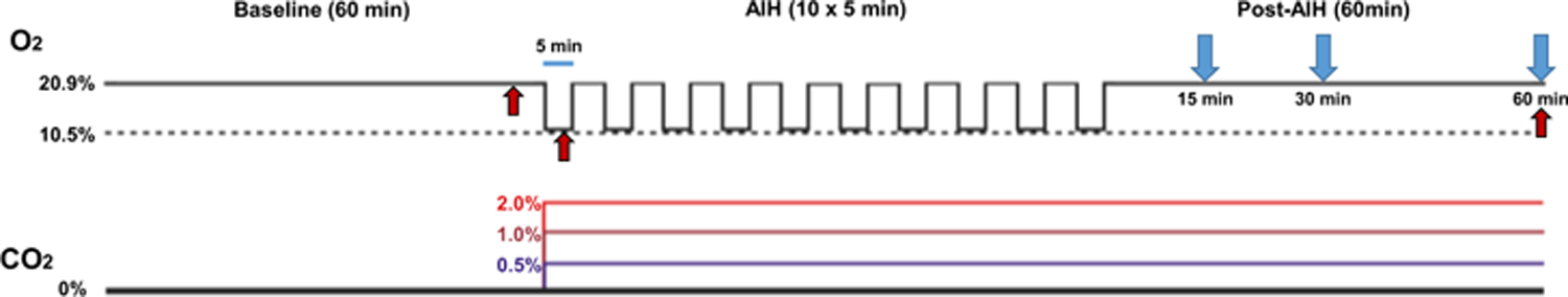
Acute, intermittent hypoxia (AIH) induces ventilatory long-term facilitation (vLTF) in awake, freely behaving rats under poikilocapnic and isocapnic experimental conditions. Establishing pre-clinical methods for vLTF induction that more closely align with successful protocols in humans and anesthetized rats would minimize dissonance in experimental findings and improve translational aspects of vLTF. Here, we tested several levels of low-dose CO2 supplementation during and after AIH to determine 1) the lowest amount of inspired CO2 that would maintain isocapnia in rats during a vLTF protocol, and 2) the net impact of supplemental CO2 on vLTF expression. Rats received one of four levels of inspired CO2 (0%, 0.5%, 1% or 2%) administered during AIH and for the 60 min following AIH to quantify vLTF. Our findings indicated that 2% inspired CO2 was sufficient to maintain isocapnia across the AIH protocol and reveal significant vLTF. These findings provide evidence-based support for using 2% supplemental CO2 during and after AIH when assessing vLTF in rats.

Keywords: Isocapnia; Long-term facilitation; Unrestrained Barometric Plethysmography; Ventilation.


Figures

Figure 1.
Figure 1.. Barometric plethysmography protocol for vLTF.
Unrestrained barometric plethysmography was used to assess ventilation and the development of vLTF in unanaesthetized, freely behaving rats. Rats were acclimated to the plethysmography chambers and breathed normal room air (i.e. normoxia) during the 60 min baseline period (BL; 20.9% O2, balance N2). Rats were then given AIH consisting of 10, 5-min exposures to 10.5% O2 (balance N2) and returned to baseline conditions for 60 min (Post-AIH). Experimental groups received supplemental inspired CO2 of either 0.5%, 1%, or 2% beginning at the start of AIH and continuing for the duration of the plethysmography session. A separate set of rats with indwelling femoral artery catheters underwent identical plethysmography testing. Blood samples (denoted with red arrows) were taken during baseline, the first hypoxic exposure, and 60 min post-AIH to assess for changes in PaCO2 across time during the protocol. Ventilation parameters were compared at 15 min, 30 min, and 60 min post-AIH (blue arrows).
Figure 2.
Figure 2.. Supplemental CO2 did not alter the hypoxic ventilatory response.
The hypoxic ventilatory response (HVR) was assessed during the first AIH hypoxic episode. All groups experienced similar increases in VE (A), VT (B), and respiratory frequency (C) in response to hypoxia. One-way ANOVAs. All comparisons p>0.05. All groups: n=6. Data represent means ± SEM.
Figure 3.
Figure 3.. CO2 supplementation revealed robust vLTF.
Ventilation was assessed at 15 min, 30 min, and 60 min post-AIH to determine the magnitude of vLTF with supplemental CO2. Rats receiving 2% CO2 showed significantly higher VE at all post-AIH time points relative to baseline (BL), while the 1% group showed significant elevations in VE at 15 min and 30 min (A). The 2% CO2 group also showed significantly higher VE compared with the 0% group at both 30 min and 60 min (A). 2% CO2 also enhance VT production above BL at all post-AIH time points; and this increased VT was higher than the 0% group at 15 min and 60 min post-AIH (B). Respiratory frequency remained similar to BL levels in all groups after AIH with one exception: 2% CO2 rats breathed faster at 30 min post-AIH relative to BL and to the 0% group (C). Two-way ANOVAs with repeated measures. *p<0.05 from BL; #p<0.05 from 0%; ^p<0.05 from 0.5%. All groups: n=6. Data represent means ± SEM.
Figure 4.
Figure 4.. vLTF remains with CO2 supplementation when controlling for metabolism.
Measures of CO2 content in the expired gas permitted calculations of VCO2 across groups. All groups had similar baseline VCO2 (A) and baseline VE standardized to VCO2 (B) following one-way ANOVA. When vLTF was recalculated using two-way ANOVA with repeated measures including VCO2 standardization, 1% and 2% CO2 groups remained separate from others in terms of vLTF magnitude across time (C). Both 1% and 2% CO2 increased VE at 15 min, and 1% remained elevated at 60 min post-AIH (C). The 1% CO2 group also had significantly elevated VE at 15 min relative to the 0% CO2 group, and this difference remained for the 1% group at 60 min. The 1% and 2% groups did not differ from each other at any time. *: p<0.05 from BL; #: p<0.05 from 0%; ^: p<0.05 from 0.5%. All groups: n=6. Data represent means ± SEM.
Figure 5.
Figure 5.. 2% inspired CO2 maintained isocapnia throughout the plethysmography protocol.
In a separate set of rats (n=15 total), blood samples were drawn from an indwelling femoral artery catheter during BL, during the first hypoxic episode, and 60 min post AIH to assess for PaCO2 levels. Our goal was to find the lowest level of inspired CO2 that would maintain isocapnia. Only the 2% CO2 group showed consistent PaCO2 during hypoxia and for the 60 min after. Other groups (0% CO2 and 1% CO2) showed significant declines in PaCO2 during the hypoxic challenge relative to BL and 60 min. Two-way ANOVA. *p<0.05; **p<0.01. All groups: n=5. Data represent means ± SEM.

References

    1. Afsharipour B, Pearcey GEP, Rymer WZ & Sandhu MS (2023). Acute intermittent hypoxia enhances strength, and modulates spatial distribution of muscle activation in persons with chronic incomplete spinal cord injury. Exp Neurol 367, 114452. - PubMed
    1. Bach KB & Mitchell GS (1996). Hypoxia-induced long-term facilitation of respiratory activity is serotonin dependent. Respir Physiol 104, 251–260. - PubMed
    1. Baker-Herman TL & Strey KA (2011). Similarities and differences in mechanisms of phrenic and hypoglossal motor facilitation. Respir Physiol Neurobiol 179, 48–56. - PMC - PubMed
    1. Barok R, Grittner JML & Dougherty BJ (2021). The long-term impact of ovariectomy on ventilation and expression of phrenic long-term facilitation in female rats. Exp Physiol 106, 2002–2012. - PMC - PubMed
    1. Bavis RW & Mitchell GS (2003). Intermittent hypoxia induces phrenic long-term facilitation in carotid-denervated rats. J Appl Physiol 94, 399–409. - PubMed

LinkOut - more resources