Review
. 2024 Jun;34(6):4142-4154.
doi: 10.1007/s00330-023-10334-7. Epub 2023 Nov 8.

Primary immunodeficiency diseases of adults: a review of pulmonary complication imaging findings

Affiliations
Review

Primary immunodeficiency diseases of adults: a review of pulmonary complication imaging findings

Philippe A Grenier et al. Eur Radiol. 2024 Jun.

Abstract

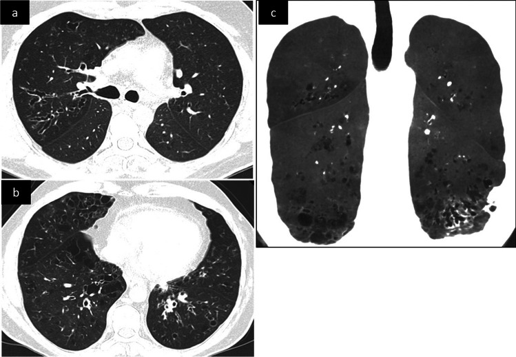
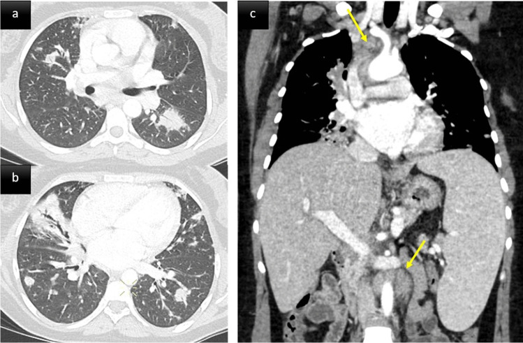
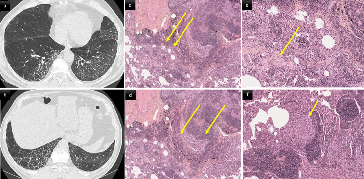
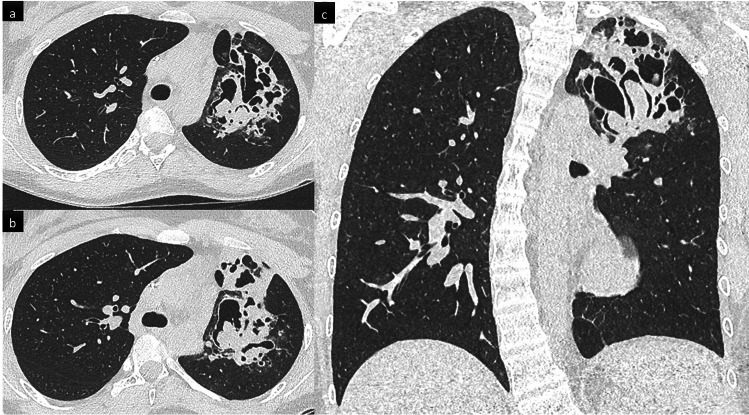
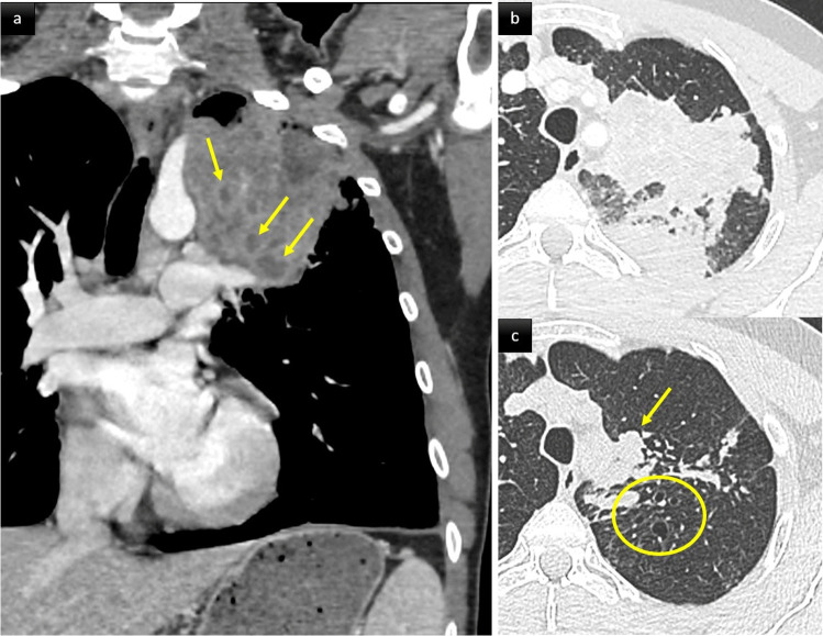
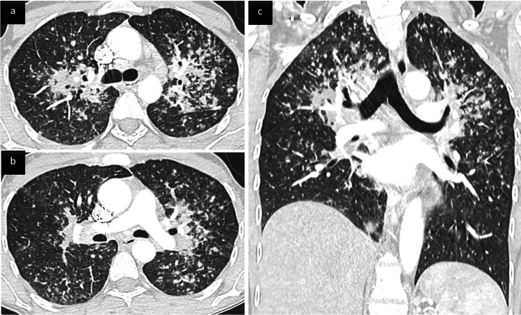
Our objective in this review is to familiarize radiologists with the spectrum of initial and progressive CT manifestations of pulmonary complications observed in adult patients with primary immunodeficiency diseases, including primary antibody deficiency (PAD), hyper-IgE syndrome (HIES), and chronic granulomatous disease (CGD). In patients with PAD, recurrent pulmonary infections may lead to airway remodeling with bronchial wall-thickening, bronchiectasis, mucus-plugging, mosaic perfusion, and expiratory air-trapping. Interstitial lung disease associates pulmonary lymphoid hyperplasia, granulomatous inflammation, and organizing pneumonia and is called granulomatous-lymphocytic interstitial lung disease (GLILD). The CT features of GLILD are solid and semi-solid pulmonary nodules and areas of air space consolidation, reticular opacities, and lymphadenopathy. These features may overlap those of mucosa-associated lymphoid tissue (MALT) lymphoma, justifying biopsies. In patients with HIES, particularly the autosomal dominant type (Job syndrome), recurrent pyogenic infections lead to permanent lung damage. Secondary infections with aspergillus species develop in pre-existing pneumatocele and bronchiectasis areas, leading to chronic airway infection. The complete spectrum of CT pulmonary aspergillosis may be seen including aspergillomas, chronic cavitary pulmonary aspergillosis, allergic bronchopulmonary aspergillosis (ABPA)-like pattern, mixed pattern, and invasive. Patients with CGD present with recurrent bacterial and fungal infections leading to parenchymal scarring, traction bronchiectasis, cicatricial emphysema, airway remodeling, and mosaicism. Invasive aspergillosis, the major cause of mortality, manifests as single or multiple nodules, areas of airspace consolidation that may be complicated by abscess, empyema, or contiguous extension to the pleura or chest wall. CLINICAL RELEVANCE STATEMENT: Awareness of the imaging findings spectrum of pulmonary complications that can occur in adult patients with primary immunodeficiency diseases is important to minimize diagnostic delay and improve patient outcomes. KEY POINTS: • Unexplained bronchiectasis, associated or not with CT findings of obliterative bronchiolitis, should evoke a potential diagnosis of primary autoantibody deficiency. • The CT evidence of various patterns of aspergillosis developed in severe bronchiectasis or pneumatocele in a young adult characterizes the pulmonary complications of hyper-IgE syndrome. • In patients with chronic granulomatous disease, invasive aspergillosis is relatively frequent, often asymptomatic, and sometimes mimicking or associated with non-infectious inflammatory pulmonary lesions.

Keywords: Bronchiectasis; Interstitial lung disease; Pulmonary infection; Pulmonary lymphoma.


Conflict of interest statement

The authors of this manuscript declare no relationships with any companies whose products or services may be related to the subject matter of the article.

Figures

Fig. 1
Fig. 1
Unenhanced CT scan in a 35-year-old patient with common variable immunodeficiency (CVID), axial (a, b) and coronal oblique with mIP reconstruction (c) images. Diffuse bronchial wall thickening, varicose and cylindrical bronchiectasis with lower and right predominance. The mIP reconstruction shows mosaicism, a few scattered cysts in the lung bases and distal traction bronchiectasis in the left lower lobe
Fig. 2
Fig. 2
A middle aged woman with CVID. Post contrast thoraco-abdominal CT scan, axial parenchymal (a, b) and coronal soft tissue (c) images. Lower zone predominant broncho centric nodules and airspace consolidation consistent with granulomatous-lymphocytic interstitial lung disease (GLILD). The patient also presented with splenomegaly, mediastinal and retroperitoneal lymphadenopathy (arrows)
Fig. 3
Fig. 3
66-year-old male with CVID and biopsy proven GLILD. CT scan (a, b) and histological (cf) correlation. a, b Smooth and irregular thickening of interlobular septa associated with intralobular reticular lines and architectural distortion in the lower lobes. c: diffuse cellular interstitial infiltrate. The alveolar interstitium is markedly expanded by a dense collection of mature lymphocytes and plasma cells (hematoxylin–eosin, original magnification × 5). d Follicular bronchiolitis. Dense airway-centered lymphoid hyperplasia with several small nodular lymphoid aggregates (hematoxylin × 5). e Polyps of organizing pneumonia (hematoxylin × 10). f Non-necrotizing granulomatous inflammation. Scattered loose aggregates of epithelioid histiocytes often found within interstitial lymphoid background (hematoxylin × 20)
Fig. 4
Fig. 4
Chronic aspergillosis in a 32-year-old patient with autosomal dominant hyper IgE syndrome (HIES). Axial (a, b) and coronal mIP (c) CT scan images. Large cavities in the left upper lobe (a, c) with evidence of intra cavitary aspergillomas. Note the thoracic scoliosis, which is a frequent finding in HIES
Fig. 5
Fig. 5
Chronic granulomatous disease (CGD) revealed by a mixed pattern of aspergillosis in a 41 yo man. Coronal post contrast (a) and axial parenchymal (b, c) CT images. Large partially necrotic consolidation in the left upper lobe. Tubular opacities less dense than airspace consolidation are consistent with plugged dilated airways (arrows). Note the presence of underlying varicose bronchiectasis in the same lobe (circle)
Fig. 6
Fig. 6
Sarcoid-like infiltrative lung disease following systemic aspergillosis in a 46 yo patient with chronic granulomatous disease (CGD). Axial (a, b) and coronal (c) CT scan images show innumerable small nodules with a peri-lymphatic distribution (peri-broncho-vascular, centrilobular and subpleural) predominant in the upper parts of the lungs, associated with perihilar confluence of nodules, and enlarged hilar and mediastinal lymph nodes. Granulomas were confirmed on transbronchial biopsies

References

    1. Tangye SG, Al-Herz W, Bousfiha A, et al. Human inborn errors of immunity: 2019 update on the classification from the International Union of Immunological Societies Expert Committe. J Clin Immunol. 2019;40:24–64. doi: 10.1007/s10875-019-00737-x. - DOI - PMC - PubMed
    1. Thalhammer J, Kindle G, Nieters A, et al. Initial presenting manifestations in 16,486 patients with inborn errors of immunity include infections and noninfectious manifestations. J Allergy Clin Immunol. 2021;148:1332–1341. doi: 10.1016/j.jaci.2021.04.015. - DOI - PubMed
    1. Schussler E, Beasley MB, Maglione PJ. Lung disease in primary antibody deficiencies. J Allergy Clin Immunol Pract. 2016;4:1039–1052. doi: 10.1016/j.jaip.2016.08.005. - DOI - PMC - PubMed
    1. Gennery AR, Holland SM. Primary immunodeficiencies: not just paediatric diseases. Eur Respir J. 2015;45:1521–1523. doi: 10.1183/09031936.00020215. - DOI - PubMed
    1. Baumann U, Routes JM, Soler-Palacín P, Jolles, The lung in primary immunodeficiencies: new concepts in infection and inflammation. Front Immunol. 2018;9:1837. doi: 10.3389/fimmu.2018.01837. - DOI - PMC - PubMed

MeSH terms