. 2023 Nov 7;30(1):91.
doi: 10.1186/s12929-023-00978-4.

Spatiotemporal roles of AMPK in PARP-1- and autophagy-dependent retinal pigment epithelial cell death caused by UVA

Affiliations

Spatiotemporal roles of AMPK in PARP-1- and autophagy-dependent retinal pigment epithelial cell death caused by UVA

Anthony Yan-Tang Wu et al. J Biomed Sci. .

Abstract

Background: Although stimulating autophagy caused by UV has been widely demonstrated in skin cells to exert cell protection, it remains unknown the cellular events in UVA-treated retinal pigment epithelial (RPE) cells.

Methods: Human ARPE-19 cells were used to measure cell viability, mitochondrial reactive oxygen species (ROS), mitochondrial membrane potential (MMP), mitochondrial mass and lysosomal mass by flow cytometry. Mitochondrial oxygen consumption rate (OCR) was recorded using Seahorse XF flux analyzer. Confocal microscopic images were performed to indicate the mitochondrial dynamics, LC3 level, and AMPK translocation after UVA irradiation.

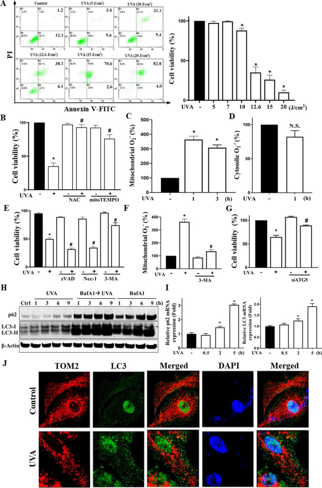
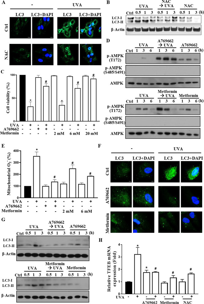
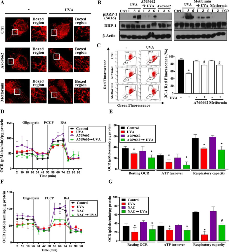
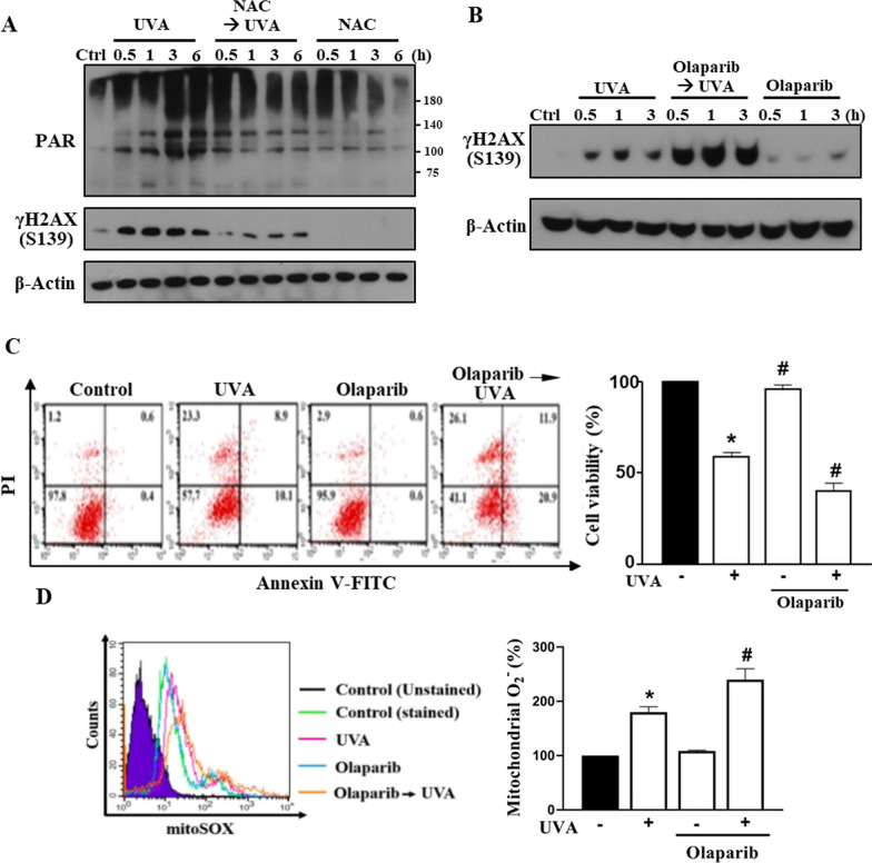
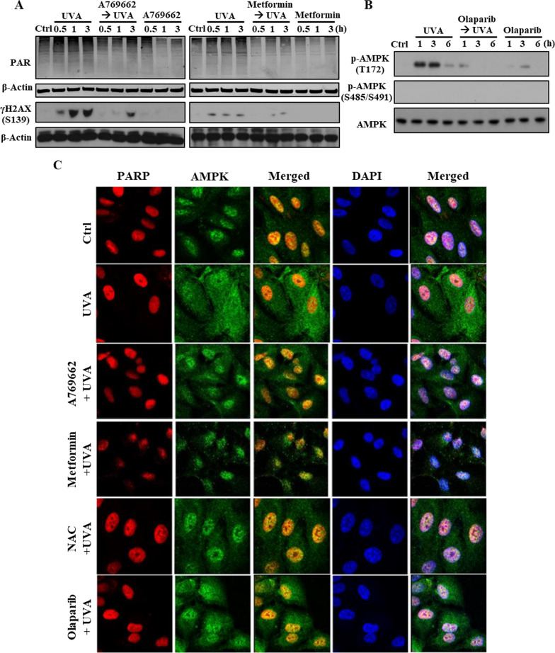
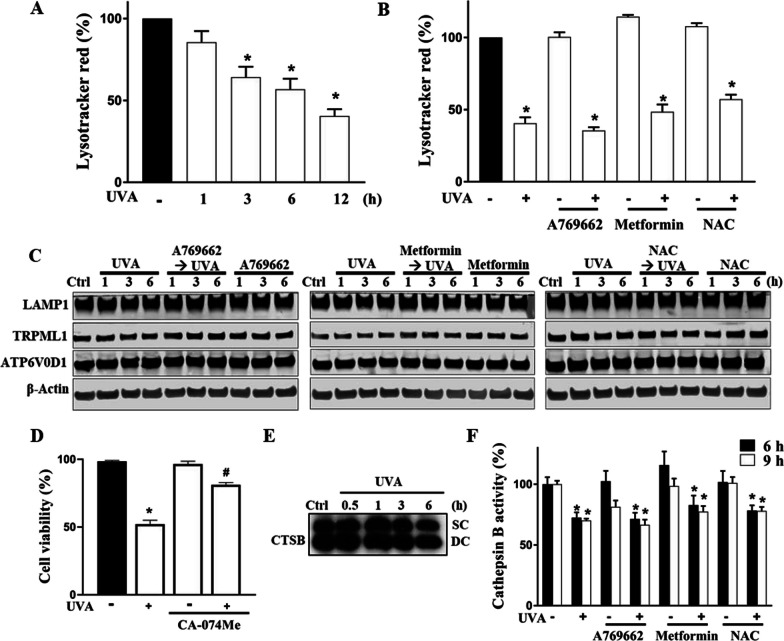
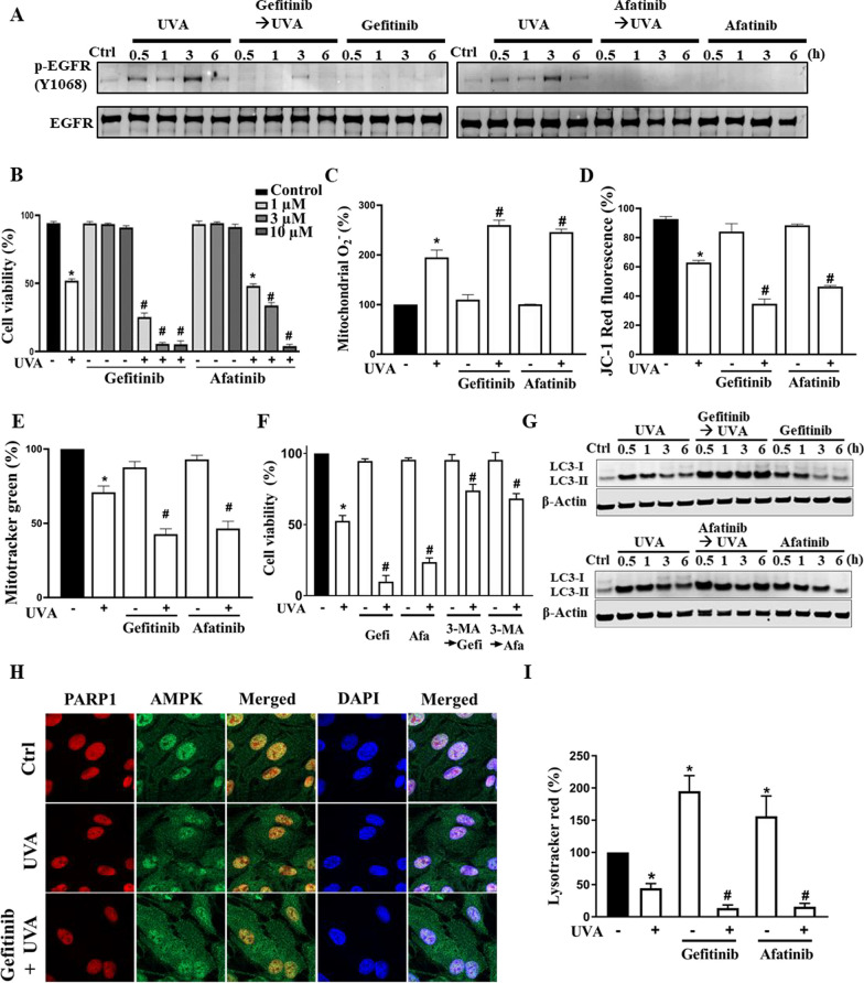
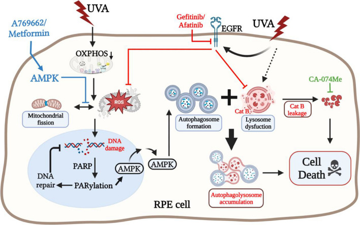
Results: We confirmed mitochondrial ROS production and DNA damage are two major features caused by UVA. We found the cell death is prevented by autophagy inhibitor 3-methyladenine and gene silencing of ATG5, and UVA induces ROS-dependent LC3II expression, LC3 punctate and TFEB expression, suggesting the autophagic death in the UVA-stressed RPE cells. Although PARP-1 inhibitor olaparib increases DNA damage, ROS production, and cell death, it also blocks AMPK activation caused by UVA. Interestingly we found a dramatic nuclear export of AMPK upon UVA irradiation which is blocked by N-acetylcysteine and olaparib. In addition, UVA exposure gradually decreases lysosomal mass and inhibits cathepsin B activity at late phase due to lysosomal dysfunction. Nevertheless, cathepsin B inhibitor, CA-074Me, reverses the death extent, suggesting the contribution of cathepsin B in the death pathway. When examining the role of EGFR in cellular events caused by UVA, we found that UVA can rapidly transactivate EGFR, and treatment with EGFR TKIs (gefitinib and afatinib) enhances the cell death accompanied by the increased LC3II formation, ROS production, loss of MMP and mass of mitochondria and lysosomes. Although AMPK activation by ROS-PARP-1 mediates autophagic cell death, we surprisingly found that pretreatment of cells with AMPK activators (A769662 and metformin) reverses cell death. Concomitantly, both agents block UVA-induced mitochondrial ROS production, autophagic flux, and mitochondrial fission without changing the inhibition of cathepsin B.

Conclusion: UVA exposure rapidly induces ROS-PARP-1-AMPK-autophagic flux and late lysosomal dysfunction. Pre-inducing AMPK activation can prevent cellular events caused by UVA and provide a new protective strategy in photo-oxidative stress and photo-retinopathy.

Keywords: AMPK; Autophagic cell death; EGFR; Lysosome dysfunction; PARP; ROS; RPE; UVA.


Conflict of interest statement

The authors declare that they have no competing interests.

Figures

Fig. 1
Fig. 1
UVA-induced mitochondrial ROS production contributes to autophagic cell death in RPE cells. A Human ARPE-19 cells were subjected to UVA irradiation at different doses (5–20 J/cm2). B–F Cells were pretreated with vehicle, NAC (5 mM), mitoTEMPO (100 µM), zVAD (10 µM), necrostatin-1 (10 µM), or 3-MA (5 mM) 30 min prior to UVA (12.6 J/cm2) irradiation. G Cells were treated with ATG5 siRNA before UVA irradiation. Cell viability in A, B, E, G was determined by Annexin V-FITC/PI staining followed by flow cytometry analysis at 18 h post-UVA irradiation. C, D, F After 1 h (C, D, F) and/or 3 h (C) post-UVA irradiation, mitochondrial ROS (C, F) and cytosolic ROS (D) were determined using mitoSOX and DHE staining, respectively. H ARPE-19 cells were pretreated with bafilomycin A1 (100 nM) 60 min prior to UVA (12.6 J/cm2) irradiation. Immunoblotting of p62/SQSTM1 and LC3-I/-II expression was determined at the indicated times (1, 3, 6, and 9 h) post-UVA irradiation. I Real time PCR was used to determine p62/SQSTM1 and LC3 gene expression at 0.5, 2, and 5 h post-UVA irradiation. J Confocal laser microscopic images of TOM20, LC3 and DAPI in ARPE-19 cells at 1 h post-UVA (12.6 J/cm2) irradiation. Data were the mean ± S.E.M. of at least three independent experiments. *p < 0.05, indicating the significant effects of UVA; #p < 0.05, indicating the significant effect of pretreatment to either reduce or enhance the effect of UVA; N.S., not significant
Fig. 2
Fig. 2
A769662 and metformin protect RPE cells from UVA-induced cell death. ARPE-19 cells were pretreated with NAC (5 mM) (A, B, H), A769662 (25 µM) or metformin (6 mM unless otherwise indicated) (CH) 30 min prior to UVA (12.6 J/cm2) irradiation. A, F Confocal microscopic images of LC3 and DAPI in cells 1 h post-UVA irradiation. B, D, G At 0.5, 1, 3 and 6 h post-UVA irradiation, cell lysates were harvested for immunoblotting. C Cell viability was determined at 18 h post-UVA irradiation by Annexin V-FITC/PI staining. E Mitochondrial ROS level was detected by MitoSOX staining at 1 h post-UVA irradiation. H Real time PCR was used to determine TFEB gene expression at 5 h post-UVA irradiation. Data were the mean ± S.E.M. of at least 3 independent experiments. *p < 0.05, indicating the significant effect of UVA; #p < 0.05, indicating the significant effects of pretreatment agents on the effects of UVA
Fig. 3
Fig. 3
AMPK activators inhibit UVA-induced mitochondria fragmentation and MMP loss without affecting the inhibition on mitochondrial oxidative phosphorylation. ARPE-19 cells were pretreated with AMPK activators A769662 (25 µM), metformin (6 mM), or NAC (5 mM) 30 min prior to UVA (12.6 J/cm2) irradiation. A Confocal microscopic images were performed at 1 h post-UVA irradiation. Mitotracker-Red CMXRos was used to detect the morphology of the mitochondria. B At the indicated time points after UVA irradiation cells were harvested by sample loading buffer followed by immunoblotting. C Mitochondrial membrane potential was determined by JC-1 staining at 1 h-post UVA irradiation. D–G Seahorse assay was performed for measuring mitochondrial OXPHOS in RPE cells after 1 h treatment with UVA. Data were the mean ± S.E.M. of 3 independent experiments. *p < 0.05, indicating the significant effects of UVA. #p < 0.05, indicating the significant effects of A769662 and metformin to reverse UVA actions
Fig. 4
Fig. 4
UVA-induced PARP-1 activation can reduce DNA damage and subsequent cell death. ARPE-19 cells were pre-treated with NAC (5 mM) (A) or olaparib (10 µM) (BD) 30 min prior to UVA (12.6 J/cm2) irradiation. A, B After UVA exposure at the indicated time points cell lysates were collected for immunoblotting. C Cell viability was determined by Annexin V-FITC/PI at 18 h post-UVA irradiation. D Mitochondrial ROS level was detected by MitoSOX staining at 1 h after UVA. Data were the mean ± S.E.M. of 3 independent experiments. *p < 0.05, indicating the significant effects of UVA; #p < 0.05, indicating the significant effects of olaparib to enhance cell death and increase ROS production
Fig. 5
Fig. 5
A769662 and metformin inhibit UVA-induced DNA damage and AMPK nuclear export. A, B ARPE-19 cells were pre-treated with AMPK activators A769662 (25 µM),  metformin (6 mM) (A) or olaparib (10 µM) (B) 30 min prior to UVA (12.6 J/cm2) irradiation. Cells lysates were collected at the indicated time points post-UVA irradiation for immunoblotting analysis. C ARPE-19 cells were pretreated with A769662 (25 µM), metformin (6 mM), NAC (5 mM), or olaparib (10 µM) 30 min prior to UVA irradiation. Cells were fixed at 1 h post-UVA irradiation for confocal microscopic analysis of PARP-1 and AMPK. Data were the representative of 3 independent experiments
Fig. 6
Fig. 6
Cathepsin B is involved in cell death and UVA-induced gradual lysosome dysfunction is independent of ROS-AMPK axis. A At the indicated times (1, 3, 6, and 12 h) post-UVA irradiation lysosomes in ARPE cells were determined by flow cytometry with LysoTracker. B–D, F ARPE-19 cells were pretreated with A769662 (25 µM), metformin (6 mM), NAC (5 mM) or CA-074Me (10 µM) 30 min prior to UVA (12.6 J/cm2) irradiation. In B cells were harvested at 12 h post-UVA irradiation for flow cytometry analysis with LysoTracker. In C at 1, 3, and 6 h after UVA, immunoblotting was conducted. In D cell viability was determined by Annexin V-FITC/PI at 18 h after UVA. In F cathepsin B activity was determined using flow cytometry analysis with MagicRed at 6 and 9 h after UVA. E Immunoblotting of cathepsin B expression was determined at the indicated times (0.5, 1, 3, and 6 h) post-UVA irradiation. Data were the mean ± S.E.M. of 3 independent experiments. *p < 0.05, indicating the significant effects of UVA. #p < 0.05, indicating the significant effect of CA-074Me to protect UVA-induced cell death
Fig. 7
Fig. 7
UVA-induced EGFR transactivation reduces cell death by exerting dual actions in balancing autophagic flux and lysosomal dysfunction. Cells were pretreated with gefitinib (gefi; 1 µM), afatinib (afa; 3 µM), or 3-MA (5 mM) 30 min prior to UVA (12.6 J/cm2) irradiation. A, G At 0.5, 1, 3, and 6 h after UVA immunoblotting was performed. B, F Cell viability was determined by Annexin V-FITC/PI staining at 18 h post-UVA irradiation. C, D Mitochondrial ROS level and MMP were determined at 1 h after UVA by flow cytometry with MitoSOX and JC-1 staining, respectively. E, I Mitochondrial mass and lysosomal mass were determined at 6 h after UVA by flow cytometry with MitoTracker and LysoTracker staining, respectively. Data were the mean ± S.E.M. of 3 independent experiments. *p < 0.05, indicating the significant effects of UVA; #p < 0.05, indicating the significant effects of drug pretreatments on UVA-induced responses as compared to vehicle-treated cells. H ARPE-19 cells were pretreated with gefitinib (1 µM) 30 min prior to UVA irradiation. Cells were fixed at 1 h post-UVA irradiation for confocal microscopic analysis of PARP-1 and AMPK. Data were the representative of 3 independent experiments
Fig. 8
Fig. 8
Spatiotemporal role of AMPK in regulating UVA-induced autophagy cell death in RPE cells. UVA-irradiation rapidly increases mitochondrial ROS production and DNA damage, leading to AMPK nuclear export and overactivated autophagic flux. At the mid to late phase, UVA also induces lysosome dysfunction (i.e. lysosomal rupture and leakage of cathepsin B), causing incomplete autophagy and autophagolysosome accumulation. Pre-activating AMPK by AMPK activators, on the other hand, protects RPE cells from UVA stress by reducing mitochondrial ROS production and the following signal cascades. EGFR transactivation by UVA also exerts balanced effects on autophagic flux and lysosome dysfunction. As such, EGFR TKI can deteriorate the UVA-induced cell death

References

    1. Glickman RD. Ultraviolet phototoxicity to the retina. Eye Contact Lens. 2011;37(4):196–205. doi: 10.1097/ICL.0b013e31821e45a9. - DOI - PubMed
    1. Chalam KV, Khetpal V, Rusovici R, Balaiya S. A review: role of ultraviolet radiation in age-related macular degeneration. Eye Contact Lens. 2011;37(4):225–232. doi: 10.1097/ICL.0b013e31821fbd3e. - DOI - PubMed
    1. Bellezza I. Oxidative stress in age-related macular degeneration: Nrf2 as therapeutic target. Front Pharmacol. 2018;9:1280. doi: 10.3389/fphar.2018.01280. - DOI - PMC - PubMed
    1. Chen Y, McMillan-Ward E, Kong J, Israels SJ, Gibson SB. Oxidative stress induces autophagic cell death independent of apoptosis in transformed and cancer cells. Cell Death Differ. 2008;15(1):171–182. doi: 10.1038/sj.cdd.4402233. - DOI - PubMed
    1. Ohsumi Y. Historical landmarks of autophagy research. Cell Res. 2014;24(1):9–23. doi: 10.1038/cr.2013.169. - DOI - PMC - PubMed

MeSH terms