. 2023 Nov 7;14(1):319.
doi: 10.1186/s13287-023-03547-6.

Comparison of osteoclast differentiation protocols from human induced pluripotent stem cells of different tissue origins

Affiliations

Comparison of osteoclast differentiation protocols from human induced pluripotent stem cells of different tissue origins

Alexander Blümke et al. Stem Cell Res Ther. .

Abstract

Background: Ever since their discovery, induced pluripotent stem cells (iPSCs) have been extensively differentiated into a large variety of cell types. However, a limited amount of work has been dedicated to differentiating iPSCs into osteoclasts. While several differentiation protocols have been published, it remains unclear which protocols or differentiation methods are preferable regarding the differentiation of osteoclasts.

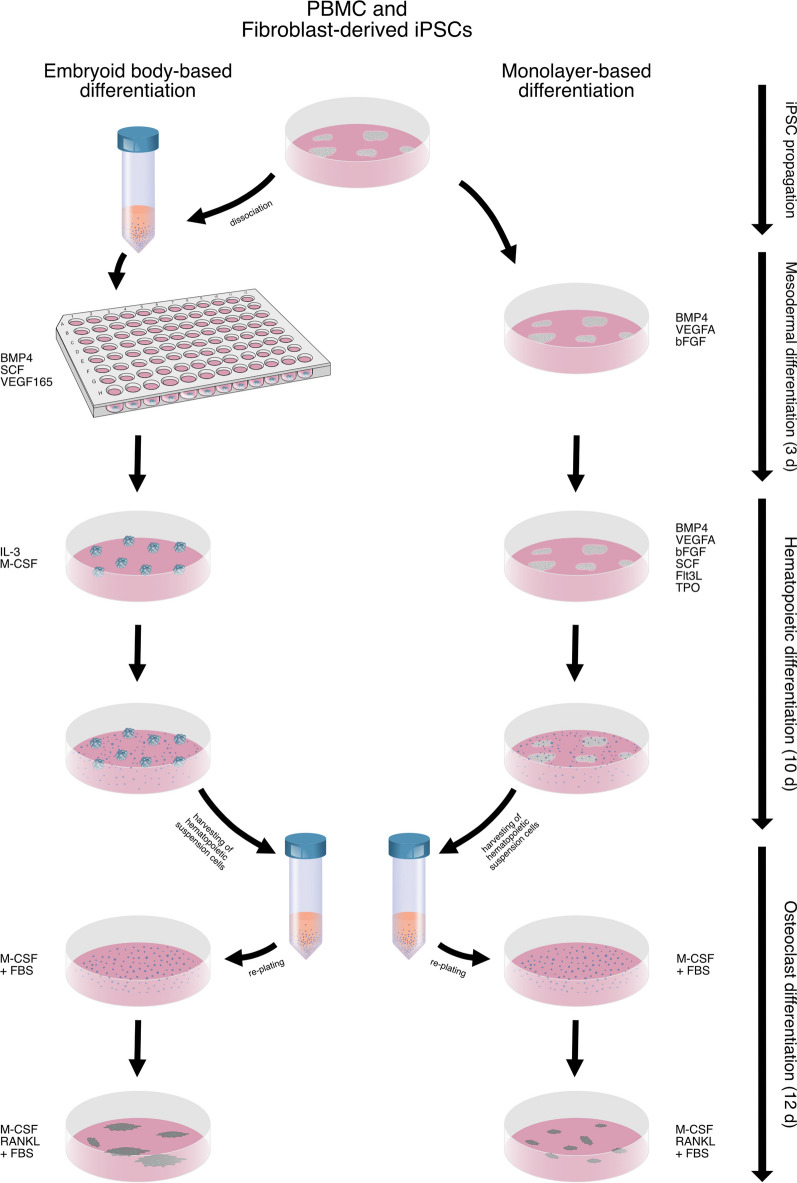
Methods: In this study, we compared the osteoclastogenesis capacity of a peripheral blood mononuclear cell (PBMC)-derived iPSC line to a fibroblast-derived iPSC line in conjunction with either embryoid body-based or monolayer-based differentiation strategies. Both cell lines and differentiation protocols were investigated regarding their ability to generate osteoclasts and their inherent robustness and ease of use. The ability of both cell lines to remain undifferentiated while propagating using a feeder-free system was assessed using alkaline phosphatase staining. This was followed by evaluating mesodermal differentiation and the characterization of hematopoietic progenitor cells using flow cytometry. Finally, osteoclast yield and functionality based on resorptive activity, Cathepsin K and tartrate-resistant acid phosphatase (TRAP) expression were assessed. The results were validated using qRT-PCR throughout the differentiation stages.

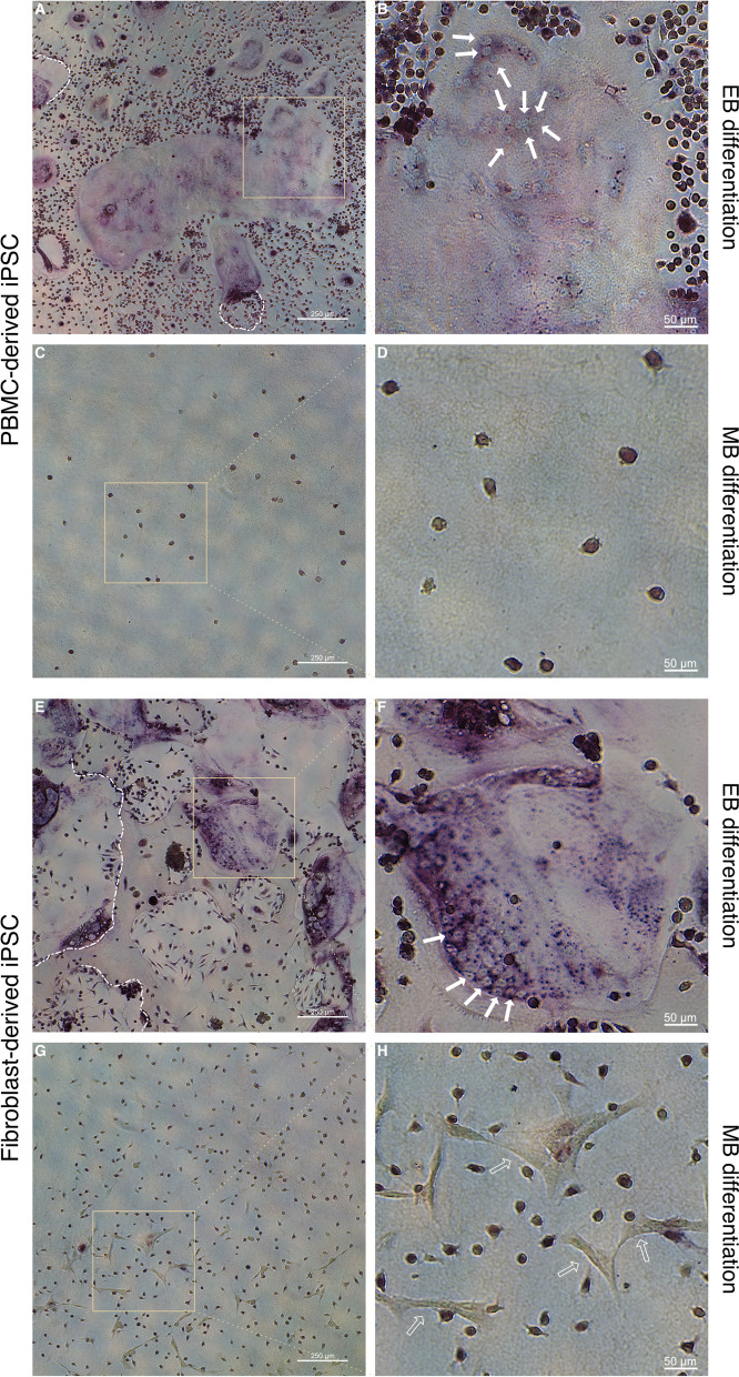
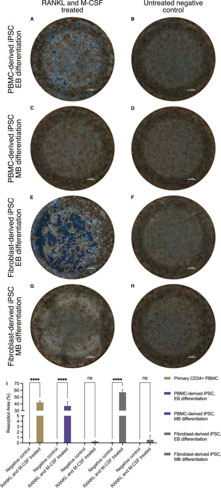
Results: Embryoid body-based differentiation yielded CD45+, CD14+, CD11b+ subpopulations which in turn differentiated into osteoclasts which demonstrated TRAP positivity, Cathepsin K expression and mineral resorptive capabilities. This was regardless of which iPSC line was used. Monolayer-based differentiation yielded lower quantities of hematopoietic cells that were mostly CD34+ and did not subsequently differentiate into osteoclasts.

Conclusions: The outcome of this study demonstrates the successful differentiation of osteoclasts from iPSCs in conjunction with the embryoid-based differentiation method, while the monolayer-based method did not yield osteoclasts. No differences were observed regarding osteoclast differentiation between the PBMC and fibroblast-derived iPSC lines.

Keywords: Hematopoietic differentiation; Human induced pluripotent stem cells; Mesodermal differentiation; Mineral resorption; Osteoclastogenesis; Osteoclasts.


Conflict of interest statement

The authors declare no competing interests.

Figures

Fig. 1
Fig. 1
Schematic outline of the differentiation process and the comparison between embryoid body-based (EB) and monolayer-based (MB) differentiation. Illustration drawn by Hannah and Alexander Blümke using Affinity Designer 2.1.1
Fig. 2
Fig. 2
Assessment of alkaline phosphatase (ALP) expression in iPSC lines prior and throughout mesodermal and hematopoietic differentiation. A-L Representative images of 3 replicates display ALP-stained colonies and cell-forming complexes of a PBMC-derived cell line (A-F) and a fibroblast-derived cell line (G-L), showing high expression of ALP while expanding (A, B, G, H). Following mesodermal and hematopoietic differentiation according to either the embryoid body-based (C, D, I, J) or monolayer-based protocol (E, F, K,L), cell-forming complexes retained ALP expression, especially in centrally located cells (solid arrows). An abundance of ALP-negative cells can be observed following hematopoietic differentiation (empty arrows). Scale bar = 500 µm
Fig. 3
Fig. 3
Analysis of cell-forming complexes following mesodermal differentiation using CLSM. A-L Representative images of 3 replicates show PBMC-derived iPSCs (A-F) or fibroblast-derived iPSCs (G-L) differentiated according to either an embryoid body-based (EB) protocol (A, B, C, G, H, I) or a monolayer-based (MB) protocol (D, E, F, J, K, L) and stained for ectodermal (A, D, G, J), mesodermal (B, E, H, K), and endodermal markers (C, F, I, L). Expression patterns of cell-forming complexes of PBMC-derived iPSCs demonstrate a higher degree of organization than complexes formed by fibroblast-derived iPSCs. Scale bar = 300 µm
Fig. 4
Fig. 4
Assessment of hematopoietic cells using flow cytometry. A, D Undifferentiated iPSCs were used as a reference for marker expression. B, C, E, F Hematopoietic cells differentiated according to the embryoid body-based (EB) protocol show a higher expression of later hematopoietic markers CD43 and CD45 (B, E) in comparison with monolayer-based (MB) differentiated cells (C, F). Additionally, the monocyte markers CD14 and CD11b were elevated in the EB group (B, E). No differences in RANK expression could be observed between either differentiation protocol
Fig. 5
Fig. 5
Morphology assessment of cells following osteoclast differentiation using CLSM. AH Representative CLSM images of hematopoietic cells from a PBMC-derived iPSC line AD or a fibroblast-derived iPSC line (EH) that had been differentiated either according to an embryoid body-based (EB) (A, B, E, F) or a monolayer-based (MB) protocol (C, D, G, H) were further subjected to osteoclast differentiation and stained for Cathepsin K (turquoise), F-actin (red) and counterstained with DAPI nuclear stain (blue). Cells differentiated according to the EB protocol showed large multinucleated polykaryons (solid arrows in A, E) which also demonstrated Cathepsin K expression (arrow tips in B, F). MB differentiated cells on the other hand showed a low number of cells with up to 5 nuclei (solid arrow in D) in the PBMC-derived iPSC line and cells with a stellar-like morphology in the fibroblast-iPSC line (chevron arrows in H). A limited number of cells expressing Cathepsin K can be seen in both groups (arrow heads in D, H). Mononuclear cells with some degree of Cathepsin K expression can be seen throughout all groups (empty arrows in A, D, E, H). I-K Image quantitation shows a significant difference in osteoclast number (3 or more nuclei) between the EB and MB protocols (I). No significant differences were observed in osteoclast size or number of nuclei when the EB protocol was used with the different iPSC lines (J, K). Scale bars: A, C, E, G = 100 µm, B, D, F, H = 25 µm. Statistics are based on ANOVA followed by Tukey’s multiple comparison post hoc test (I n = 3 well replicates, J, K n = 50 analyzed cells, **p < 0.01, ***p < 0.001)
Fig. 6
Fig. 6
TRAP staining of cells following osteoclast differentiation in conjunction with methyl green nuclear counterstaining. AH Hematopoietic cells from an PBMC-derived iPSC line AD or a fibroblast-derived iPSC line (EH) that had been differentiated either according to an embryoid body-based (EB) (A, B, E, F) or a monolayer-based (MB) protocol (C, D, G, H) were seeded onto calcium-phosphate coated wells and further subjected to osteoclast differentiation conditions. Representative images of cells derived from EB protocols show large TRAP positive cells (A, B, E, F) with multiple nuclei (solid arrows in B, F). Resorption pits are also visible in both groups (white dashed lines in A, E). Cells derived from the MB protocol did not give rise to osteoclasts. Cells with a stellar-like cell morphology can be seen in the fibroblast-derived iPSC MB differentiation group (empty arrows in H). Scale bars: A, C, E, G = 250 µm, B, D, F, H = 50 µm
Fig. 7
Fig. 7
Assessment and quantification of the mineral resorption activity of osteoclasts. AH Following hematopoietic differentiation of PBMC-derived iPSCs AD or fibroblast-derived iPSCs (EH), either according to an embryoid body based (EB) A, B, E, F or a monolayer-based (MB) protocol (C, D, G, H), hematopoietic cells were matured with M-CSF and differentiated into OCs with RANKL on calcium-phosphate coated wells. Osteoclasts differentiated from PBMC-derived iPSCs using the EB protocol showed clearly visible resorption pits on tiled full-well images acquired with a widefield microscope in phase-contrast mode A in comparison with undifferentiated negative controls (B). Similarly, osteoclasts from fibroblast-derived iPSCs differentiated with the EB protocol also showed clearly visible pits, albeit the total resorption area appeared much larger E than that of the negative control (F). In comparison, cells differentiated according to the MB protocol did not show visible resorption pits for either cell line C, G when compared to negative controls (D, H). Scale bar = 1 mm. I Image quantification demonstrates comparable resorption levels of PBMC-derived iPSC osteoclasts differentiated according to the EB protocol to osteoclasts differentiated from primary CD34+ PBMCs. The highest level of mineral resorption was observed in osteoclasts differentiated from the fibroblast-derived iPSC line using the EB protocol. Quantification confirms the absence of mineral resorption in cells differentiated according to the MB protocol for either iPSC line. Statistics are based on ANOVA followed by Tukey’s multiple comparison post hoc test (n = 3 well replicates, ****p < 0.0001)
Fig. 8
Fig. 8
Relative gene expression of iPSCs throughout the differentiation process. Gene expression of POU5F1 decreased in all groups significantly from the mesodermal to the hematopoietic stage (A). CSF1R increased significantly after the mesodermal stage in relation to the hematopoietic stage in the embryoid body-based (EB) differentiation protocol B independent of the iPSC line. Monolayer-based (MB) differentiation showed either no significant increase or an initial Ct value below the detection threshold. Osteoclast markers all showed a significant increase in the EB protocol for both iPSC lines while MB differentiation did not yield sufficient RNA for analyses (CF). Statistics are based on multiple comparisons using the Holm-Šídák method (n = 3 replicates, except POU5F1: OC stage of PBMC-derived iPSC EB differentiation and hematopoietic stage of PBMC-derived iPSC MB differentiation, as well as CA2: mesodermal stage of fibroblast-derived iPSC MB differentiation, where all replicates were close to, while one replicate was below the detection threshold, *p < 0.05, **p < 0.01, ***p < 0.001, ****p < 0.0001)

Update of

References

    1. Takahashi K, Yamanaka S. Induction of pluripotent stem cells from mouse embryonic and adult fibroblast cultures by defined factors. Cell. 2006;126(4):663–676. doi: 10.1016/j.cell.2006.07.024. - DOI - PubMed
    1. Singh VK, Kalsan M, Kumar N, Saini A, Chandra R. Induced pluripotent stem cells: applications in regenerative medicine, disease modeling, and drug discovery. Front Cell Dev Biol. 2015;3(FEB):2. - PMC - PubMed
    1. Wernig M, Meissner A, Foreman R, Brambrink T, Ku M, Hochedlinger K, et al. In vitro reprogramming of fibroblasts into a pluripotent ES-cell-like state. Nature. 2007;448(7151):318–324. doi: 10.1038/nature05944. - DOI - PubMed
    1. Takahashi K, Tanabe K, Ohnuki M, Narita M, Ichisaka T, Tomoda K, et al. Induction of pluripotent stem cells from adult human fibroblasts by defined factors. Cell. 2007;131(5):861–872. doi: 10.1016/j.cell.2007.11.019. - DOI - PubMed
    1. Yu J, Vodyanik MA, Smuga-Otto K, Antosiewicz-Bourget J, Frane JL, Tian S, et al. Induced pluripotent stem cell lines derived from human somatic cells. Science (1979) 2007;318(5858):1917–1920. doi: 10.1126/science.1151526. - DOI - PubMed