Case Reports
. 2023 Nov 8;8(21):e173066.
doi: 10.1172/jci.insight.173066.

An IKBKE variant conferring functional cGAS/STING pathway deficiency and susceptibility to recurrent HSV-2 meningitis

Affiliations
Case Reports

An IKBKE variant conferring functional cGAS/STING pathway deficiency and susceptibility to recurrent HSV-2 meningitis

Azadeh Reyahi et al. JCI Insight. .

Abstract

The mechanisms underlying susceptibility to recurrent herpes simplex virus type 2 (HSV-2) meningitis remain incompletely understood. In a patient experiencing multiple episodes of HSV-2 meningitis, we identified a monoallelic variant in the IKBKE gene, which encodes the IKKε kinase involved in induction of antiviral IFN genes. Patient cells displayed impaired induction of IFN-β1 (IFNB1) expression upon infection with HSV-2 or stimulation with double-stranded DNA (dsDNA) and failed to induce phosphorylation of STING, an activation marker of the DNA-sensing cyclic GMP-AMP synthase/stimulator of IFN genes (cGAS/STING) pathway. The patient allele encoded a truncated IKKε protein with loss of kinase activity and also capable of exerting dominant-negative activity. In stem cell-derived microglia, HSV-2-induced expression of IFNB1 was dependent on cGAS, TANK binding kinase 1 (TBK1), and IKBKE, but not TLR3, and supernatants from HSV-2-treated microglia exerted IKBKE-dependent type I IFN-mediated antiviral activity upon neurons. Reintroducing wild-type IKBKE into patient cells rescued IFNB1 induction following treatment with HSV-2 or dsDNA and restored antiviral activity. Collectively, we identify IKKε to be important for protection against HSV-2 meningitis and suggest a nonredundant role for the cGAS/STING pathway in human antiviral immunity.

Keywords: Immunology; Infectious disease; Innate immunity.


Figures

Figure 1
Figure 1. Identification of a monoallelic IKBKE variant in a patient with HSV-2 meningitis.
(A) Number of disease episodes in the patients with recurrent HSV-2 meningitis. (B) Genetic information and characteristics of the IKBKE c.312delC variant identified in the patient. (C) Domain organization of WT IKKε and illustration of the localization of the mutation in the patient variant, including the resulting truncated F105f*19 protein. Key residues in the active site (shown in gray) and the phosphorylation target (shown in blue) are highlighted. ULD, ubiquitin-like domain; LZ, leucine zipper; HLH, helix-loop-helix. (D) Pedigree for the patient’s family revealed by Sanger sequencing. Family members heterozygous for the mutation are indicated by blue and red color. (E) Whole cell lysate of PBMCs from the patient and from healthy controls analyzed for IKKε protein level by immunoblotting using antibodies targeting epitopes in the N- and C-terminal parts of the protein.
Figure 2
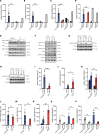
Figure 2. Impaired IFNB1 response and STING activation following HSV-2 infection in PBMCs from P1.
(AD) PBMCs were infected with HSV-2 with MOI 9. Total RNA was isolated after 6 hours for measurement of IFNA2 (A), IFNB1 (B), ISG54 (C), and TNFA (D) mRNA by reverse transcription quantitative PCR (RT-qPCR). (E and F) Whole-cell lysates from PBMCs treated for 18 hours with HSV-2, as indicated, were examined for expression of IKKε (N- and C-terminal–targeting antibody), p-IKKε (S172), TBK1, p-TBK1 (S172), IRF3, p-IRF3 (S386), STING, and p-STING (S366). Data shown are representative of 4 (E) and 2 (F) independently performed experiments. (G and H) Fibroblasts were transfected with cGAMP or dsDNA (4 μg/mL) for 4 hours, and lysates were immunoblotted for p-STING (S366), STING, and Vinculin. (IK) PBMCs were stimulated with dsDNA (4 μg/mL) or poly(IC) (4 μg/mL intracellular, 50 μg/mL extracellular). (LP) PBMCs were infected for 6 hours with Sendai virus (SeV), influenza A virus (IAV) (MOI 0.1), encephalomyocarditis virus (EMCV) (MOI 0.1), HSV-1 (MOI 3), and HSV-2 (MOI 3), and IFNB1 mRNA was quantified by qPCR. All measurements were done in triplicates, relative to housekeeping genes GAPDH or BACTIN and normalized to the pooled uninfected PBMCs from 10 (AD), 9 (I, J, and LN), 4 (K), 2 young (control 1), and 4 around 50 years old (control 2) (O and P) healthy controls. Results were obtained from 3 (AD) (except for mother and son PBMCs, which were examined once) and (IN) or 2 (O and P) independent experiments. The nonparametric Mann-Whitney (AD) and unpaired t test (IP) were used to evaluate statistical significance between groups. Error bars represent standard error of mean (SEM). *, P ≤ 0.05; **, P ≤ 0.01; ***, P ≤ 0.001; ****, P ≤0.0001.
Figure 3
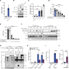
Figure 3. Impaired IFNB1 expression and increased viral load in HSV-2–infected P1 fibroblasts.
(A and B) Fibroblasts were infected with HSV-2. Total RNA was harvested after 6 and 24 hours and subjected to RT-qPCR for measurement of IFNA2 (A) and IFNB1 (B) mRNA. Cytokine mRNA levels were normalized to GAPDH, and the patient was compared with either 4 controls (for 6-hour stimulation) or 5 controls (for 24-hour infection). Unpaired t test was used for statistical analysis. (C) Fibroblasts were infected with HSV-1. Total RNA was harvested after 6 hours for IFNB1 mRNA quantification. (D) A plaque assay was used to measure HSV-2 titers in supernatants after 24 hours of infection. Nonparametric Mann-Whitney rank-sum test was used for statistical analysis. (E) Fibroblasts from P1 and 2 healthy controls were infected with measles virus (MeV) for 48 hours. A TCID50 (50% tissue culture infectious dose) assay was used to determine MeV titers in supernatants. Unpaired t test was used for statistical analysis. (F) Fibroblasts from P1 and patients functionally deficient in IRF3, IFNAR2, or TLR3 and fibroblasts from 2 healthy controls were infected with HSV-2 (MOI 1) for 24 hours, and a TCID50 assay was performed to quantify viral titers. Data were analyzed with 1-way ANOVA, and Dunnett’s multiple comparisons test was used for statistical analysis. Data shown in A and B are representative of 3 independent experiments, while the data in C represent 1 of 2 independent experiments. Data shown in DF are pooled from 3 independent experiments. Error bars representing SEM and *, P ≤ 0.05; **, P ≤ 0.01; ****, P ≤ 0.0001.
Figure 4
Figure 4. The IKKε F105f*19 protein is unable to stimulate IFN induction.
(A) HEK293T cells were transiently transfected with IFN-β promoter firefly, β-actin promoter renilla reporter plasmids, and 100 ng of vector and plasmids encoding the full-length (WT) and truncated P1 F105fs*19 IKKε to measure IFN-β gene expression indicated by luciferase activity after 16 hours. (B) p-IRF3 (S396) and total IRF3 in whole-cell lysates from HEK293T cells expressing empty vector, WT, and P1 variant IKKε. IKKε (N/C-term epi), antibodies targeting epitopes in the N- and C-terminal parts of IKKε. (C) Lysates from HEK293T cells transfected with the indicated expression plasmids were immunoprecipitated with anti-FLAG and subjected to assays determining IKKε kinase activity. Data are presented as normalized levels of luciferase signals from the ATP measurements. (D and E) HEK293T cells were transfected with IFN-β promoter firefly, β-actin promoter renilla reporter plasmids, WT IKBKE, WT TBK1, and the indicated amounts of P1 IKBKE. (F) HEK293T cells were transfected with P1 IKBKE, and the indicated amounts of plasmids encoding FLAG-TBK1 and FLAG-IKKε. Cells were lysed 24 hours later and immunoblotted for p-TBK1 (S172), p-IKKε (S172), FLAG, IKKε (probing of only low molecular range of blotted membrane), and Vinculin. (G) HEK293T cells were transfected with 6x-His-WT or P1 IKBKE, FLAG-TBK1, and FLAG-IKBKE. Lysates were immunoprecipitated with anti-His and immunoblotted with anti-FLAG. (HJ) Control fibroblasts were transduced with lentiviral vectors encoding patient IKKε variant F105fs*19 (MOI: 12), infected with HSV-2 at the indicated MOI for 6 hours (H) or 24 hours (J), or transfected with dsDNA (I) for 6 hours. Data presented are from 1 representative of 6 (A, D, and E), 2 (F and G), and 3 (B, C, and HJ) independently performed experiments. Nonparametric Mann-Whitney rank-sum test was used for statistical analysis. Error bars represent SEM. **, P ≤ 0.01; ***, P ≤ 0.001; ****, P ≤ 0.0001. NU = (Lucmax – Lucmeasured)/Lucmax (see Methods).
Figure 5
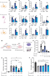
Figure 5. IKBKE, TBK1, and cGAS are important for IFNB1 induction in microglia after HSV-2 exposure.
(AH) iPSC derived microglia (AD) and neurons (EH) were transfected with IKBKE or control siRNA. (A and E) The efficiency of transfection was examined by measuring IKBKE mRNA levels. Cells were infected with HSV-2 for 6 hours, and IFNA2, IFNB1, and TNFA mRNA levels were quantified. (IL) iPSC-derived microglia were transfected with siRNA targeting cGAS (I and J) or TLR3 (K and L). Microglia were infected with HSV-2, and IFNB1 mRNA was quantified (J and L). Unpaired t test was used for statistical analysis. (M) Graphical illustration of experimental setup for microglia-neuron crosstalk experiments. Created with BioRender.com. (N) iPSC-derived neurons were treated with Human Type 1 IFN Neutralizing Antibody Mixture or control IgG (both 1:100) 30 minutes before addition of supernatants from HSV-2–infected microglia or treatment with IFN-β (10 ng/mL). Six hours later, the medium was removed, and the neurons were infected with HSV-2. Supernatants were collected after 16 hours for plaque assay (O). Microglia subjected to IKBKE or control knockdown with siRNA were infected with HSV-2. The cells were washed, and the medium was replaced after 1 hour. Supernatants were collected from the microglia after 24 hours and added to the neurons. Six hours later, the medium was removed, and the neurons were infected with HSV-2. Supernatants were collected 16 hours later for plaque assay. Data presented are pooled from 2 independently performed experiments. (P) Cells were treated as in panel N, and culture supernatants were analyzed for cell viability. Data presented are from 1 of 2 experiments performed. (NP) Groups were compared with Brown-Forsythe and Welch ANOVA with Dunnett’s T3 multiple comparisons test. Error bars represent SEM (AH) and SD (NP), and *, P ≤ 0.05; **, P ≤ 0.01; ***, P ≤ 0.001; ****, P ≤ 0.0001.
Figure 6
Figure 6. Reconstitution of WT IKBKE in patient fibroblasts rescues anti–HSV-2 response.
Fibroblasts from P1 and controls were transduced with lentiviral vectors encoding WT IKKε or GFP (MOI of 12). (A and B) Transduced fibroblasts were infected with HSV-2 at MOI of 1 and 3 for 6 hours or (C) were transfected with dsDNA (4 μg/mL) for 6 hours. Total RNA was harvested from the cell lysates and subjected to RT-qPCR for measurement of IFNB1 mRNA level. Data presented are from 1 representative of 3 independent experiments performed. (D) Supernatants were collected from the cells 24 hours after infection for quantification of viral load by plaque assay. Data presented are merged of 2 independent experiments performed. (A and D) Unpaired t test was used for statistical analysis. (B and C) Groups were compared with Brown-Forsythe and Welch’s ANOVA with Dunnett’s T3 multiple comparisons test. Error bars represent SD and *, P ≤ 0.05; **, P ≤ 0.01; ****, P ≤ 0.0001.

References

    1. Whitley R, et al. Pathogenesis and disease. In: Arvin A, et al., eds. Human Herpesviruses: Biology, Therapy, and Immunoprophylaxis. Cambridge University Press; 2007:Chapter 32. - PubMed
    1. Whitley RJ, Roizman B. Herpes simplex virus infections. Lancet. 2001;357(9267):1513–1518. doi: 10.1016/S0140-6736(00)04638-9. - DOI - PubMed
    1. Mollaret P. La meningite endothelio-leucocytaire multirecurrente benigne. Syndrome nouveau ou maladie nouvelle? Documents cliniques. [French] Rev Neurol. 1944;76:57–76.
    1. Tedder DG, et al. Herpes simplex virus infection as a cause of benign recurrent lymphocytic meningitis. Ann Intern Med. 1994;121(5):334–338. doi: 10.7326/0003-4819-121-5-199409010-00004. - DOI - PubMed
    1. Dylewski JS, Bekhor S. Mollaret’s meningitis caused by herpes simplex virus type 2: case report and literature review. Eur J Clin Microbiol Infect Dis. 2004;23(7):560–562. doi: 10.1007/s10096-004-1164-y. - DOI - PubMed

Publication types