Review
. 2023 Dec 20;36(4):e0003323.
doi: 10.1128/cmr.00033-23. Epub 2023 Nov 8.

Human strongyloidiasis: complexities and pathways forward

Affiliations
Review

Human strongyloidiasis: complexities and pathways forward

Dora Buonfrate et al. Clin Microbiol Rev. .

Abstract

Strongyloidiasis is a World Health Organization neglected tropical disease usually caused by Strongyloides stercoralis, a parasitic worm with a complex life cycle. Globally, 300-600 million people are infected through contact with fecally contaminated soil. An autoinfective component of the life cycle can lead to chronic infection that may be asymptomatic or cause long-term symptoms, including malnourishment in children. Low larval output can limit the sensitivity of detection in stool, with serology being effective but less sensitive in immunocompromise. Host immunosuppression can trigger catastrophic, fatal hyperinfection/dissemination, where large numbers of larvae pierce the bowel wall and disseminate throughout the organs. Stable disease is effectively treated by single-dose ivermectin, with disease in immunocompromised patients treated with multiple doses. Strategies for management include raising awareness, clarifying zoonotic potential, the development and use of effective diagnostic tests for epidemiological studies and individual diagnosis, and the implementation of treatment programs with research into therapeutic alternatives and medication safety.

Keywords: Strongyloides; neglected tropical diseases; soil-transmitted helminth; strongyloidiasis.


Conflict of interest statement

The authors declare no conflict of interest.

Figures

FIG 1
FIG 1
Life cycles of Strongyloides stercoralis. (a) Homogonic life cycle. The L1r larvae are passed per rectum in feces ① and then mature in the soil to longer L2r ②, followed by molting further to become L3 filariform infective (L3i) larvae ③. These L3i larvae must find a host and infect it via transdermal penetration. These larvae randomly migrate (light blue arrows) or via the cardio-pulmonary-esophagus route (red arrows) to reach the small intestine. Once in the small intestine, they further mature to become new parasitic females. (b) Heterogonic life cycle. The L1r larvae are passed per rectum in feces ① and then mature in the soil to L2r larvae ②, followed by further molting again into morphologically identical but larger L3r larvae ③, where sexual differentiation begins, and then to the final L4r stage ④. The L4r larvae further mature and differentiate into free-living adult male ⑤ and female ⑥ worms, which mate and produce eggs ⑦. These eggs hatch in the soil into a new generation of L1r larvae ⑧, which molt into L2r larvae ⑨ and are then all transformed into L3i larvae ⑩, which must find a host to infect via transdermal penetration or perish. Once within the host, these larvae migrate and develop into new parasitic females as per the homogonic life cycle (blue and red arrows). (c) Autoinfective life cycle. The parasitic female S. stercoralis ① passes eggs ②, which typically develop in the intestinal crypts of Lieberkűhn ③ before hatching into L1 rhabditiform (L1r) larvae ④. These molt within the intestine to L2 rhabditiform (L2r) larvae ⑤ and further molt into L3 filariform autoinfective (L3a) larvae ⑥. These larvae randomly migrate (light blue arrows) or via the cardio-pulmonary-esophagus route (red arrows) to reach the small intestine. Once in the small intestine, they further mature to become new parasitic females.
FIG 2
FIG 2
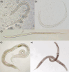
Major life stages of Strongyloides stercoralis—light micrographs. (a) Rhabditiform larva; (b) autoinfective filariform larva; (c) infective filariform larva [reproduced from reference (12)]; (d) parasitic female; (e) mating free-living female and male.
FIG 3
FIG 3
Anatomy of Strongyloides stercoralis adult life stages. (A) Parasitic adult; (B) free-living adult female; (C) free-living adult male. An, anus; Eg, eggs; Es, esophagus; Gu, gubernaculum; In, intestine; Nr, nerve ring; Rt, reproductive tubule; Sp, spicule; Te, testis; Ut, uterus; Vo, vaginal opening. (Image licensed under the terms of the Creative Commons Attribution 4.0 International license.)
FIG 4
FIG 4
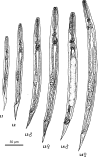
Anatomy of Strongyloides stercoralis larval life stages. (A) Larvated egg; (B) L1 rhabditiform larva; (C) L3 autoinfective filariform larvae; (D) L3 infective filariform larvae. An, anus; Bc, buccal cavity; Eg, egg; Es/In, esophageal-intestinal junction; Gp, genital primordium; Nr, nerve ring. (Image licensed under the terms of the Creative Commons Attribution 4.0 International license.)
FIG 5
FIG 5
Development of the four stages of rhabditiform larvae of Strongyloides stercoralis in the heterogonic (free-living) life cycle. (Adapted from original drawings provided by Emeritus Professor Richard “Rick” Speare.)
FIG 6
FIG 6
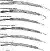
Morphological comparison of rhabditiform larvae that may be recovered from human fecal samples. (Reproduced from reference .)
FIG 7
FIG 7
Morphological comparison of filariform larvae that may be cultured from human fecal samples. (Reproduced from reference .)
FIG 8
FIG 8
Various life stages of Strongyloides stercoralis in the histological section of the duodenum. Active chronic duodenitis. (a) eggs (Eg); (b) L1 rhabditiform larvae (Rh) developing within eggs; (c) parasitic adult female, sectioned at the midline and demonstrating the two branches of the reflected uterus (Ut) and the single intestinal tube (In); all 400× magnification; (d) larvated eggs (white darts) and parasitic females in transverse (black darts) and longitudinal (arrows) section; 100× magnification. (Photographs courtesy of K. Hart, reproduced with permission.)
FIG 9
FIG 9
Sensitivity ranges of Strongyloides diagnostic tests. Limited studies have indicated that mini-FLOTAC and filtration stool concentration methods have sensitivities similar to direct smear. Formalin-ether concentration includes modified methods. Baermann sedimentation and agar plate culture require live larvae. PCR sensitivity is dependent on the amount of stool extracted and the effectiveness of the stool extraction method. Serology false-negative results may occur in immunosuppression/hyperinfection.

References

    1. Nutman TB. 2017. Human infection with Strongyloides stercoralis and other related Strongyloides species. Parasitology 144:263–273. doi:10.1017/S0031182016000834 - DOI - PMC - PubMed
    1. Schad GA. 1989. Morphology and life history of Strongyloides stercoralis, p 85–104. In Grove DI (ed), Strongyloidiasis: a major roundworm infection of man. Taylor & Francis Ltd, London.
    1. Buonfrate D, Fittipaldo A, Vlieghe E, Bottieau E. 2021. Clinical and laboratory features of Strongyloides stercoralis infection at diagnosis and after treatment: a systematic review and meta-analysis. Clin Microbiol Infect 27:1621–1628. doi:10.1016/j.cmi.2021.07.016 - DOI - PubMed
    1. Forrer A, Khieu V, Schär F, Hattendorf J, Marti H, Neumayr A, Char MC, Hatz C, Muth S, Odermatt P. 2017. Strongyloides stercoralis is associated with significant morbidity in rural Cambodia, including stunting in children. PLoS Negl Trop Dis 11:e0005685. doi:10.1371/journal.pntd.0005685 - DOI - PMC - PubMed
    1. Jember TH, Amor A, Nibret E, Munshea A, Flores-Chavez M, Ta-Tang T-H, Saugar JM, Benito A, Anegagrie M. 2022. Prevalence of Strongyloides stercoralis infection and associated clinical symptoms among schoolchildren living in different altitudes of Amhara National Regional State, northwest Ethiopia. PLoS Negl Trop Dis 16:e0010299. doi:10.1371/journal.pntd.0010299 - DOI - PMC - PubMed

Publication types

LinkOut - more resources