Review
. 2023 Dec 20;36(4):e0015922.
doi: 10.1128/cmr.00159-22. Epub 2023 Nov 8.

Invasive fusariosis

Affiliations
Review

Invasive fusariosis

Marcio Nucci et al. Clin Microbiol Rev. .

Abstract

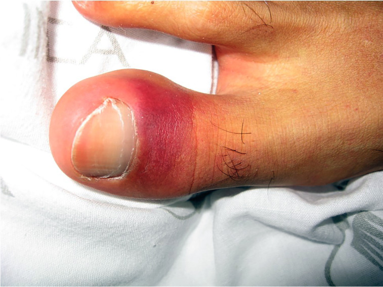
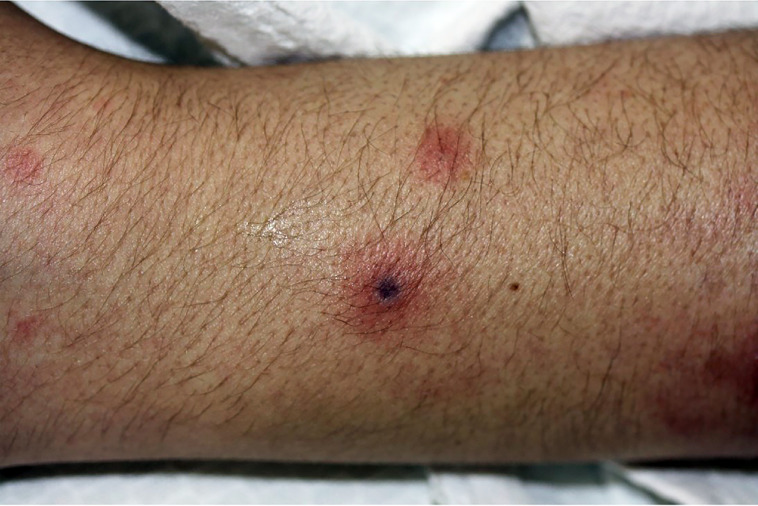
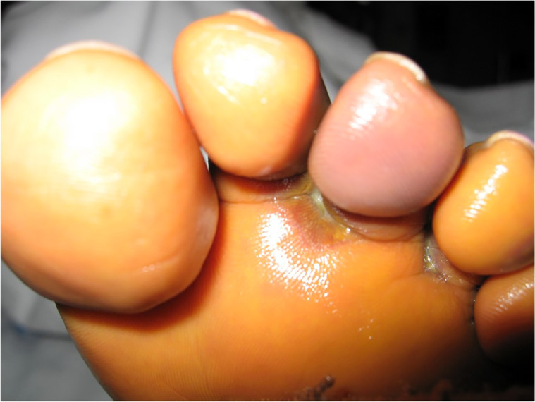
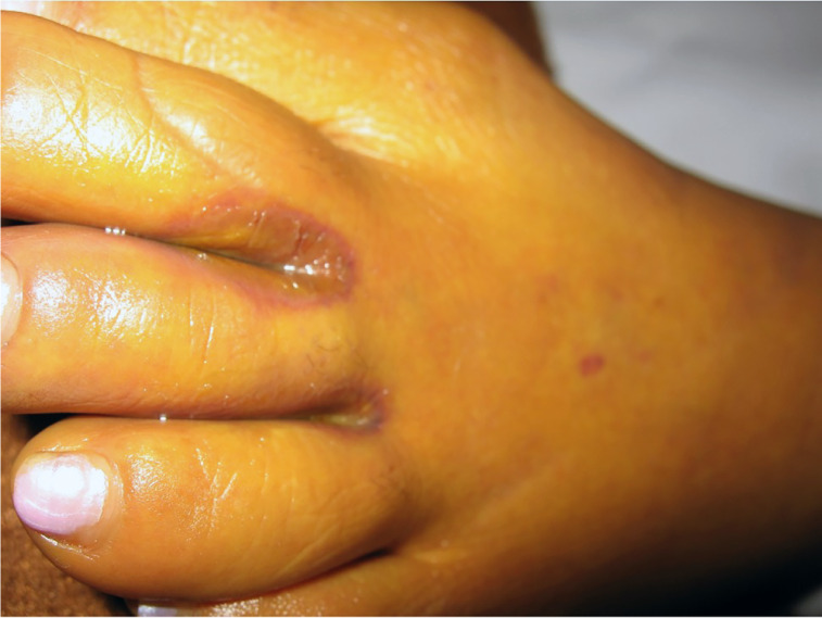
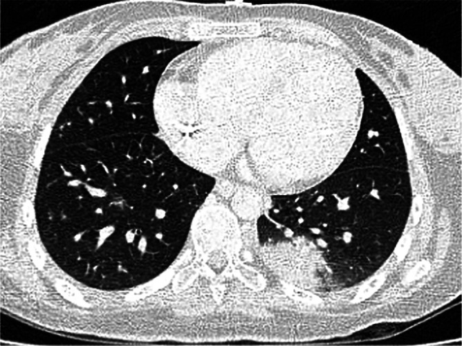
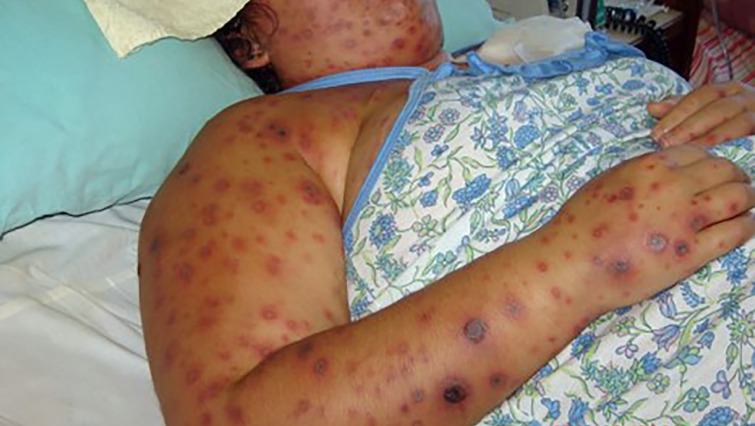
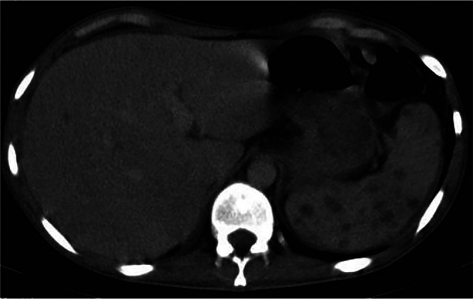
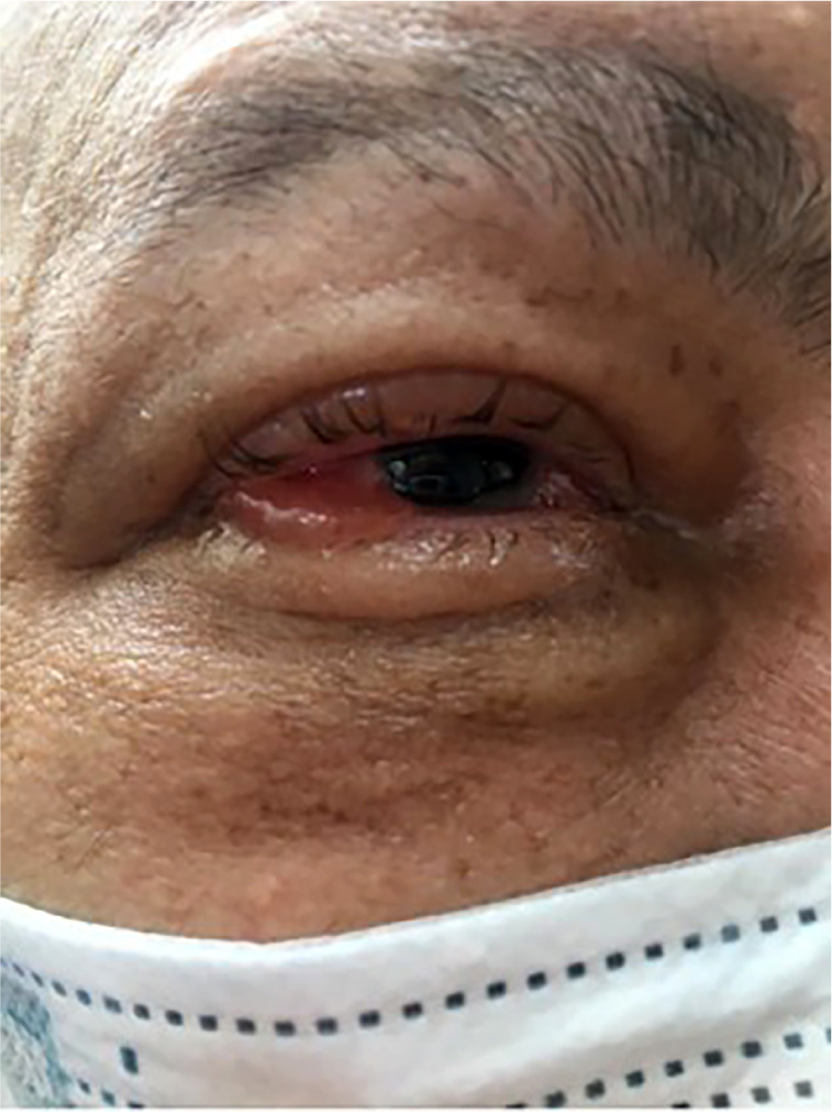
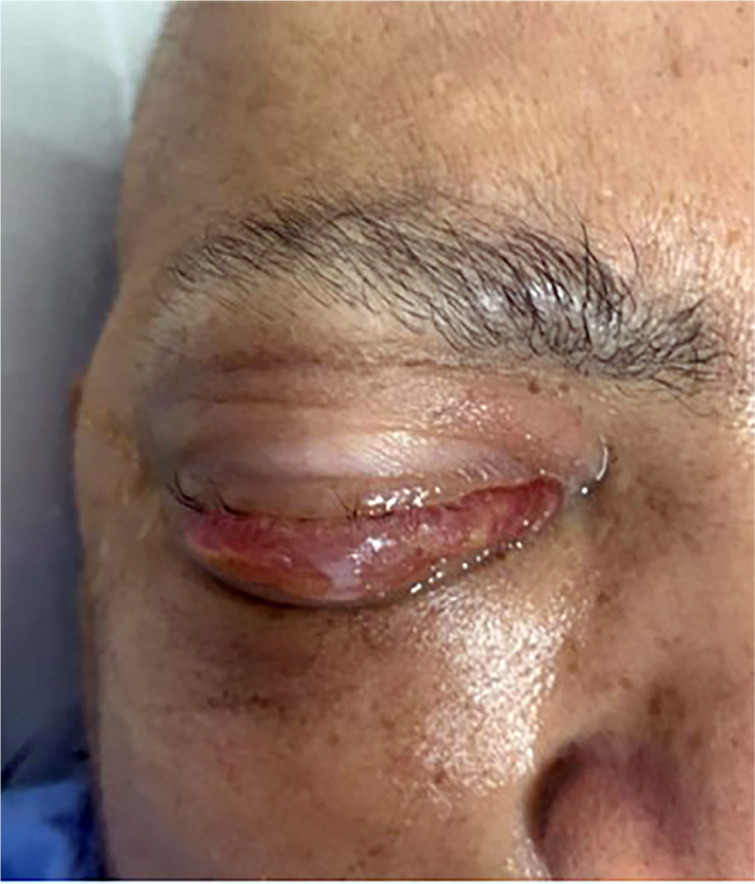
Invasive fusariosis is a serious invasive fungal disease, affecting immunocompetent and, more frequently, immunocompromised patients. Localized disease is the typical clinical form in immunocompetent patients. Immunocompromised hosts at elevated risk of developing invasive fusariosis are patients with acute leukemia receiving chemotherapeutic regimens for remission induction, and those undergoing allogeneic hematopoietic cell transplant. In this setting, the infection is usually disseminated with positive blood cultures, multiple painful metastatic skin lesions, and lung involvement. Currently available antifungal agents have poor in vitro activity against Fusarium species, but a clear-cut correlation between in vitro activity and clinical effectiveness does not exist. The outcome of invasive fusariosis is largely dependent on the resolution of immunosuppression, especially neutrophil recovery in neutropenic patients.

Keywords: Fusarium; fungal infection; fusariosis; immunocompromised.


Conflict of interest statement

M.N. has received honoraria as a speaker or consultant from Pfizer, MSD, Cidara, Knight, F2G, Teva, and Astellas. E.A. has no competing interests.

Figures

Fig 1
Fig 1
Culture of Fusarium species on potato dextrose agar after 7 days showing colony with white cottony margins and velvety center with shades of gray. (Courtesy of Dr. Geovanni Breda, reproduced with permission.)
Fig 2
Fig 2
Microscopic morphology (×400) showing hyaline septate hyphae with banana-shaped macroconidia. (Courtesy of Dr. Geovanni Breda, reproduced with permission.)
Fig 3
Fig 3
Periungueal cellulitis. (Courtesy of Dr. Marcia Matos, reproduced with permission.)
Fig 4
Fig 4
Periungueal cellulitis with tissue destruction. (Courtesy of Dr. Marcia Matos, reproduced with permission.)
Fig 5
Fig 5
Nodular skin lesion with an area of central necrosis, with a typical appearance of ecthyma gangrenosum. (Courtesy of Dr. Marcia Matos, reproduced with permission.)
Fig 6
Fig 6
Interdigital intertrigo with cellulitis. (Courtesy of Dr. Marcia Matos, reproduced with permission.)
Fig 7
Fig 7
Interdigital intertrigo with cellulitis. (Courtesy of Dr. Hugo Morales, reproduced with permission.)
Fig 8
Fig 8
Interdigital intertrigo with cellulitis. (Courtesy of Dr. Marcia Matos, reproduced with permission.)
Fig 9
Fig 9
Chest computed tomography showing macronodules in both lungs.
Fig 10
Fig 10
Chest computed tomography showing a large nodule with a halo sign in the left lung.
Fig 11
Fig 11
Disseminated skin lesions and toxemic appearance. Lesions at various stages of evolution: papular and nodular lesions with and without central necrosis. (Courtesy of Dr. Marilza Campos Magalhães, reproduced with permission.)
Fig 12
Fig 12
Disseminated skin lesions and toxemic appearance. Lesions at various stages of evolution: papular and nodular lesions with and without central necrosis. (Courtesy of Dr. Marilza Campos Magalhães, reproduced with permission.)
Fig 13
Fig 13
Nodules in the spleen.
Fig 14
Fig 14
Endophthalmitis with periorbital cellulitis. (Courtesy of Dr. Clara Rosemberg, reproduced with permission.)
Fig 15
Fig 15
Endophthalmitis with periorbital cellulitis. (Courtesy of Dr. Clara Rosemberg, reproduced with permission.)
Fig 16
Fig 16
Endophthalmitis with periorbital cellulitis. (Courtesy of Dr. Clara Rosemberg, reproduced with permission.)

References

    1. Kadaifciler DG, Demirel R 2. 2018. Fungal contaminants in man-made water systems connected to municipal water. J Water Health 16:244–252. doi:10.2166/wh.2018.272 - DOI - PubMed
    1. Anaissie EJ, Kuchar RT, Rex JH, Francesconi A, Kasai M, Müller FC, Lozano‐Chiu M, Summerbell RC, Dignani MC, Chanock SJ, Walsh TJ. 2001. Fusariosis associated with pathogenic Fusarium species colonization of a hospital water system: a new paradigm for the epidemiology of opportunistic mold infections . CLIN INFECT DIS 33:1871–1878. doi:10.1086/324501 - DOI - PubMed
    1. Dean R, Van Kan JAL, Pretorius ZA, Hammond-Kosack KE, Di Pietro A, Spanu PD, Rudd JJ, Dickman M, Kahmann R, Ellis J, Foster GD. 2012. The top 10 fungal pathogens in molecular plant pathology. Mol Plant Pathol 13:414–430. doi:10.1111/j.1364-3703.2011.00783.x - DOI - PMC - PubMed
    1. Sáenz V, Alvarez-Moreno C, Pape PL, Restrepo S, Guarro J, Ramírez AMC. 2020. A one health perspective to recognize Fusarium as important in clinical practice. JoF 6:235. doi:10.3390/jof6040235 - DOI - PMC - PubMed
    1. Uemura EVG, Barbosa M dos S, Simionatto S, Al-Harrasi A, Al-Hatmi AMS, Rossato L. 2022. Onychomycosis caused by Fusarium species. JoF 8:360. doi:10.3390/jof8040360 - DOI - PMC - PubMed

Substances

LinkOut - more resources