. 2023 Nov 8;7(11):e0274.
doi: 10.1097/HC9.0000000000000274. eCollection 2023 Nov 1.

A pig model of chronic hepatitis E displaying persistent viremia and a downregulation of innate immune responses in the liver

Affiliations

A pig model of chronic hepatitis E displaying persistent viremia and a downregulation of innate immune responses in the liver

Nancy León-Janampa et al. Hepatol Commun. .

Abstract

Background: Hepatitis E virus (HEV) is a zoonotic virus transmitted by pig meat and responsible for chronic hepatitis E in immunocompromised patients. It has proved challenging to reproduce this disease in its natural reservoir. We therefore aimed to develop a pig model of chronic hepatitis E to improve the characterization of this disease.

Methods: Ten pigs were treated with a tacrolimus-based regimen and intravenously inoculated with HEV. Tacrolimus trough concentration, HEV viremia, viral diversity, innate immune responses, liver histology, clinical disease and biochemical markers were monitored for 11 weeks post-infection (p.i.).

Results: HEV viremia persisted for 11 weeks p.i. HEV RNA was detected in the liver, small intestine, and colon at necropsy. Histological analysis revealed liver inflammation and fibrosis. Several mutations selected in the HEV genome were associated with compartmentalization in the feces and intestinal tissues, consistent with the hypothesis of extrahepatic replication in the digestive tract. Antiviral responses were characterized by a downregulation of IFN pathways in the liver, despite an upregulation of RIG-I and ISGs in the blood and liver.

Conclusions: We developed a pig model of chronic hepatitis E that reproduced the major hallmarks of this disease. This model revealed a compartmentalization of HEV genomes in the digestive tract and a downregulation of innate immune responses in the liver. These original features highlight the relevance of our model for studies of the pathogenesis of chronic hepatitis E and for validating future treatments.


Conflict of interest statement

The authors have no conflicts to report.

Figures

None
Graphical abstract
FIGURE 1
FIGURE 1
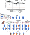
Monitoring of HEV RNA levels in 10 immunosuppressed HEV-infected pigs. HEV RNA levels in the serum (A) and feces during follow-up (B) in immunocompromised HEV-infected pigs. Individual values (gray) and means with the SD (black). *Pig 15 experienced spontaneously resolving HEV infection. †Pig 12 was euthanized 10 weeks after infection due to a severe leg injury.
FIGURE 2
FIGURE 2
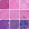
Discreet to moderate inflammation and fibrosis in the liver of immunosuppressed HEV-infected pigs 11 weeks p.i. Steatosis and inflammatory activity were scored by hematoxylin-eosin staining (A–E). Most liver samples (7/10) from the control group revealed no steatosis or inflammation (A). Half the liver samples (5/10) from the immunosuppressed group displayed discreet steatosis (B). Most liver samples (42/50) from the HEV-infected immunosuppressed pigs displayed discreet (D, n=8/50), moderate (E, n=33/50), or severe (F, n=1/50) inflammatory activity. Fibrosis was scored with Masson’s trichrome stain (G–I). Most liver samples from the control group (8/10) and the immunosuppressed group (8/10) displayed no fibrosis (G). By contrast, most liver samples from the HEV-infected immunosuppressed group displayed portal and periportal fibrosis (45/50), without (37/50, H) or with (4/50, I) centrilobular perisinusoidal fibrosis. Abbreviations: p.i., post-infection. This figure was formatted with ImageJ, using the Scientifig plug-in.
FIGURE 3
FIGURE 3
Quantification of HEV RNA and characterization of HEV diversity in tissues. (A) HEV RNA levels in tissue samples from immunocompromised HEV-infected pigs on necropsy. Individual follow-up data are indicated with gray lines for each pig. The black line and error bars indicate the mean and SD. Pig 15 experienced spontaneously resolving HEV infection and all tissue samples from this pig tested negative for HEV RNA (not shown). Weeks p.i., weeks after infection. (B) Prevalence of HEV genetic changes at position Y590 in ORF1, 11 weeks p.i. in HEV-infected pigs 14, 17, 19, and 20. Y590C (orange), Y590H (gray), and wild-type Y590 (blue) are shown. Data are missing for the liver of pig 14 due to insufficient sequencing depth. Arrows represent a proposed model of HEV transit between the liver and intestines in immunocompromised hosts, based on our observations and published in vitro data. Abbreviations: LL, lateral left; LR, lateral right; MR, medial right; ORF, open reading frame; p.i., post-infection.
FIGURE 4
FIGURE 4
Modulation of host innate immune responses in immunocompromised HEV-infected pigs. (A, B) Gene expression, assessed by high-throughput RT-qPCR (BioMark HD, Fluidigm), in (A) blood at 0, 4, 8, and 10 weeks p.i. and (B) the liver, at necropsy (11 wk p.i.). Gene expression levels are shown relative to the control group at 0 week p.i. for the blood samples and the control group at 11 weeks p.i. for the liver. The significance of differences between groups was analyzed with Mann-Whitney Wilcoxon tests corrected for multiple testing by the false discovery rate method. Adjusted p values were calculated with the Benjamini and Hochberg method. Data are shown for 5 animals for the control and immunocompromised groups and 9 of the 10 animals in the immunocompromised HEV-infected group (pig 15 was excluded due to nonchronic infection). (C, D) Host innate immune responses are represented for which significant (p<0.05) modulations of gene expression were observed in the blood (C) and liver (D) of immunocompromised HEV-infected pigs relative to immunocompromised pigs. Increase (up arrow) or decrease (down arrow) in expression levels at 4 (in blue) and 10 weeks p.i. (in red). Abbreviations: ER, endosplasmic reticulum; HSPG, heparan sulfate proteoglycan; IFIT, interferon-induced protein with tetratricopeptide repeats; MAVS, mitochondrial antiviral-signaling; NF-kB, nuclear factor-kappa B; p.i., post-infection; PRR, pattern recognition receptor; RIG-I, retinoic acid–inducible gene I. Created with BioRender.com.

References

    1. Khuroo MS, Khuroo MS, Khuroo NS. Hepatitis E: discovery, global impact, control and cure. World J Gastroenterol. 2016;22:7030. - PMC - PubMed
    1. Kamar N, Garrouste C, Haagsma EB, Garrigue V, Pischke S, Chauvet C, et al. . Factors associated with chronic hepatitis in patients with hepatitis E virus infection who have received solid organ transplants. Gastroenterology. 2011;140:1481–1489. - PubMed
    1. Kamar N, Selves J, Mansuy J-M, Ouezzani L, Péron J-M, Guitard J, et al. . Hepatitis E virus and chronic hepatitis in organ-transplant recipients. N Engl J Med. 2008;358:811–817. - PubMed
    1. Dalton HR, Kamar N, Baylis SA, Moradpour D, Wedemeyer H, Negro F. EASL Clinical Practice Guidelines on hepatitis E virus infection. J Hepatol. 2018;68:1256–1271. - PubMed
    1. Lenggenhager D, Pawel S, Honcharova-Biletska H, Evert K, Wenzel JJ, Montani M, et al. . The histologic presentation of hepatitis E reflects patients’ immune status and pre-existing liver condition. Mod Pathol. 2021;34:233–248. - PMC - PubMed

Publication types