. 2024 Apr;20(4):863-882.
doi: 10.1080/15548627.2023.2281126. Epub 2023 Nov 17.

Autophagy captures the retromer-TBC1D5 complex to inhibit receptor recycling

Affiliations

Autophagy captures the retromer-TBC1D5 complex to inhibit receptor recycling

Julian M Carosi et al. Autophagy. 2024 Apr.

Abstract

Retromer prevents the destruction of numerous receptors by recycling them from endosomes to the trans-Golgi network or plasma membrane. This enables retromer to fine-tune the activity of many signaling pathways in parallel. However, the mechanism(s) by which retromer function adapts to environmental fluctuations such as nutrient withdrawal and how this affects the fate of its cargoes remains incompletely understood. Here, we reveal that macroautophagy/autophagy inhibition by MTORC1 controls the abundance of retromer+ endosomes under nutrient-replete conditions. Autophagy activation by chemical inhibition of MTOR or nutrient withdrawal does not affect retromer assembly or its interaction with the RAB7 GAP protein TBC1D5, but rather targets these endosomes for bulk destruction following their capture by phagophores. This process appears to be distinct from amphisome formation. TBC1D5 and its ability to bind to retromer, but not its C-terminal LC3-interacting region (LIR) or nutrient-regulated dephosphorylation, is critical for retromer to be captured by autophagosomes following MTOR inhibition. Consequently, endosomal recycling of its cargoes to the plasma membrane and trans-Golgi network is impaired, leading to their lysosomal turnover. These findings demonstrate a mechanistic link connecting nutrient abundance to receptor homeostasis.Abbreviations: AMPK, 5'-AMP-activated protein kinase; APP, amyloid beta precursor protein; ATG, autophagy related; BafA, bafilomycin A1; CQ, chloroquine; DMEM, Dulbecco's minimum essential medium; DPBS, Dulbecco's phosphate-buffered saline; EBSS, Earle's balanced salt solution; FBS, fetal bovine serum; GAP, GTPase-activating protein; MAP1LC3/LC3, microtubule associated protein 1 light chain 3; LIR, LC3-interacting region; LANDO, LC3-associated endocytosis; LP, leupeptin and pepstatin; MTOR, mechanistic target of rapamycin kinase; MTORC1, MTOR complex 1; nutrient stress, withdrawal of amino acids and serum; PDZ, DLG4/PSD95, DLG1, and TJP1/zo-1; RPS6, ribosomal protein S6; RPS6KB1/S6K1, ribosomal protein S6 kinase B1; SLC2A1/GLUT1, solute carrier family 2 member 1; SORL1, sortillin related receptor 1; SORT1, sortillin 1; SNX, sorting nexin; TBC1D5, TBC1 domain family member 5; ULK1, unc-51 like autophagy activating kinase 1; WASH, WASH complex subunit.

Keywords: Autophagy; MTOR; MTORC1; TBC1D5; VPS35; retromer.


Conflict of interest statement

No potential conflict of interest was reported by the author(s).

Figures

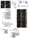
Figure 1.
Figure 1.
Autophagy inhibition by MTORC1 controls the abundance and distribution of retromer+ endosomes. (A) nutrient stress reduces the abundance of retromer-decorated endosomes. Cells were treated with or without EBSS for 6 h prior to immunofluorescence with a VPS35 antibody. Blue, DAPI stained nuclei. Cell outlines are indicated. Scale bar: 10 µm. (B) VPS35+ vesicles per cell from (A). Values are the mean ± SEM from n = 10–11 fields/condition from three independent experiments (unpaired t-test). (C) MTORC1 inhibition reduces the abundance of retromer-decorated endosomes. Cells were treated with or without AZD8055 or rapamycin for 6 h prior to immunofluorescence with a VPS35 antibody. Blue, DAPI-stained nuclei. Cell outlines are indicated. Scale bar: 10 µm. (D) VPS35+ vesicles per cell from (C). Values are the mean ± SEM from n = 10–25 fields/condition from three independent experiments (one-way ANOVA with Tukey’s multiple comparisons test). (E) the impact of nutrient stress and pharmacological inhibitors on MTOR and AMPK signalling. Immunoblot of lysates from cells treated with or without EBSS, AZD8055 or rapamycin for 6 h. Blots were probed for the indicated proteins and phosphoproteins. (F) MTORC1 inhibition downstream of nutrient stress reduces the abundance of retromer-decorated endosomes. Cells transfected with RHEBY35N-FLAG or an empty vector were treated with or without EBSS for 6 h prior to immunofluorescence with VPS35 and FLAG antibodies. Blue, DAPI stained nuclei. Scale bar: 10 µm. (G) VPS35+ vesicles per cell from (F). Values are the mean ± SEM from n = 41–115 cells/condition from three independent experiments (two-way ANOVA with Tukey’s multiple comparisons test). (H) loss of ATG5 impairs lipidation of ATG8 proteins following autophagy activation. Immunoblot of lysates from wild-type and ATG5-null cells treated with or without AZD8055 or EBSS for 6 h. Blots were probed for the indicated proteins. (I) loss of ATG5 prevents nutrient stress and MTOR inhibition from regulating the abundance of retromer-decorated endosomes. Wild-type and ATG5-null cells were treated with or without AZD8055 or EBSS for 6 h prior to immunofluorescence with a VPS35 antibody. Blue, DAPI stained nuclei. Scale bar: 10 µm. (J) VPS35+ vesicles per cell from (I). Values are the mean ± SEM from n = 44–51 cells/condition from three independent experiments (two-way ANOVA with Tukey’s multiple comparisons test).
Figure 2.
Figure 2.
The retromer-TBC1D5 interaction is not nutrient-sensitive. (A) experimental workflow to determine VPS35-FLAG interactome rewiring by MTOR inhibition and nutrient stress. VPS35-null cells stably expressing VPS35-FLAG were treated with or without either AZD8055 or EBSS for 18 h prior to anti-FLAG immunoprecipitation and quantitative determination of the VPS35-FLAG interactome. VPS35-null cells without VPS35-FLAG rescue served as negative control (background). (B) volcano plot of the VPS35-FLAG interactome under basal conditions (treated with vehicle for 18 h). Log2 fold-change over background (VPS35-null cells without VPS35-FLAG rescue) is shown. Significant enrichment was attributed for P < 0.05, indicated as data points above the horizontal line (corrected for multiple testing using the benjamini – Hochberg method). Data are the representative of four independent experiments. (C) the retromer complex and its interactions with TBC1D5 or other core machinery is not influenced by MTOR inhibition and nutrient stress. Scatter plot comparing VPS35-FLAG interactome changes with or without AZD8055 for 18 h. Log2 fold-change over background (VPS35-null cells without VPS35-FLAG rescue) is shown. Data are the representative of four independent experiments. (D) scatter plot comparing VPS35-FLAG interactome changes with or without EBSS for 18 h. Log2 fold-change over background (VPS35-null cells without VPS35-FLAG rescue) is shown. Data are the representative of four independent experiments. (E) the retromer-TBC1D5 interaction is not nutrient-sensitive. Anti-FLAG immunoprecipitation of lysates from VPS35-null cells stably expressing VPS35-FLAG were treated with or without AZD8055 or EBSS for 18 h. Immunoprecipitated and co-immunoprecipitated proteins were analyzed by immunoblot. VPS35-null cells without VPS35-FLAG rescue served as negative control (background). (F) loss of TBC1D5 or VPS35 does not affect the stability of the other. Immunoblot of lysates from wild-type, TBC1D5-null, and VPS35-null cells probed for the indicated proteins. (G) loss of TBC1D5 does not affect retromer stability. Immunoblot of lysates from wild-type or TBC1D5-null cells stably expressing either HA-TBC1D5 or HA-GST (negative control protein) were probed for the indicated proteins. (H) VPS35 is required for endosomal recruitment of TBC1D5 but not vice versa. Immunofluorescence of wild-type, TBC1D5-null, and VPS35-null cells with TBC1D5 and VPS35 antibodies. Nuclear staining of TBC1D5 is an artifact. Blue, DAPI stained nuclei. Scale bar: 10 µm. Insets show 2.5× magnification. (I) only a small fraction of the total TBC1D5 pool binds to retromer. Anti-FLAG and -HA immunoprecipitations of lysates from VPS35-null cells stably co-expressing VPS35-FLAG and HA-TBC1D5 were performed in parallel. Immunoprecipitated and co-immunoprecipitated proteins were analyzed by immunoblot. Non-transgenic cells served as a negative control. The high amount of anti-HA immunoprecipitate was 8× greater than the lower amount. Long exposure (L.E.). (J) VPS35 and TBC1D5 remain colocalized following MTOR inhibition or in the absence of ATG5. Wild-type and ATG5-null cells were treated with or without AZD8055 for 6 h. AZD8055-treated cells were supplemented with BafA for the final 3 h prior to immunofluorescence with VPS35 and TBC1D5 antibodies. Blue, DAPI stained nuclei. Scale bar: 10 µm. Insets show 2.5× magnification. A representative of three independent experiments is shown.
Figure 3.
Figure 3.
MTORC1 prevents capture and turnover of retromer-TBC1D5 decorated endosomes by autophagosomes. (A) VPS35 colocalizes with LC3B following MTOR inhibition but not in the absence of ATG5. Wild-type and ATG5-null cells were treated with or without AZD8055 for 6 h. AZD8055-treated cells were supplemented with BafA for the final 3 h prior to immunofluorescence with VPS35 and LC3B antibodies. Blue, DAPI stained nuclei. Scale bar: 10 µm. Insets show 2.5× magnification. A representative of three independent experiments is shown. (B) compared to EEA1+ endosomes, VPS26+ endosomes are preferentially targeted to LC3B+ vesicles following MTOR inhibition. Cells stably expressing FLAG-mTurbo-LC3B were treated with or without AZD8055 for 6 h. AZD8055-treated cells were supplemented with BafA for the final 3 h prior to immunofluorescence with EEA1, VPS26 and FLAG antibodies. Scale bar: 10 µm. Insets show 5× magnification. A representative of three biological replicates is shown. (C) colocalization of EEA1, VPS26, and EEA1 and VPS26 double-positive vesicles with FLAG-mTurbo-LC3B from (B). Values are the mean ± SEM from n = 29–33 cells/condition from three biological replicates (two-way ANOVA with Tukey’s multiple comparisons test). (D) VPS35 is recruited to autophagosomes following MTOR inhibition. Cells stably expressing RFP-GFP-LC3B to distinguish autophagosomes (GFP+ and RFP+) from autolysosomes or amphisomes (GFP and RFP+) were treated with or without AZD8055 for 6 h prior to immunofluorescence with a VPS35 antibody. Blue, DAPI stained nuclei. Scale bar: 10 µm. Insets show 2.5× magnification. A representative of two independent experiments is shown. (E) autophagosomes (GFP+ and RFP+) and autolysosomes/amphisomes (GFP and RFP+) per cell from (D). Values are the mean ± SEM from n = 19–20 cells/condition from two independent experiments (two-way ANOVA with Tukey’s multiple comparisons test). (F) VPS35+ autophagosomes (GFP+/RFP+) per cell from (D). Values are the mean ± SEM from n = 19–20 cells/condition from two independent experiments (mann-whitney U test). (G) the retromer-TBC1D5 complex becomes membrane-protected following MTOR inhibition. Post-nuclear lysates from cells treated with or without a combination of AZD8055 and BafA for 18 h were digested with proteinase K in the presence of absence of Triton X-100 to lyse intracellular membranes or were left untreated. Immunoblots were probed for the indicated proteins to indicate whether they resided within or outside intracellular vesicles/membranes. TAX1BP1 and SQSTM1 served as membrane-protected control proteins, and RPS6KB1 served as a non-membrane protected control protein. A representative of three independent experiments is shown.
Figure 4.
Figure 4.
The TBC1D5 interaction is critical for retromer to be trafficked to autophagosomes following MTOR inhibition. (A) loss of TBC1D5 prevents VPS35 colocalization with LC3B following MTOR inhibition. Wild-type and TBC1D5-null cells were treated with or without AZD8055 for 6 h. AZD8055-treated cells were supplemented with BafA for the final 3 h prior to immunofluorescence with VPS35 and LC3B antibodies. Blue, DAPI stained nuclei. Scale bar: 10 µm. Insets show 2.5× magnification. A representative of three independent experiments is shown. (B) VPS35 colocalization with LC3B from (A). Values are the mean ± SEM from n = 28–38 cells/condition from three independent experiments (kruskal-wallis test corrected for multiple comparisons with Dunn’s method). (C) predicted structure and topology of TBC1D5. The positions of its LIRs, TBC/GAP domain, retromer binding regions (ins #1 and ins #2), and phosphorylated residues are indicated. The structure shown was adapted from AlphaFold (uniprot ID: Q92609). (D) the ability of retromer to bind to TBC1D5 variants. Anti-FLAG immunoprecipitation of lysates from VPS35-null cells stably co-expressing VPS35-FLAG and HA-TBC1D5 variants. Immunoprecipitated and co-immunoprecipitated proteins were analyzed by immunoblot. HA-GST served as a negative control protein. A representative of two independent experiments is shown. (E) TBC1D5 must bind to retromer to enable delivery of VPS35 to LC3B+ compartments following MTOR inhibition. TBC1D5-null cells stably expressing HA-TBC1D5 or variants were treated with or without AZD8055 for 6 h. AZD8055-treated cells were supplemented with BafA for the final 3 h prior to immunofluorescence with VPS35 and LC3B antibodies. Blue, DAPI stained nuclei. Scale bar: 10 µm. Insets show 2.5× magnification. A representative of three independent experiments is shown. (F) VPS35 colocalization with LC3B from (E). Values are the mean ± SEM from n = 32–45 cells/condition from three independent experiments (kruskal-wallis test corrected for multiple comparisons with Dunn’s method). (G) TBC1D5 does not interact with endogenous ATG8 proteins or ULK1. Anti-HA immunoprecipitation of lysates from VPS35-null cells stably co-expressing VPS35-FLAG and HA-TBC1D5. Immunoprecipitated and co-immunoprecipitated proteins were analyzed by immunoblot. Non-transgenic cells served as a negative control. ULK1 nonspecifically binds to anti-HA beads. (H) TBC1D5 does not interact with overexpressed LC3B. Anti-GFP immunoprecipitation of lysates from VPS35-null cells stably co-expressing VPS35-FLAG and HA-TBC1D5 transfected with either GFP or GFP-LC3B. Immunoprecipitated and co-immunoprecipitated proteins were analyzed by immunoblot. GFP served as a negative control protein and anti-LMNB1 served as an IgG control immunoprecipitation. With long exposure (L.E.) of the immunoblot a nonspecific interaction between HA-TBC1D5 and GFP becomes apparent.
Figure 5.
Figure 5.
MTORC1 supports the trafficking of retromer cargoes. (A) the agonist isoproterenol stimulates ADRB2 endocytosis. Cells transfected with HA-FLAG-ADRB2 were treated with or without isoproterenol for 1 h prior to immunofluorescence with a FLAG antibody. Blue, DAPI stained nuclei. Scale bar: 10 µm. Insets show 2.5× magnification. (B) ADRB2 accumulates within lysosomes in cells lacking VPS35. Cells transfected with HA-FLAG-ADRB2 were treated with VPS35 or non-targeting control siRnas. Cells were treated with BafA for 2 h and isoproterenol for the final 30 min prior to immunofluorescence with a FLAG and LAMP1 antibodies. Blue, DAPI stained nuclei. Scale bar: 10 µm. Insets show 2.5× magnification. (C) HA-FLAG-ADRB2 colocalization with LAMP1 from (B). Values are the mean ± SEM from n = 6–8 cells/condition (unpaired t-test). (D) ADRB2 accumulates moderately within lysosomes following MTOR inhibition. Cells transfected with HA-FLAG-ADRB2 treated with or without AZD8055 for 6 h. Cells were treated with leupeptin and pepstatin (LP) for the final 4 h and isoproterenol for the final 1 h to immunofluorescence with FLAG and LAMP1 antibodies. Blue, DAPI stained nuclei. Scale bar: 10 µm. Insets show 2.5× magnification. (E) HA-FLAG-ADRB2 colocalization with LAMP1 from (D). Values are the mean ± SEM from n = 26–31 fields/condition from three independent experiments (unpaired t-test). (F) reduced association of ADRB2 with retromer following MTOR inhibition. Cells transfected with HA-FLAG-ADRB2 treated with or without AZD8055 for 6 h, and isoproterenol for the final 1 h prior to immunofluorescence with FLAG and VPS35 antibodies. Blue, DAPI stained nuclei. Scale bar: 10 µm. Insets show 2.5× magnification. (G) HA-FLAG-ADRB2 colocalization with VPS35 from (F). Values are the mean ± SEM from n = 25 fields/condition from three independent experiments (unpaired t-test). (H) MTOR inhibition impairs HA-FLAG-ADRB2 recycling back to the plasma membrane following agonist-induced endocytosis. Cells transfected with HA-FLAG-ADRB2 receptor were treated with or without AZD8055 for 6 h, then were pulsed with isoproterenol for 30 min to induce endocytosis and then chased for 10 min to allow recycling to occur. Cell surface HA-FLAG-ADRB2 was measured by flow cytometry. Recycling index refers to the frequency of transfected cells that recycled the receptor back to the cell surface. Values are the mean ± SEM from three independent experiments (One-sample t and Wilcoxon test compared to 1). (I) model showing how nutrients (such as amino acids) activate MTORC1 to inhibit capture of retromer-TBC1D5 decorated endosomes by autophagosomes to promote receptor recycling.

References

    1. Carosi JM, Denton D, Kumar S, et al. Receptor recycling by Retromer. Mol Cell Biol. 2023;43(7):317–334. doi: 10.1080/10985549.2023.2222053 - DOI - PMC - PubMed
    1. Seaman MN. Cargo-selective endosomal sorting for retrieval to the Golgi requires retromer. J Cell Biol. 2004;165(1):111–122. doi: 10.1083/jcb.200312034 - DOI - PMC - PubMed
    1. Seaman MN, Marcusson EG, Cereghino JL, et al. Endosome to Golgi retrieval of the vacuolar protein sorting receptor, Vps10p, requires the function of the VPS29, VPS30, and VPS35 gene products. J Cell Biol. 1997;137(1):79–92. doi: 10.1083/jcb.137.1.79 - DOI - PMC - PubMed
    1. Harbour ME, Breusegem SYA, Antrobus R, et al. The cargo-selective retromer complex is a recruiting hub for protein complexes that regulate endosomal tubule dynamics. J Cell Sci. 2010;123(21):3703–3717. doi: 10.1242/jcs.071472 - DOI - PMC - PubMed
    1. Hesketh GG, Pérez-Dorado I, Jackson L, et al. VARP is recruited on to endosomes by direct interaction with retromer, where together they function in export to the cell surface. Dev Cell. 2014;29(5):591–606. doi: 10.1016/j.devcel.2014.04.010 - DOI - PMC - PubMed

Publication types

MeSH terms

Substances