. 2023 Nov 8;14(11):726.
doi: 10.1038/s41419-023-06253-6.

SCFβTrCP-mediated degradation of SHARP1 in triple-negative breast cancer

Affiliations

SCFβTrCP-mediated degradation of SHARP1 in triple-negative breast cancer

Juliana Haydeé Enriqué Steinberg et al. Cell Death Dis. .

Abstract

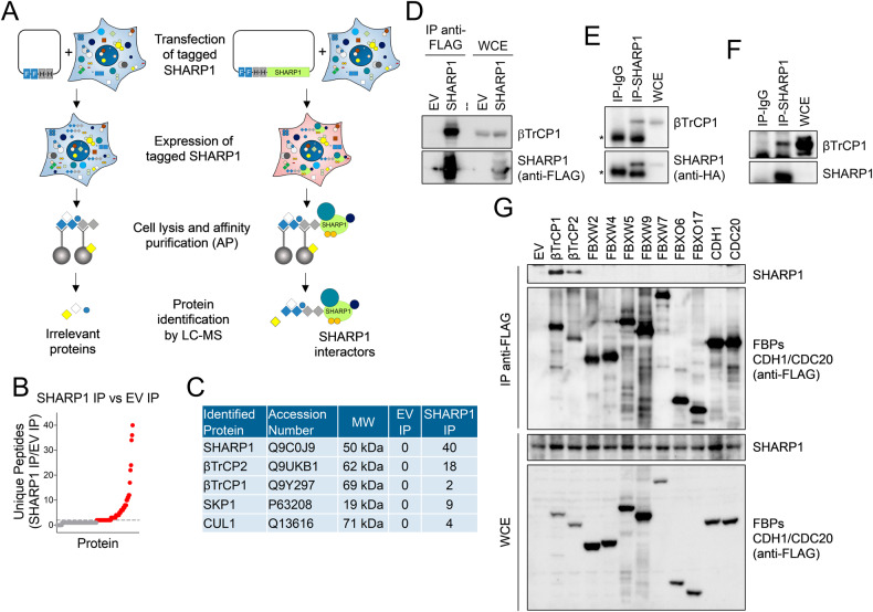
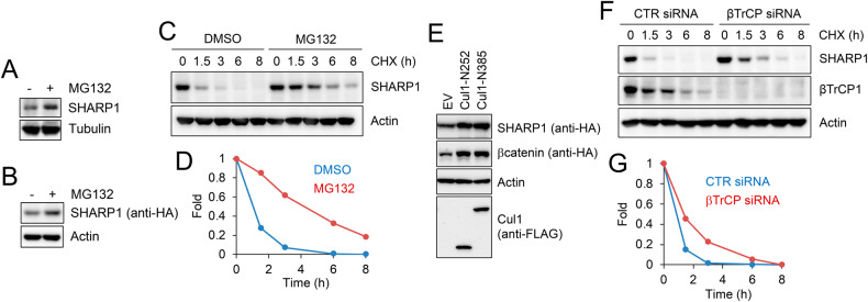
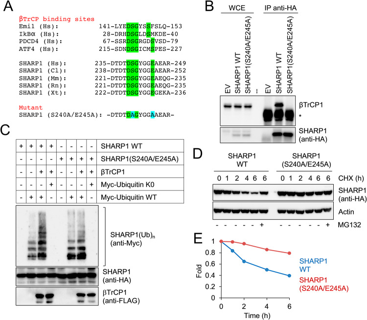
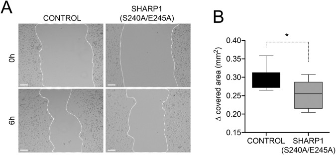
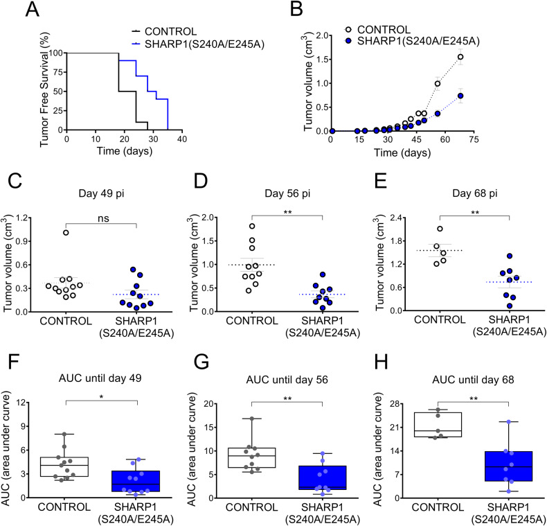
Triple-negative breast cancer (TNBC) is a subtype of breast cancer associated with metastasis, high recurrence rate, and poor survival. The basic helix-loop-helix transcription factor SHARP1 (Split and Hairy-related Protein 1) has been identified as a suppressor of the metastatic behavior of TNBC. SHARP1 blocks the invasive phenotype of TNBC by inhibiting hypoxia-inducible factors and its loss correlates with poor survival of breast cancer patients. Here, we show that SHARP1 is an unstable protein that is targeted for proteasomal degradation by the E3 ubiquitin ligase complex SCFβTrCP. SHARP1 recruits βTrCP via a phosphodegron encompassing Ser240 and Glu245 which are required for SHARP1 ubiquitylation and degradation. Furthermore, mice injected with TNBC cells expressing the non-degradable SHARP1(S240A/E245A) mutant display reduced tumor growth and increased tumor-free survival. Our study suggests that targeting the βTrCP-dependent degradation of SHARP1 represents a therapeutic strategy in TNBC.


Conflict of interest statement

The authors declare no competing interests.

Figures

Fig. 1
Fig. 1. SHARP1 interacts with βTrCP1 and βTrCP2.
A Overview of the experimental workflow aimed at the identification of SHARP1 interactors. FLAG-HA-tagged SHARP1 was expressed in HEK293T cells. Cells were lysed and whole-cell extracts were subjected to immunoprecipitation using FLAG and HA beads. SHARP1 immunoprecipitates were then analyzed by mass spectrometry. B Dot plot showing the number of unique peptides detected by mass spectrometry after SHARP1 immunoprecipitation (IP) compared to the control immunoprecipitation (EV: empty vector). C The number of unique peptides, accession numbers, and molecular weights (MW) for selected proteins recovered by mass spectrometry in control (EV: empty vector) and SHARP1 immunopurifications (IP) are shown. D HEK293T cells were transfected with an empty vector (EV) or FLAG-tagged SHARP1. Whole-cell extracts (WCE) were immunoprecipitated (IP) with anti-FLAG resin and immunocomplexes were probed with the indicated antibodies. E, F Whole-cell extracts (WCE) from MDA-MB231 cells stably expressing HA-tagged SHARP1 E or parental MDA-MB231 cells F were immunoprecipitated with an anti-SHARP1 antibody. SHARP1 immunocomplexes were probed with antibodies to the indicated proteins. G HEK293T cells, transfected with the indicated FLAG-tagged F-box proteins and the APC/C subunits CDH1 and CDC20, were lysed. WCEs were immunoprecipitated (IP) with anti-FLAG resin and analyzed by immunoblotting with antibodies for the indicated proteins.
Fig. 2
Fig. 2. Characterization of SHARP1 degradation.
A, B Parental MDA-MB231 cells A or MDA-MB231 cells stably expressing HA-tagged SHARP1 B were treated with the proteasome inhibitor MG132 for 6 hours. Cells were lysed and whole-cell extracts were subjected to immunoblotting with antibodies for the indicated proteins. C MDA-MB231 cells were treated with cycloheximide (CHX) with or without the proteasome inhibitor MG132, collected at the indicated times, and lysed. Whole-cell extracts were subjected to immunoblotting with the indicated antibodies. D The graph represents the quantification of SHARP1 levels (shown in C) normalized to the Actin loading control. E Cells were transfected with an empty vector (EV) or FLAG-tagged CUL1 dominant-negative mutants (CUL1-N252 or CUL1-N385) along with HA-tagged SHARP1 or HA-tagged β-catenin. Cells were collected and lysed. Whole-cell extracts were analyzed by immunoblotting with the indicated antibodies. F Cells were transfected with a siRNA targeting both βTrCP1 and βTrCP2 or a control siRNA and treated with cycloheximide (CHX) for the indicated times. Cells were lysed and whole-cell extracts were analyzed by immunoblotting with the indicated antibodies. G The graph represents the quantification of SHARP1 levels (shown in F) normalized to the Actin loading control.
Fig. 3
Fig. 3. Ser240 and Glu245 in SHARP1 are required for SHARP1-βTrCP1 interaction, SHARP1 ubiquitylation, and proteasomal degradation.
A Alignment of the amino acid regions encompassing the βTrCP-binding motif (highlighted in green) in previously reported βTrCP substrates and SHARP1 orthologs. The amino acid sequence of the SHARP1 phosphodegron mutant is shown at the bottom, with the altered amino acids highlighted in blue. Hs, Homo sapiens; Cl, Canis lupus familiaris; Mm, Mus musculus; Rn, Rattus norvegicus; Xt, Xenopus tropicalis. B MDA-MB231 parental cells or expressing HA-tagged wild-type SHARP1 or the SHARP1(S240A/E245A) mutant were lysed. Whole-cell extracts were subjected to immunoblotting with HA resin, followed by immunoblotting. Asterisk indicates the immunoglobulin heavy chain. C HEK293T cells were transfected with HA-tagged SHARP1 (wild type or S240A/E245A) and MYC-tagged ubiquitin [wild type or lysine-less (K0)], with or without FLAG-tagged βTrCP1. Cells were treated with the proteasome inhibitor MG132 for 5 hours and lysed. Whole-cell extracts were prepared in denaturing conditions and immunoprecipitated with an anti-HA antibody. Immunoprecipitates were then immunoblotted with the indicated antibodies. D MDA-MB231 cells expressing wild-type (WT) SHARP1 or the SHARP1(S240A/E245A) mutant were treated with cycloheximide (CHX). Cells were collected at the indicated times and lysed. Whole-cell extracts were subjected to immunoblotting with antibodies specific to the indicated proteins. E The graph represents the quantification of SHARP1 levels shown in D normalized to the Actin loading control.
Fig. 4
Fig. 4. SHARP1 degradation controls migration of triple-negative breast cancer cells.
A MDA-MB231 control cells or expressing the SHARP1(S240A/E245A) mutant were grown to confluence. Cell monolayers were wounded with a micropipette tip and photographed immediately after wounding (0 h) and after 6 hours (6 h). Representative areas at the indicated time points are shown. Scale bar = 100 μm. B The graph represents the Δ covered area (mm2) after 6 h (n = 9, Student’s t test, p = 0.0121).
Fig. 5
Fig. 5. Expression of the SHARP1(S240A/E245A) mutant inhibits triple-negative breast cancer in vivo.
A MDA-MB231 control cells or expressing the SHARP1(S240A/E245A) mutant were inoculated subcutaneously into the mammary fat pads of female NOD/SCID mice. Kaplan–Meier curves were built for Tumor-Free Survival (TFS) over time (n ≥ 10, Log-Rank (Mantel–Cox) test, p value = 0.0024). Hazard ratio (logrank): 2.625 [95% CI of ratio = 0.9845–6.997]. Number of mice used in each treatment: 11 control, 10 SHARP1(S240A/E245A) mutant. B Tumor volume was calculated at the indicated time points (mean value ± S.E.). CE Tumor volume at the indicated time points post-injection (pi). Unpaired two-tailed t test. P values: 0.1104 (ns), 0.0012 (**), and 0.0045 (**) for tumor volume differences at days 49, 56, or 68 post-injection, respectively. FH Area under the curve (AUC) until the indicated time points. Unpaired two-tailed t test. P values: 0.0100 (*), 0.0031 (**), and 0.0050 (**) for the area under curve differences (curves ending at day 49, 56, or 68 post-injection, respectively).

References

    1. Jiang YZ, Ma D, Suo C, Shi J, Xue M, Hu X, et al. Genomic and transcriptomic landscape of triple-negative breast cancers: subtypes and treatment strategies. Cancer Cell. 2019;35:428–440.e5. doi: 10.1016/j.ccell.2019.02.001. - DOI - PubMed
    1. Montagner M, Enzo E, Forcato M, Zanconato F, Parenti A, Rampazzo E, et al. SHARP1 suppresses breast cancer metastasis by promoting degradation of hypoxia-inducible factors. Nature. 2012;487:380–4. doi: 10.1038/nature11207. - DOI - PubMed
    1. Adorno M, Cordenonsi M, Montagner M, Dupont S, Wong C, Hann B, et al. A mutant-p53/smad complex opposes p63 to empower TGFβ-induced metastasis. Cell. 2009;137:87–98. doi: 10.1016/j.cell.2009.01.039. - DOI - PubMed
    1. Bragado P, Estrada Y, Parikh F, Krause S, Capobianco C, Farina HG, et al. TGF-β2 dictates disseminated tumour cell fate in target organs through TGF-β-RIII and p38α/β signalling. Nat Cell Biol. 2013;15:1351–61. doi: 10.1038/ncb2861. - DOI - PMC - PubMed
    1. Honma S, Kawamoto T, Takagi Y, Fujimoto K, Sato F, Noshiro M, et al. Dec1 and Dec2 are regulators of the mammalian molecular clock. Nature. 2002;419:841–4. doi: 10.1038/nature01123. - DOI - PubMed

Publication types

Substances