. 2022 Jun 3;2(1):47.
doi: 10.1038/s43705-022-00130-7.

The plant rhizosheath-root niche is an edaphic "mini-oasis" in hyperarid deserts with enhanced microbial competition

Affiliations

The plant rhizosheath-root niche is an edaphic "mini-oasis" in hyperarid deserts with enhanced microbial competition

Ramona Marasco et al. ISME Commun. .

Abstract

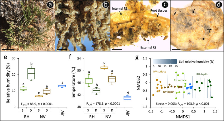
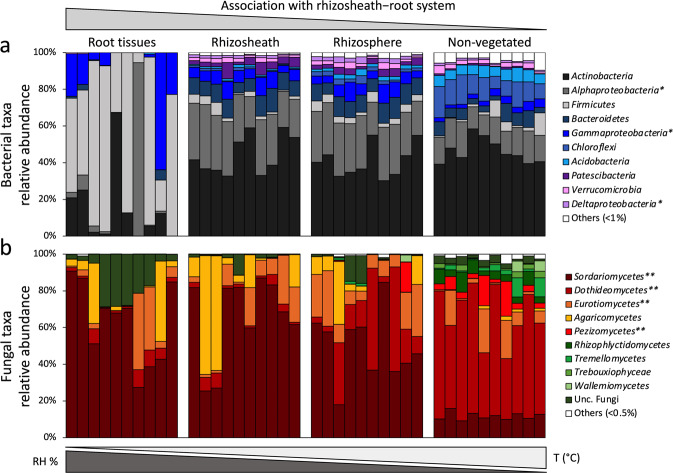
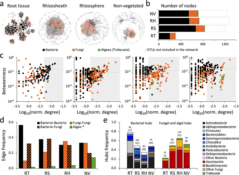
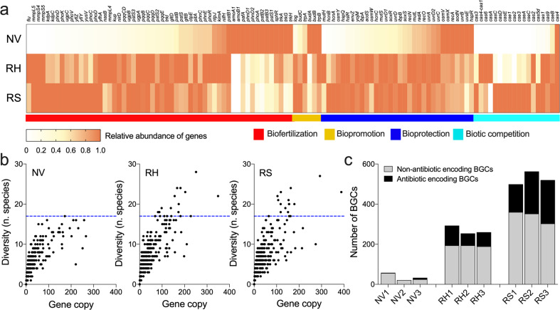
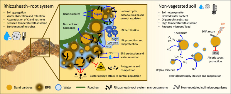
Plants have evolved unique morphological and developmental adaptations to cope with the abiotic stresses imposed by (hyper)arid environments. Such adaptations include the formation of rhizosheath-root system in which mutualistic plant-soil microbiome associations are established: the plant provides a nutrient-rich and shielded environment to microorganisms, which in return improve plant-fitness through plant growth promoting services. We hypothesized that the rhizosheath-root systems represent refuge niches and resource islands for the desert edaphic microbial communities. As a corollary, we posited that microorganisms compete intensively to colonize such "oasis" and only those beneficial microorganisms improving host fitness are preferentially selected by plant. Our results show that the belowground rhizosheath-root micro-environment is largely more hospitable than the surrounding gravel plain soil with higher nutrient and humidity contents, and cooler temperatures. By combining metabarcoding and shotgun metagenomics, we demonstrated that edaphic microbial biomass and community stability increased from the non-vegetated soils to the rhizosheath-root system. Concomitantly, non-vegetated soil communities favored autotrophy lifestyle while those associated with the plant niches were mainly heterotrophs and enriched in microbial plant growth promoting capacities. An intense inter-taxon microbial competition is involved in the colonization and homeostasis of the rhizosheath zone, as documented by significant enrichment of antibiotic resistance genes and CRISPR-Cas motifs. Altogether, our results demonstrate that rhizosheath-root systems are "edaphic mini-oases" and microbial diversity hotspots in hyperarid deserts. However, to colonize such refuge niches, the desert soil microorganisms compete intensively and are therefore prepared to outcompete potential rivals.


Conflict of interest statement

The authors declare no competing interests.

Figures

Fig. 1
Fig. 1. Rhizosheath–root system niche of S. ciliata in Namib Desert gravel plain.
a The speargrass S. ciliata growing in the gravel plain as shown by the new green leaves developing from the basal portion of the plant (bar, 2 cm). b Close-up photograph of the S. ciliata rhizosheath–root system extracted from the soil (bar, 1 cm). The rhizosheath (RS) is composed of sand grains physically attached to the root, along with trapped stones and sand grains. c Stereomicroscope image of the rhizosheath–root system structure shows the external RS layer of the matrix with long root hairs developing from the epidermis (internal layer of RS; i.e., outermost cells of the root) that entrap sand grains and stones, as well as the central root tissue of vascular plants (bar, 1 mm). d Magnification of a stone detached from the RS; biological mineral weathering is indicated by black arrows (bar, 1 mm). e Relative humidity (%) and f temperature (°C) measured (n = 10; ±standard deviation) in NV soils (surface and in-depth) and soils under S. ciliata plants (surface and in-depth RH); S, surface, and D, in-depth. Values from air are also reported. Results of the ANOVA main test are indicated, along with lowercase letters referring to results of the post-hoc multiple comparison Tukey’s tests. g Non-parametric multidimensional scaling (NMDS) ordination plot showing the relative distribution of humidity and temperature measured; the relative humidity trend is plotted onto the ordination space. The result of the PERMANOVA main test is reported.
Fig. 2
Fig. 2. Diversity and dynamics of microbial communities associated with the S. ciliata rhizosheath–root system.
Principal coordinate analysis (PCoA) of (a) bacterial and (b) microeukaryotic communities associated with the rhizosheath–root system (root tissue (RT), rhizosheath (RS) and rhizosphere (RH)) and non-vegetated (NV) soil. Arrows indicate the “horseshoes” shape distribution of microbial communities associated to different compartments, starting from the RT, and ending into the NV soils. Decay relationships among microbial communities’ similarities (BC: Bray–Curtis) and compartment relative distance (cm) for (c) bacteria and (d) microeukaryotes; red regression lines indicate the significant correlation among BC’ similarity and distance considering all the compartments. We note that when excluding the root tissues from the analyses, the correlation coefficients increased; blue regression lines, bacteria: p < 0.0001, R2 = 0.81; microeukaryotes: p < 0.0001, R2 = 0.73. Ternary plot presenting the variation in species composition among sites (beta-diversity) as result of its three components: similarity, replacement and richness difference [51]; the indices decomposing beta-diversity are visualized for the (e) bacterial and (f) microeukaryotic communities. Each point represents a pair of samples, and its position is determined by a triplet of values from the similarity, replacement, and richness difference. In each ternary plot, the large central dots where the lines start are the centroid of the points for each beta-diversity component; the lines represent their mean values.
Fig. 3
Fig. 3. Taxonomical composition of microbial communities associated with the S. ciliata rhizosheath–root system and non-vegetated soil.
Relative abundance of (a) bacterial and (b) microeukaryotic phyla/classes associated with rhizosheath–root system compartments (root tissue (RT), rhizosheath (RS) and rhizosphere (RH)) and non-vegetated (NV) soil. Relative abundances are expressed as percentages; star (*) indicates classes belonging to the Proteobacteria phylum; stars (**) indicate classes belonging to the Ascomycota phylum. Gradients of relative humidity and temperature are also schematically reported on the top.
Fig. 4
Fig. 4. Bacterial and fungal co-occurrence network in S. ciliata rhizosheath–root system compartments.
a Significant interactions (edge, p < 0.05) between bacterial and fungal OTUs in root tissue (RT), rhizosheath (RS) and rhizosphere (RH) of S. ciliate and non-vegetated (NV) soil were visualized by co-occurrence network. Circles (nodes) represent individual OTUs (bacteria, fungi, and algae); size of circles indicates the number of connections of such node (degree); nodes were colored according to their taxonomic affiliation: black, orange and green indicate bacteria, fungi and algae, respectively. For co-occurrence networks’ properties refer to Table 2. b Proportion of bacterial, fungal and algae OTUs included in co-occurrence networks. Portion of OTUs not included in the network is also reported (white portion of bars). c Relationship between node-normalized degree (log10) and betweenness centrality in networks of rhizosheath–root system and non-vegetated soil compartments. Colors indicate the taxonomic affiliation of nodes (bacterial, fungal and algal OTUs); gray-box indicate the nodes with high degrees defined as hubs; red-dashed boxes delineate the keystone species of each network (high degree and high betweenness). d Frequency of edges (connections) in the rhizosheath–root system compartments’ networks. Connections among the three components of the network are showed: bacteria–bacteria, fungi–fungi, bacteria–fungi, and algae with all the others (* = bacteria, fungi and algae). e Taxonomic affiliation of bacterial and fungal hubs detected for each network (c) expressed as normalized frequency; total number of bacterial and fungal hubs are reported on each bar, along with the number of keystone species accounted among them (numbers in square brackets). Details regarding taxonomy are reported in Supplementary Data S2.
Fig. 5
Fig. 5. Functional potential of microbial communities from root-associated edaphic niches of the Namib Desert.
a Heatmap showing the relative normalized abundances of metabolic marker genes from the metagenome assemblies in rhizosheath (RS), rhizosphere (RH) and non-vegetated (NV) soil. Relative abundances are calculated by first normalizing by sequencing depth and then scaling against the highest proportion for each marker gene; genes within the PGP categories biofertilization, biopromotion and bioprotection (abiotic and biotic stresses), along with genes involved in biotic competition, are used in this analysis. b Relationship between the copies of PGP/biotic competition genes (x-axis) and the number of microbial species presenting these genes (y-axis) is visualized in NV, RH and RS. c Number of biosynthetic gene clusters (BGCs) detected across RS, RH and NV soil metagenomes; antibiotic-encoding BGCs are indicate in black, while the remaining BGCs in gray.
Fig. 6
Fig. 6. Ecological race and interactions of microbial partners attracted by the rich root–rhizosphere niche under limited available resources in the hyperarid desert.
Schematic illustration of microbial communities associated with S. ciliata rhizosheath–root system (RS and RH) and NV soil and their contribute to niche-ecosystem functioning. Environmental, ecological, and functional characteristics of microbial communities associated with S. ciliata rhizosheath–root system and NV soil are reported considering both plant/soil–microbe and microbe–microbe interactions. Trophic modes and functions from metagenomes are resumed. More detailed explanation is reported in the “Discussion” section of the manuscript.

References

    1. Laity JJ. Deserts and desert environments. John Wiley & Sons; UK, 2009.
    1. Huang J, Yu H, Guan X, Wang G, Guo R. Accelerated dryland expansion under climate change. Nat Clim Chang. 2015;6:166–71. doi: 10.1038/nclimate2837. - DOI
    1. Berdugo M, Delgado-Baquerizo M, Soliveres S, Hernández-Clemente R, Zhao Y, Gaitán JJ, et al. Global ecosystem thresholds driven by aridity. Science. 2020;367:787–90. doi: 10.1126/science.aay5958. - DOI - PubMed
    1. Danin A. Plant adaptations to environmental stresses in desert dunes. In: Cloudsley-Thompson J, Punzo F, editors. Adaptations of desert organisms. Plant of desert dunes. Springer; Verlag Berlin Heidelberg, 1996.
    1. Makhalanyane TP, Valverde A, Gunnigle E, Frossard A, Ramond J-B, Cowan DA. Microbial ecology of hot desert edaphic systems. FEMS Microbiol Rev. 2015;39:203–21. doi: 10.1093/femsre/fuu011. - DOI - PubMed