Review
. 2024 Mar;25(3):196-210.
doi: 10.1038/s41576-023-00663-0. Epub 2023 Nov 8.

Scrambling the genome in cancer: causes and consequences of complex chromosome rearrangements

Affiliations
Review

Scrambling the genome in cancer: causes and consequences of complex chromosome rearrangements

Ksenia Krupina et al. Nat Rev Genet. 2024 Mar.

Abstract

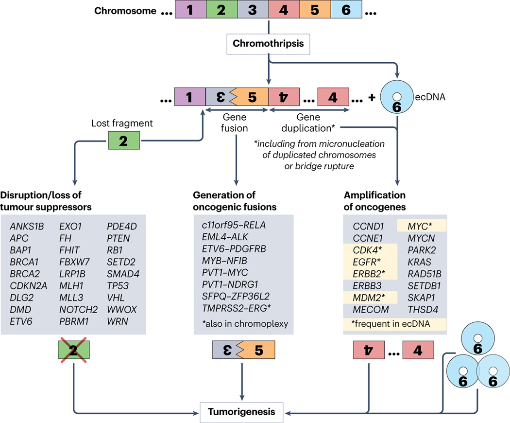
Complex chromosome rearrangements, known as chromoanagenesis, are widespread in cancer. Based on large-scale DNA sequencing of human tumours, the most frequent type of complex chromosome rearrangement is chromothripsis, a massive, localized and clustered rearrangement of one (or a few) chromosomes seemingly acquired in a single event. Chromothripsis can be initiated by mitotic errors that produce a micronucleus encapsulating a single chromosome or chromosomal fragment. Rupture of the unstable micronuclear envelope exposes its chromatin to cytosolic nucleases and induces chromothriptic shattering. Found in up to half of tumours included in pan-cancer genomic analyses, chromothriptic rearrangements can contribute to tumorigenesis through inactivation of tumour suppressor genes, activation of proto-oncogenes, or gene amplification through the production of self-propagating extrachromosomal circular DNAs encoding oncogenes or genes conferring anticancer drug resistance. Here, we discuss what has been learned about the mechanisms that enable these complex genomic rearrangements and their consequences in cancer.


Figures

Fig. 1.
Fig. 1.. Methods to detect chromothripsis with different spatial and sequence resolution.
Complementary techniques applied for the detection of chromothripsis vary in the precision of sequence resolution (ranging from the megabases to single nucleotides, horizontal axis) and spatial resolution (from clones or tissue samples to single cells, vertical axis). Top left square: examples of high-throughput techniques with relatively low spatial/sequence resolution: low coverage whole genome sequencing (WGS) and in situ hybridization probe-based methods; top right square: the highest sequence resolution with low spatial resolution is achieved by high coverage WGS (might be combined with whole exome sequencing); bottom left square: advanced fluorescent-in-situ-hybridization techniques provide the highest (single-cell) spatial/relatively low (within kilobases to megabases) sequence resolution; bottom right square: single-cell WGS aims at achieving the highest spatial/sequence resolution, but the scarcity of genomic material remains a significant challenge.
Fig. 2.
Fig. 2.. Mechanisms of micronuclei and chromosome bridges formation and rupture.
Errors in cell division, which involve physical separation of a chromosome or chromosome region from the rest of the chromosomes (such as a lagging chromosome or acentric chromosome fragments), lead to the formation of a micronucleus in the subsequent interphase. Dicentric chromosomes result in the formation of a chromosome bridge when two centromeres are pulled apart to the opposite poles in mitosis; subsequently, chromosome fragments generated following the breakage of the chromosome bridge become micronucleated. Nuclear envelope assembly in micronuclei and chromosome bridges is delayed due to aberrant chromosome separation in anaphase and spindle inhibition. Furthermore, nuclear envelopes of both micronuclei and chromosome bridges are prone to rupture due to reduced levels of some nuclear envelope proteins (including lamin B1), suboptimal numbers of nuclear pores, mechanical tension, and stress due to small radius of curvature, endoplasmic reticulum (ER) invasion and insufficient repair of the rupture sites by the ESCRTIII complex.
Fig. 3.
Fig. 3.. Molecular mechanisms initiating chromosome fragmentation.
Chromosomes in ruptured micronuclei are prone to the accumulation of DNA damage and subsequent chromosome fragmentation. (1) Entry of cytoplasmic nuclease(s) upon nuclear envelope rupture including (a) endonucleases that cause generation of multiple nicks and double strand breaks and can lead to chromosome shattering; (b) the entry of endoplasmic reticulum- and nuclear envelope-associated exonuclease TREX1 (the Three-prime Repair EXonuclease 1) induces DNA damage, however, additional factors are likely to be involved, including endonucleases/nickases which generate the initial nicks serving as the substrates for TREX1; (2) Loss of nuclear components and entry of cytosolic factors in ruptured micronuclei leads to altered DNA replication and repair, enzyme inhibition, accumulation of unrepaired DNA crosslinks and nucleotide misincorporation. As a result, a micronucleated chromosome enters mitosis with unrepaired DNA breaks, which may contribute to its fragmentation; or (3) Ruptured micronuclei-associated attenuation of transcription and accumulation of RNA-DNA hybrids triggers DNA base excision repair. RNA-DNA hybrids are edited by adenine deaminases acting on RNA (ADAR) transforming deoxyadenosine in these hybrids into deoxyinosine; next, N-methyl-purine DNA glycosylase (MPG) removes deoxyinosine to produce an abasic site. The abasic sites are next cleaved by apurinic/apyrimidinic endonuclease (APE1), which generates single-stranded DNA nicks.
Fig. 4.
Fig. 4.. Molecular mechanisms for chromosome reassembly.
Chromosome fragments from a ruptured micronucleus are held together in mitosis, which prevents their loss and is crucial for the subsequent reassembly of a derivative chromothriptic chromosome. This tethering is mediated by a three-protein complex which includes Mediator of DNA damage checkpoint 1 (MDC1) directly binding to γH2AX at double strand DNA breaks, Cellular Inhibitor of Protein phosphatase 2A (CIP2A) and DNA Topoisomerase II Binding Protein 1 (TOPBP1) or a two-protein complex of just CIP2A and TOPBP1. Reassembly of the random fragments into a chromothriptic chromosome occurs in a main nucleus in the next G1 phase. Non-homologous end-joining (NHEJ) is considered a major pathway involved in fragment re-ligation, followed by Microhomology-mediated end-joining, MMEJ (a type of alternative end-joining, Alt-EJ) which repairs clusters of DNA double strand breaks. Microhomology-mediated break-induced replication (MMBIR) and fork stalling and template switching (FoSTeS) are other replication-associated pathways which rely on microhomology.
Fig. 5.
Fig. 5.. Mechanisms through which chromothripsis drives tumorigenesis.
Three chromothripsis-associated mechanisms are thought to be capable of driving tumorigenesis: 1) disruption or loss of tumor suppressors, 2) generation of oncogenic fusions, and 3) amplification of oncogenes, with the most prevalent pathway being through the generation of circular, self-propagating and oncogene-bearing extrachromosomal DNA (ecDNA). Chromothripsis-affected genes, identified in patient-derived tumors, are listed for each mechanism.
Fig. 6.
Fig. 6.. Pan-cancer genome analysis reveals frequent co-occurrence of chromo-thripsis and ecDNA in 25 cancer types.
Left, blue: percentages of chromothripsis-positive samples per cancer type are from Cortes-Ciriano et al., Nat Genet 2020; descending from top to bottom. Right, red: percentages of ecDNA-positive samples per cancer type are from Kim et al., Nat Genet 2020. Cancer types, similar or matching between the two studies are shown. Fractions show the number of chromothripsis-positive (left, blue) or ecDNA-positive (right, red) samples per total number of samples analyzed per given cancer type in the corresponding study. Left (blue): the cancer types abbreviations (top to bottom) are: SoftTissue-Liposarc, liposarcoma, soft tissue; CNS-GBM, central nervous system glioblastoma; Eso-AdenoCA, esophagus adenocarcinoma; Skin-Melanoma, skin melanoma; Lung-AdenoCA, lung adenocarcinoma; Breast-AdenoCA, breast adenocarcinoma; Prost-AdenoCA, prostate adenocarcinoma; Lung-SCC, lung squamous cell carcinoma; Panc-AdenoCA, pancreatic adenocarcinoma; Ovary-AdenoCA, ovary adenocarcinoma; Stomach-AdenoCA, stomach adenocarcinoma; Biliary-AdenoCA, biliary adenocarcinoma; Bladder-TCC, bladder transitional cell carcinoma; Kidney-RCC, kidney renal cell carcinoma; ColoRect-AdenoCA, colorectal adenocarcinoma; Liver-HCC, liver hepatocellular carcinoma; Bone-Epith, bone neoplasm, epithelioid; Cervix-SCC, cervix squamous cell carcinoma; Lymph-BNHL, lymphoid mature B-cell lymphoma; Head-SCC, head-and-neck squamous cell carcinoma; CNS-Medullo, CNS medulloblastoma; Thy-AdenoCA, thyroid low-grade adenocarcinoma; Lymph-CLL, lymphoid chronic lymphocytic leukemia; Myeloid-MDS, myeloid myelodysplastic syndrome; Myeloid-MPN, myeloid myeloproliferative neoplasm; Myeloid-AML, myeloid acute myeloid leukemia.

References

    1. Nowell PC & Hungerford DA Minute Chromosome in Human Chronic Granulocytic Leukemia. Science 132, 1497–1497 (1960).
    1. Kaung DT & Swartzendruber AA Effect of chemotherapeutic agents on chromosomes of patients with lung cancer. Dis Chest 55, 98–100, doi:10.1378/chest.55.2.98 (1969). - DOI - PubMed
    1. Van Steenis H. Chromosomes and cancer. Nature 209, 819–821, doi:10.1038/209819a0 (1966). - DOI - PubMed
    1. Ishihara T, Kikuchi Y. & Sandberg AA Chromosomes of twenty cancer effusions: correlation of karyotypic, clinical, and pathologic aspects. J Natl Cancer Inst 30, 1303–1361 (1963). - PubMed
    1. Rowley JD Letter: A new consistent chromosomal abnormality in chronic myelogenous leukaemia identified by quinacrine fluorescence and Giemsa staining. Nature 243, 290–293, doi:10.1038/243290a0 (1973). - DOI - PubMed

LinkOut - more resources