. 2023 Nov 22;145(46):25120-25133.
doi: 10.1021/jacs.3c04991. Epub 2023 Nov 8.

In Situ Structural Observation of a Substrate- and Peroxide-Bound High-Spin Ferric-Hydroperoxo Intermediate in the P450 Enzyme CYP121

Affiliations

In Situ Structural Observation of a Substrate- and Peroxide-Bound High-Spin Ferric-Hydroperoxo Intermediate in the P450 Enzyme CYP121

Romie C Nguyen et al. J Am Chem Soc. .

Abstract

The P450 enzyme CYP121 from Mycobacterium tuberculosis catalyzes a carbon-carbon (C-C) bond coupling cyclization of the dityrosine substrate containing a diketopiperazine ring, cyclo(l-tyrosine-l-tyrosine) (cYY). An unusual high-spin (S = 5/2) ferric intermediate maximizes its population in less than 5 ms in the rapid freeze-quenching study of CYP121 during the shunt reaction with peracetic acid or hydrogen peroxide in acetic acid solution. We show that this intermediate can also be observed in the crystalline state by EPR spectroscopy. By developing an on-demand-rapid-mixing method for time-resolved serial femtosecond crystallography with X-ray free-electron laser (tr-SFX-XFEL) technology covering the millisecond time domain and without freezing, we structurally monitored the reaction in situ at room temperature. After a 200 ms peracetic acid reaction with the cocrystallized enzyme-substrate microcrystal slurry, a ferric-hydroperoxo intermediate is observed, and its structure is determined at 1.85 Å resolution. The structure shows a hydroperoxyl ligand between the heme and the native substrate, cYY. The oxygen atoms of the hydroperoxo are 2.5 and 3.2 Å from the iron ion. The end-on binding ligand adopts a near-side-on geometry and is weakly associated with the iron ion, causing the unusual high-spin state. This compound 0 intermediate, spectroscopically and structurally observed during the catalytic shunt pathway, reveals a unique binding mode that deviates from the end-on compound 0 intermediates in other heme enzymes. The hydroperoxyl ligand is only 2.9 Å from the bound cYY, suggesting an active oxidant role of the intermediate for direct substrate oxidation in the nonhydroxylation C-C bond coupling chemistry.


Conflict of interest statement

The authors declare no competing interests.

Figures
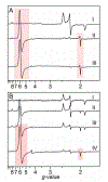
Figure 1.
Figure 1.
A high-spin intermediate was observed by rapid freeze-quench (RFQ) EPR of the solution state and single-crystal slurry EPR of the crystalline state after the reaction of the cYY-CYP121 complex with peracetic acid (PAA). (A) RFQ-EPR of the solution state CYP121. The top spectrum represents ES complex (I), the middle spectrum is from the enzyme-substrate (ES) complex after 5 ms reaction with PAA (II) as reported previously, and the bottom spectrum is the ES complex reaction with peracetic acid after 500 ms of mixing time (III). No low- to high-spin transition is observed from E-to-ES complex. After a 5-ms reaction, a new high-spin ferric heme is observed, which persists up to 500 ms before decay begins to occur. The signals pertaining to the rhombic high-spin species are highlighted with a red box. (B) Single-crystal EPR with a slurry of large-sized ES complex crystals. The top spectrum (ES, I) represents crystals measured without PAA. Subsequent time points used parallel samples of ES complex single-crystal slurry reacting with 5 mM PAA. The ES complex spectrum is magnified 2.5x, so the relative size of high-spin and low-spin signals are comparable to other spectra. Upon reaction of the single-crystal slurry with PAA, an initial axial high-spin EPR signal is observable by 30 s (II). A smaller rhombic high-spin EPR signal appears at 60 s (III) and plateaus by 120 s (IV). The high-spin ferric heme intermediate observed both in RFQ-EPR and in crystallo reaction EPR is highlighted with a colored box.
Figure 2.
Figure 2.
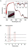
Single-crystal UV-vis monitoring of the CYP121 ES complex crystal reaction with peracetic acid and crystal reaction product analysis. (A) ES complex reaction with peracetic acid monitored by a single-crystal UV-visible microspectrophotometer in real-time. An image of the crystal and the difference spectra is shown in the inset. The initial ES complex spectrum (navy) is observed to transition to the E only state (red) by the shifting of the α/β band and decay of the charge transfer band, which indicates loss of the ligand from the active site. This ES to E transition is observed in the minutes time window depending on the size of the single-crystal, and it suggests substrate-to-product conversion. (B) HPLC analysis of the supernatant from CYP121-cYY crystal reaction with peracetic acid. (I) Mother liquor in which crystals are stored in, (II) mother liquor with 2 mM peracetic acid, and (III) reaction of ES complex crystals with peracetic acid after 60 min. The product, mycocyclosin, and substrate, cYY, elutions are highlighted with red boxes.
Figure 3.
Figure 3.
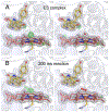
A hydroperoxyl ligand observed in situ by time-resolved SFX-XFEL of CYP121 cocrystallized with cYY microcrystal slurry reacted with peracetic acid for 200 ms. (A) The ES complex at 1.65 Å resolution (8TDQ.pdb) and (B) the 200 ms intermediate at 1.85 Å resolution (8TDP.pdb). The 2Fo-Fc electron density map is contoured at 1 σ (gray), and the Fo-Fc electron density map is contoured at 3 and −3 σ (colored green and red, respectively). The left panels show the omit map of the ES complex and the 200 ms intermediate without modeling the sixth ligand in the axial position of the heme iron. In contrast, the right panels show the electron density after modeling a ligand. For the 200 ms intermediate, the shape of the omit map is elongated rather than spherical, as expected for a water molecule. A peroxide ligand refined into the axial ligand density at 50% occupancy results in a consummate fitting which shows the ligand residing in a near side-on orientation relative to the heme iron.
Figure 4.
Figure 4.
A small fraction of the high-spin cpd 0 intermediate is generated from H2O2 in glacial acetic acid. (A) The CYP121 ES complex was rapidly mixed with the oxidant for 5 and 125 ms and hand-mixed for 30 and 90 s. In the millisecond time points, the heme remains low-spin with a small observable radical signal. In the seconds time points, a small fraction of the high-spin intermediate, different from that generated by substrate binding is observed. (B) Overlay of the RFQ-EPR 5 ms high-spin ferric heme-based intermediate observed when using H2O2 in peracetic acid (red) and glacial acetic acid (black) as the oxidant.
Figure 5.
Figure 5.
Structural comparison of the observed time-resolved SFX-XFEL ferric-hydroperoxo intermediate of P450 CYP121 (8TDP.pdb) to the hydroperoxo intermediates in heme-dependent tyrosine hydroxylase (7KQU.pdb), lactoperoxidase (7DLQ.pdb), and a trapped oxygen adduct in the resting state CYP121 without cYY (1N40.pdb). The Fe-O-O coordination angle for the end-on peroxo intermediates is more significant than 130° compared to 111.2° in CYP121 ferric-hydroperoxo, indicative of a more side-on geometry relative to the heme iron. The 2Fo-Fc electron density maps (gray) for the heme ligand are contoured at 1 σ, and the hemes are shown in pink and the hydroperxyl/peroxyl ligand in red.
Scheme 1.
Scheme 1.
Typical catalytic shunt pathway in cytochromes P450, including CYP121 with a substrate analog, cYY(O)Me (A); and proposed catalytic pathway for CYP121 with its native substrate (B). The bracket represents proposed intermediates.

References

    1. Huang X; Groves JT Oxygen activation and radical transformations in heme proteins and metalloporphyrins. Chem. Rev 2018, 118, 2491–2553. - PMC - PubMed
    1. Kühnel K; Derat E; Terner J; Shaik S; Schlichting I Structure and quantum chemical characterization of chloroperoxidase compound 0, a common reaction intermediate of diverse heme enzymes. Proc. Natl. Acad. Sci. U.S.A 2007, 104, 99–104. - PMC - PubMed
    1. Rittle J; Green MT Cytochrome P450 compound I: capture, characterization, and C-H bond activation kinetics. Science 2010, 330, 933–937. - PubMed
    1. Jin S; Bryson TA; Dawson JH Hydroperoxoferric heme intermediate as a second electrophilic oxidant in cytochrome P450-catalyzed reactions. J. Biol. Inorg. Chem 2004, 9, 644–653. - PubMed
    1. Mak PJ; Gregory MC; Denisov IG; Sligar SG; Kincaid JR Unveiling the crucial intermediates in androgen production. Proc. Natl. Acad. Sci. U.S.A 2015, 112, 15856–15861. - PMC - PubMed

Publication types