Review
. 2023 Nov 8;43(45):7575-7586.
doi: 10.1523/JNEUROSCI.1427-23.2023.

Advances in Deep Brain Stimulation: From Mechanisms to Applications

Affiliations
Review

Advances in Deep Brain Stimulation: From Mechanisms to Applications

Stephanie S Sandoval-Pistorius et al. J Neurosci. .

Abstract

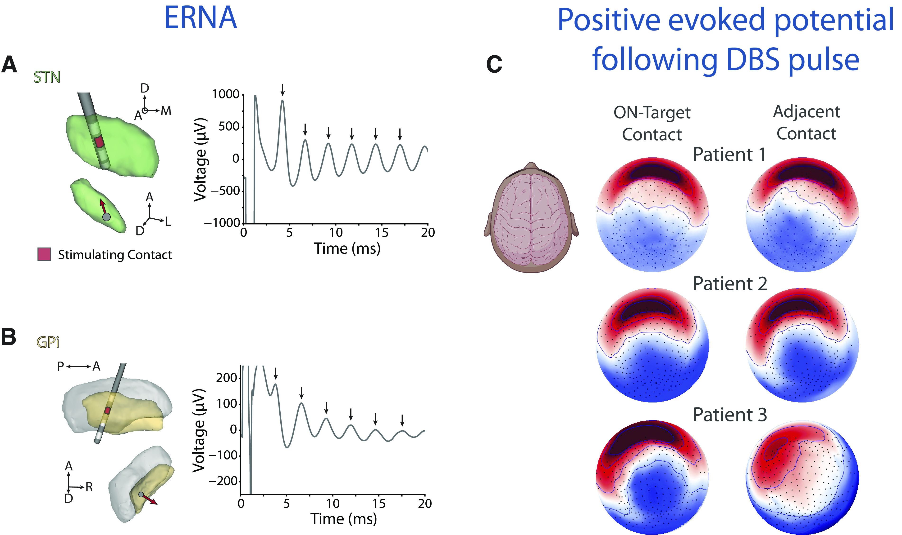
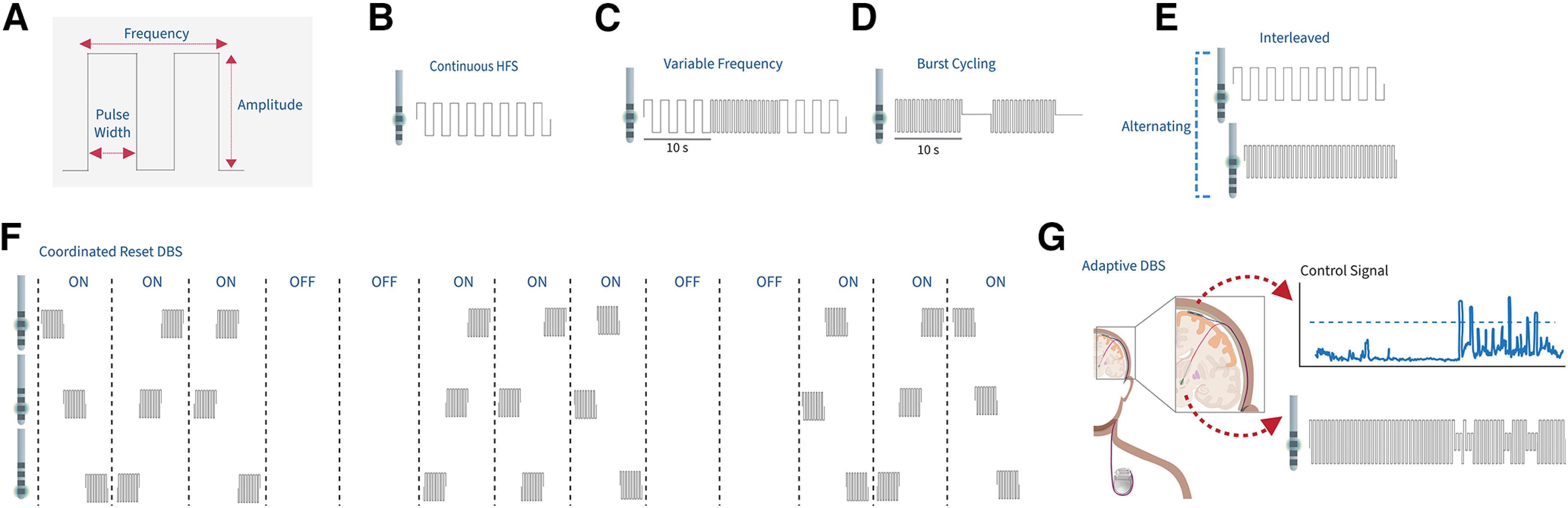
Deep brain stimulation (DBS) is an effective therapy for various neurologic and neuropsychiatric disorders, involving chronic implantation of electrodes into target brain regions for electrical stimulation delivery. Despite its safety and efficacy, DBS remains an underutilized therapy. Advances in the field of DBS, including in technology, mechanistic understanding, and applications have the potential to expand access and use of DBS, while also improving clinical outcomes. Developments in DBS technology, such as MRI compatibility and bidirectional DBS systems capable of sensing neural activity while providing therapeutic stimulation, have enabled advances in our understanding of DBS mechanisms and its application. In this review, we summarize recent work exploring DBS modulation of target networks. We also cover current work focusing on improved programming and the development of novel stimulation paradigms that go beyond current standards of DBS, many of which are enabled by sensing-enabled DBS systems and have the potential to expand access to DBS.

Keywords: coordinated reset; deep brain stimulation; evoked potentials; functional magnetic resonance imaging; sweet spot mapping.


Figures

Figure 1.
Figure 1.
Schematic of internal DBS system components and example results across targeting techniques. A, The DBS system involves internal and external components, including implanted electrodes, an IPG, extension cables connecting the electrodes and IPG. Not shown is the clinician programmer for setting and optimizing DBS parameters, and the patient programmer. Some components of this schematic were created with Biorender.com. B, An example of sweet spot mapping for motor progression and white matter tracts associated with motor progression in patients with Parkinson's disease. Adapted from Hacker et al. (2023). Top left, Coronal. Top right, Axial. Purple outlines the subthalamic nucleus. Red represents red nucleus. White dashed line indicates Bejjani line. C, An example of circuitry characteristics derived from fMRI in a patient with OCD. D, Neuroplastic reductions in OCD-related cortico-striatal hyperconnectivity are also apparent after chronic stimulation, with unique STN-frontostriatal coupling when DBS is off that may reflect disease spread. Caud = Caudate Putamen, VST = Ventral Striatum, ACC = Anterior Cingulate Cortex, STN = Subthalamic nucleus, OFC = orbitofrontal cortex.
Figure 2.
Figure 2.
EPs from DBS stimulation both within the target and on the cortex. Examples of intraoperative ERNA with (A) STN DBS and (B) GPi DBS using directional DBS leads. A, B, Left, The electrode locations and stimulation contacts. Red contact represents the contact used for stimulation on the lead. Arrow within the nuclei indicates the directional contact used for stimulation. Each arrow on the time domain plots (A, B, right) indicates an evoked response after a DBS pulse. C, Examples of cortical topography of the evoked response at 40 ms (P40) following a DBS pulse to the on-target contact (top row) and adjacent contact (bottom row) in 3 patients undergoing SCC-DBS for TRD. Saturated color represents higher amplitude (red represents positive; blue represents negative). Included components created with Biorender.com.
Figure 3.
Figure 3.
Stimulation paradigms beyond standard high-frequency DBS. A, Schematic of the DBS stimulation pulse, including the amplitude, pulse width, and frequency. B, Continuous high-frequency DBS delivers stimulation at >130 Hz with no change to stimulation amplitude, frequency, pulse width, or active contact. C, Variable frequency stimulation involves stimulating in alternating blocks of high-frequency stimulation and low-frequency stimulation (60-80 Hz). D, Burst cycling DBS delivers bursts of stimulation at the same intraburst frequency as HFS but with an interburst frequency ranging from 4 to 15 Hz. E, Interleaved stimulation alternates between two stimulation programs in which each can have independent amplitude, frequencies, pulse widths, and active contacts. F, Coordinated reset DBS in which the order of the contacts being stimulated is shuffled between each set of stimulation blocks. G, In this example of an adaptive DBS paradigm (components of the schematic were created in Biorender.com), a cortical control signal is being used to control stimulation amplitude on the depth lead within the STN. Once the control signal exceeds a predefined threshold (shown as the blue dotted line), stimulation amplitude decreases to avoid any stimulation-induced symptoms, such as dyskinesia.

Similar articles

Cited by

References

    1. Adamchic I, Hauptmann C, Barnikol UB, Pawelczyk N, Popovych O, Barnikol TT, Silchenko A, Volkmann J, Deuschl G, Meissner WG, Maarouf M, Sturm V, Freund HJ, Tass PA (2014) Coordinated reset neuromodulation for Parkinson's disease: proof-of-concept study. Mov Disord 29:1679–1684. 10.1002/mds.25923 - DOI - PMC - PubMed
    1. Allawala A, Bijanki KR, Goodman W, Cohn JF, Viswanathan A, Yoshor D, Borton DA, Pouratian N, Sheth SA (2021) A novel framework for network-targeted neuropsychiatric deep brain stimulation. Neurosurgery 89:E116–E121. 10.1093/neuros/nyab112 - DOI - PMC - PubMed
    1. Aman JE, Johnson LA, Sanabria DE, Wang J, Patriat R, Hill M, Marshall E, MacKinnon CD, Cooper SE, Schrock LE, Park MC, Harel N, Vitek JL (2020) Directional deep brain stimulation leads reveal spatially distinct oscillatory activity in the globus pallidus internus of Parkinson's disease patients. Neurobiol Dis 139:104819. 10.1016/j.nbd.2020.104819 - DOI - PMC - PubMed
    1. Arlotti M, Marceglia S, Foffani G, Volkmann J, Lozano AM, Moro E, Cogiamanian F, Prenassi M, Bocci T, Cortese F, Rampini P, Barbieri S, Priori A (2018) Eight-hours adaptive deep brain stimulation in patients with Parkinson disease. Neurology 90:e971–e976. 10.1212/WNL.0000000000005121 - DOI - PMC - PubMed
    1. Ashkan K, Rogers P, Bergman H, Ughratdar I (2017) Insights into the mechanisms of deep brain stimulation. Nat Rev Neurol 13:548–554. 10.1038/nrneurol.2017.105 - DOI - PubMed