. 2023 Nov 9;20(1):256.
doi: 10.1186/s12974-023-02940-5.

Efferocytosis is restricted by axon guidance molecule EphA4 via ERK/Stat6/MERTK signaling following brain injury

Affiliations

Efferocytosis is restricted by axon guidance molecule EphA4 via ERK/Stat6/MERTK signaling following brain injury

Eman Soliman et al. J Neuroinflammation. .

Abstract

Background: Efferocytosis is a process that removes apoptotic cells and cellular debris. Clearance of these cells alleviates neuroinflammation, prevents the release of inflammatory molecules, and promotes the production of anti-inflammatory cytokines to help maintain tissue homeostasis. The underlying mechanisms by which this occurs in the brain after injury remain ill-defined.

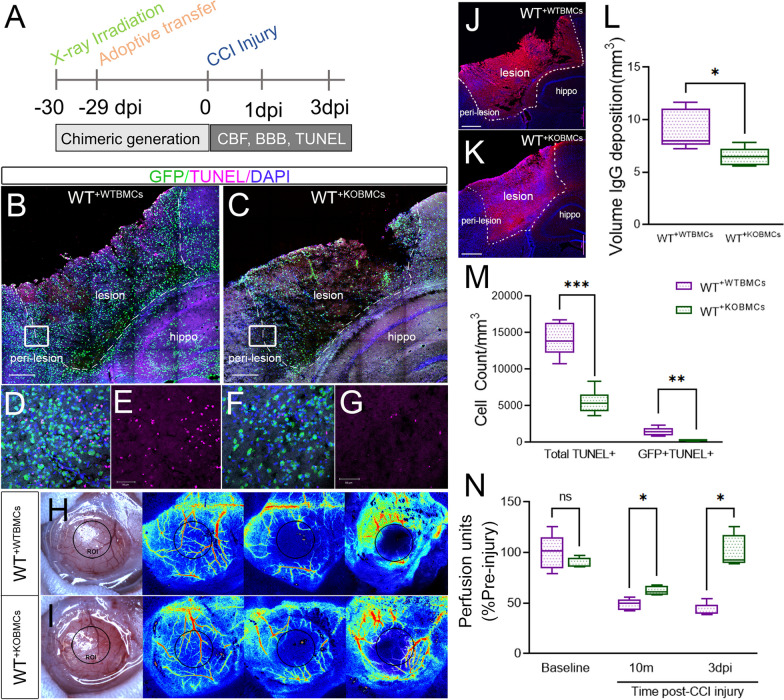
Methods: We used GFP bone marrow chimeric knockout (KO) mice to demonstrate that the axon guidance molecule EphA4 receptor tyrosine kinase is involved in suppressing MERTK in the brain to restrict efferocytosis of resident microglia and peripheral-derived monocyte/macrophages.

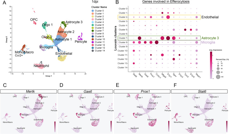
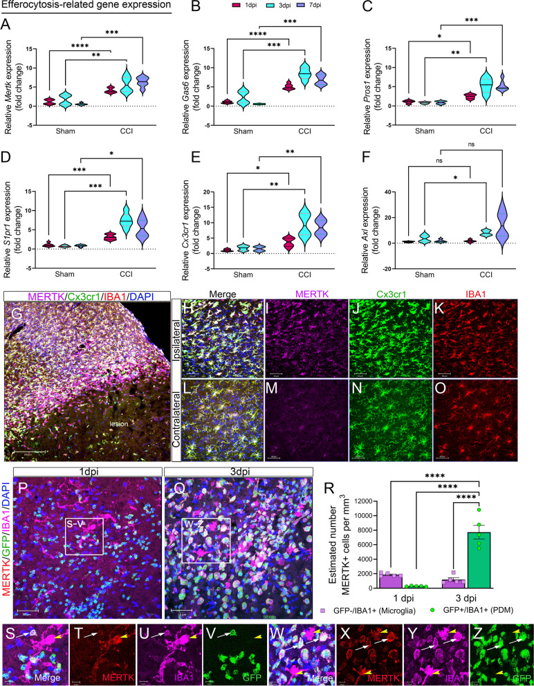
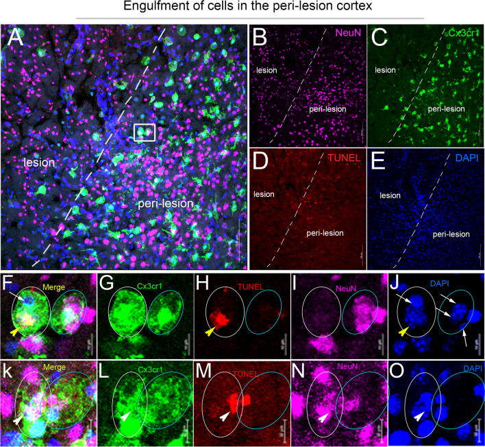
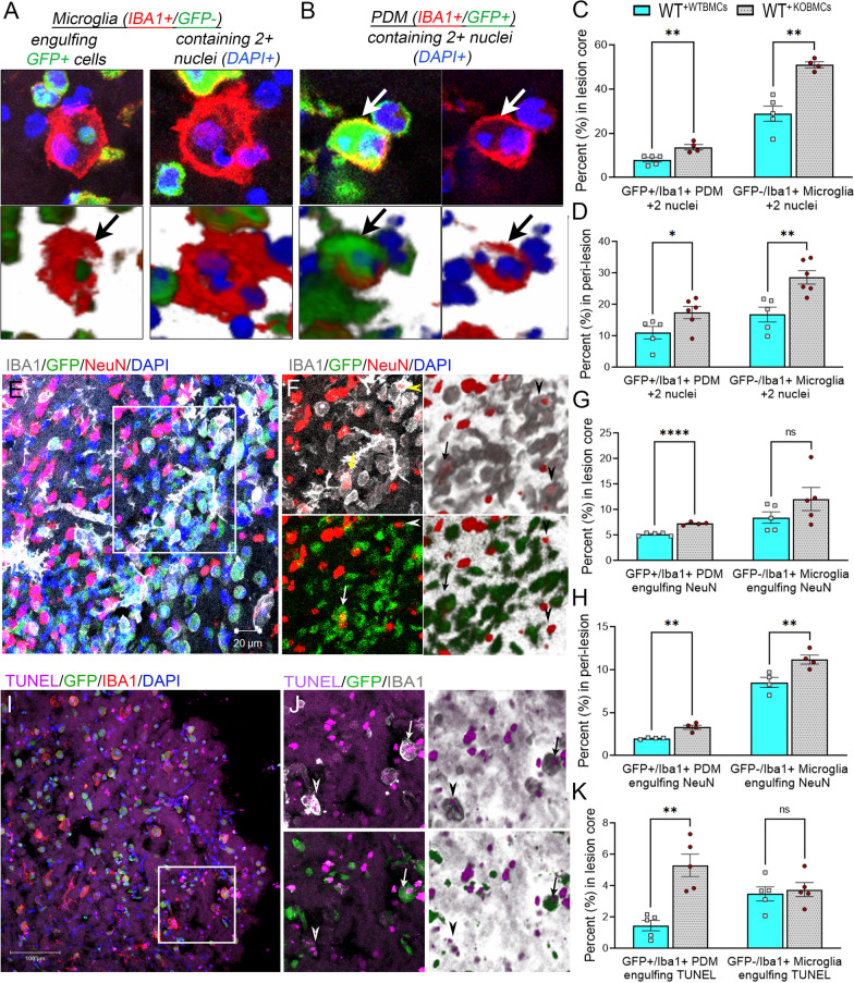
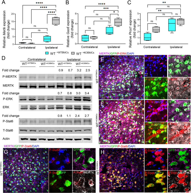
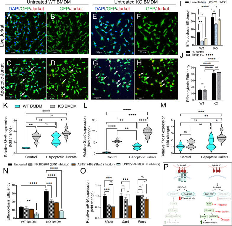
Results: Single-cell RNAseq identified MERTK expression, the primary receptor involved in efferocytosis, on monocytes, microglia, and a subset of astrocytes in the damaged cortex following brain injury. Loss of EphA4 on infiltrating GFP-expressing immune cells improved functional outcome concomitant with enhanced efferocytosis and overall protein expression of p-MERTK, p-ERK, and p-Stat6. The percentage of GFP+ monocyte/macrophages and resident microglia engulfing NeuN+ or TUNEL+ cells was significantly higher in KO chimeric mice. Importantly, mRNA expression of Mertk and its cognate ligand Gas6 was significantly elevated in these mice compared to the wild-type. Analysis of cell-specific expression showed that p-ERK and p-Stat6 co-localized with MERTK-expressing GFP + cells in the peri-lesional area of the cortex following brain injury. Using an in vitro efferocytosis assay, co-culturing pHrodo-labeled apoptotic Jurkat cells and bone marrow (BM)-derived macrophages, we demonstrate that efferocytosis efficiency and mRNA expression of Mertk and Gas6 was enhanced in the absence of EphA4. Selective inhibitors of ERK and Stat6 attenuated this effect, confirming that EphA4 suppresses monocyte/macrophage efferocytosis via inhibition of the ERK/Stat6 pathway.

Conclusions: Our findings implicate the ERK/Stat6/MERTK axis as a novel regulator of apoptotic debris clearance in brain injury that is restricted by peripheral myeloid-derived EphA4 to prevent the resolution of inflammation.

Keywords: Apoptosis; Efferocytosis; Eph; Ephrin; MERTK; Microglia; Neuroinflammation; Peripheral-derived macrophages; Traumatic brain injury.


Conflict of interest statement

The authors have declared that no conflict of interest exists.

Figures

Fig. 1
Fig. 1
ScRNA-seq analysis displays a differential expression of efferocytosis signals in the brain following CCI injury. A Uniform Manifold Approximation and Projection (UMAP) plot showing different cell clusters and their cell-specific annotation in the injured cortex at 1dpi. B Dot plot of efferocytosis genes expressed by each cluster type. C–F Feature maps highlighting the expression across clusters of MERTK (C), Gas 6 (D), Pros1 (E), and Stat6 (F)
Fig. 2
Fig. 2
Efferocytosis-related gene changes and MERTK expression in the damaged cortex following CCI injury. A–F Relative mRNA expression of Mertk (A), Gas6 (B), Pros1 (C), S1pr1 (D), Cx3cr1 (E), and Axl (F) in the ipsilateral cortex of CCI-injured wild type mice at 1, 3, and 7 dpi. G–O Representative confocal images for MERTK expression in the wild-type Cx3cr1EYFP−CreER/+ mice at 3 dpi. G 2 × 2 tile confocal images for the ipsilateral cortex showing MERTK (purple), Cx3cr1 (green), and IBA1 (red) expression. MERTK is upregulated and co-localized with Cx3cr1 and IBA1 in the ipsilateral cortex (H–K). MERTK expression is low in Cx3cr1 + IBA1 + cells in the contralateral cortex (L–O). P–Z MERTK expression in microglia and PDM in the ipsilateral cortex of CCI-injured GFP+ bone marrow chimeric wild type (WT+WTBMCs) mice. MERTK (red) is upregulated in GFP IBA1+ microglia at 1dpi (P, S–V) and 3dpi (Q, W–Z) and in GFP+IBA1+ PDM at 3 dpi (Q, W–Z). R The total number of MERTK+GFPIBA1+ microglia and MERTK+GFP+IBA1+ PDM was counted in the ipsilateral cortex at 1- and 3- dpi using the optical fractionator probe function of Stereoinvestigator. N = 5–6 mice/group. *P < 0.05; **P < 0.01; ***P < 0.001, ****P < 0.0001. Two-way ANOVA followed by Šídák’s multiple comparisons test. Scale bar = 200 µm in G, 50 µm in H–Q and 10 µm in S–Z
Fig. 3
Fig. 3
Engulfment of apoptotic neurons by Cx3cr1-expressing microglia/macrophages in the injured cortex following CCI. A–E Representative confocal images for TUNEL (red, D)- and NeuN (purple, B)-stained coronal section of Cx3cr1EYFP−CreER/+ mice at 3 dpi. F–O Region of interest (ROI) showing Cx3cr1+ cells (green) containing TUNEL + cells (yellow arrowhead), NeuN + cells (white arrow), and TUNEL + /NeuN + cells (white arrowhead) in the peri-lesion cortex. Scale bar = 100 µm in A–E and 10 µm in F–O
Fig. 4
Fig. 4
Peripheral-immune EphA4 deficiency enhances efferocytosis of PDMs and microglia in the damaged cortex following CCI injury. Serial coronal sections of WT+WTBMCs and WT+KOBMCs mice were stained with IBA1 (red) (A, B), NeuN (red), and IBA1 (gray) (E, F), or TUNEL and IBA1 (I, J). A, B Representative images showing microglia (IBA1 + /GFP −) engulfing GFP + cells (A, left) and containing 2 + nuclei (A, right) or PDM (IBA1 + /GFP +) containing 2 + nuclei (B). C, D quantification of microglia and PDM containing 2 + nuclei in the core and peri-lesion at 3dpi. E, F Microglia (arrowhead) and PDM (arrow) engulfing NeuN + neurons. G, H Quantification of microglia and PDM engulfing NeuN + neurons. I, J Microglia (arrowhead) and PDM (arrow) engulfing TUNEL + nuclei. K Quantification of microglia and PDM containing at least one TUNEL + nuclei and one TUNEL-DAPI + nuclei in the core at 3dpi. N = 4–6 mice/group. ns = non-significant; *P < 0.05; **P < 0.01; ***P < 0.001, ****P < 0.0001. Multiple t-tests. Scale bar = 20 µm in E and 100 µm in I
Fig. 5
Fig. 5
Peripheral EphA4 deficiency promotes P-ERK/P-Stat6/MERTK signaling. A–C mRNA expression of the efferocytosis receptor (MERTK, A) and its ligands (Gas6, B) and (Pros1, C) in the contralateral and ipsilateral cortex of WT+WTBMCs and WT+KOBMCs mice at 3dpi. N = 5–6 mice/group. Ns = non-significant; *P < 0.05; **P < 0.01; ***P < 0.001, ****P < 0.0001. Two-way ANOVA followed by Šídák’s multiple comparisons test. D Western blot analysis shows increased MERTK, ERK, and Stat6 phosphorylation in the ipsilateral cortex of chimeric WT+KO BMCs mice. E–H Representative images showing peripheral-derived GFP + cells expressing MERTK (purple) and P-ERK (E, F) and P-Stat6 (G, H) in WT+WT BMCs and WT+KO BMCs mice. Scale bar = 50 µm in E, F, G, H and 10 µm in insets
Fig. 6
Fig. 6
EphA4-null BMDMs show enhanced efferocytosis regulated by ERK/ Stat6/MERTK. AI EphA4 deletion improves the efferocytosis efficiency of BMDMS in vitro. AH Representative images showing the engulfment of the pHrodo-stained apoptotic (but not live) Jurkat cells (red) by GFP + untreated WT (AD) and EphA4 KO (EH) BMDMS. I Quantification of the efferocytosis efficiency of WT and EphA4 KO BMDMS after stimulation with LPS and HMGB1 for 4 h. J Efferocytosis of EphA4 KO BMDMSs is mediated by the blockade of forward EphA4, not reverse ephrin signals. Treatment of WT and EphA4 KO BMDMS with clustered EphA4-FC to activate reverse ephrin signals did not reduce the efferocytosis of EphA4 KO BMDMS. KM mRNA expression of Mertk (K), Gas6 (L), and Pros1 (M) with and without engulfment of apoptotic Jurkat cells. EphA4 KO BMDMSs have higher expression of Mertk and Gas6 than WT. N The use of MERTK inhibitor reduced efferocytosis of both WT and EphA4 KO BMDMS; however, Stat6 and ERK inhibitors selectively reduced efferocytosis in EphA4 KO BMDMS. O Stat6 inhibitor reduced Mertk and Gas6 expression, and ERK inhibitor reduced Gas6 expression in KO BMDMS engulfing apoptotic Jurkat cells. P Suggested pathway for the regulation of efferocytosis by EphA4. N = 5–6 mice/group. Ns = non-significant; *P < 0.05; **P < 0.01; ***P < 0.001, ****P < 0.0001. Two-way ANOVA followed by Šídák’s multiple comparisons test (IN) or one-way ANOVA followed by Tukey’s multiple comparisons test (O). Scale bar = 50 µm in AH
Fig. 7
Fig. 7
Peripheral EphA4 deficiency reduces apoptotic cell count and improves cerebral blood flow and BBB stability after CCI injury. A Experimental outline. BG, M Apoptotic TUNEL + (purple) cell count is reduced in WT+KO BMCs (B, D, E) compared to WT+WT BMCs (C, F, G) mice. The total number of TUNEL + and GFP + /TUNEL + cells (M) was counted using image J in 3 serial sections using 5 × 7 tiled z-stack confocal images. H, I, N Cerebral blood flow is improved in WT+KO BMCs compared to WT+WT BMCs. Blood flow was measured using laser speckle contrast imaging (LSCI) at 10 min and 3 days post-injury and presented as a percentage perfusion of the baseline. JL Blood–brain barrier permeability is improved in WT+KO BMCs (K) compared to WT+WT BMCs (J). The volume of IgG deposition was measured at 3 dpi in serial coronal sections using Cavalieri Estimator from StereoInvestigator (L). N = 5–6 mice/group. Ns = non-significant; *P < 0.05; **P < 0.01; ***P < 0.001. t-tests (L) or multiple t-tests (M, N). Scale bar = 500 µm in B, C, J, K and 50 µm DG

Update of

Similar articles

Cited by

References

    1. Miller GF, DePadilla L, Xu L. Costs of nonfatal traumatic brain injury in the United States, 2016. Med Care. 2021;59(5):451–455. doi: 10.1097/MLR.0000000000001511. - DOI - PMC - PubMed
    1. Corrigan F, Mander KA, Leonard AV, Vink R. Neurogenic inflammation after traumatic brain injury and its potentiation of classical inflammation. J Neuroinflammation. 2016;13(1):264. doi: 10.1186/s12974-016-0738-9. - DOI - PMC - PubMed
    1. Russo MV, McGavern DB. Inflammatory neuroprotection following traumatic brain injury. Science. 2016;353(6301):783–785. doi: 10.1126/science.aaf6260. - DOI - PMC - PubMed
    1. Hellewell SC, Morganti-Kossmann MC. Guilty molecules, guilty minds? The conflicting roles of the innate immune response to traumatic brain injury. Mediators Inflamm. 2012;2012:356494. doi: 10.1155/2012/356494. - DOI - PMC - PubMed
    1. Alam A, Thelin EP, Tajsic T, Khan DZ, Khellaf A, Patani R, Helmy A. Cellular infiltration in traumatic brain injury. J Neuroinflammation. 2020;17(1):328. doi: 10.1186/s12974-020-02005-x. - DOI - PMC - PubMed