. 2023 Nov 8;16(1):31.
doi: 10.1186/s13039-023-00662-0.

Cytogenomic features of Richter transformation

Affiliations

Cytogenomic features of Richter transformation

Renata Woroniecka et al. Mol Cytogenet. .

Abstract

Background: Richter transformation (RT) is the development of aggressive lymphoma in patients with chronic lymphocytic leukemia (CLL) or small lymphocytic lymphoma (SLL). This rare disease is characterised by dismal prognosis. In recent years, there has been a deeper understanding of RT molecular pathogenesis, and disruptions of apoptosis (TP53) and proliferation (CDKN2A, MYC, NOTCH1) has been described as typical aberrations in RT.

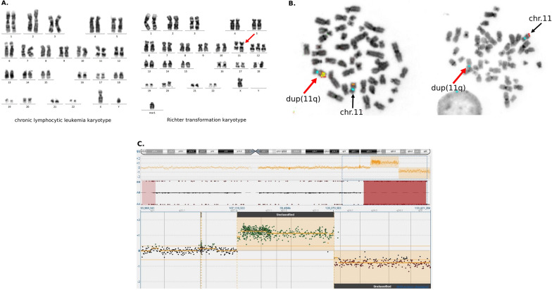
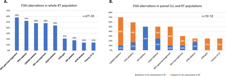
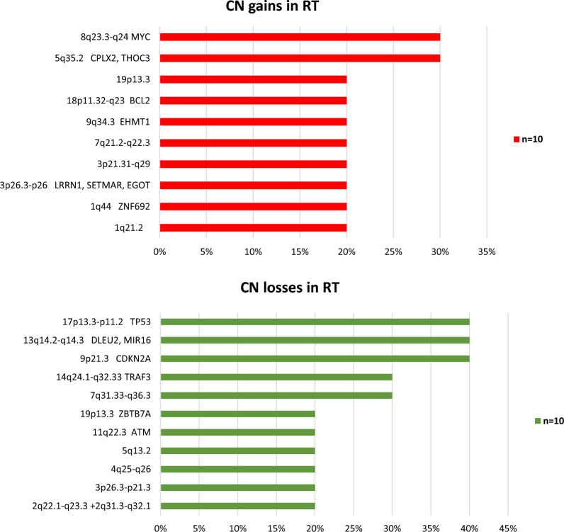
Results: A single-institution cohort of 33 RT patients were investigated by karyotyping, fluorescence in situ hybridization and single nucleotide polymorphism/copy number (CN) arrays. Most of RTs were typically manifested by diffuse large B-cell lymphoma, not otherwise specified, among the remaining cases one was classified as high-grade B-cell lymphoma with 11q aberrations. The most frequent alterations (40-60% of cases) were represented by MYC rearrangement/gain, deletions of TP53 and CDKN2A, IGH rearrangement and 13q14 deletion. Several other frequent lesions included losses of 14q24.1-q32.33, 7q31.33-q36.3, and gain of 5q35.2. Analysis of 13 CLL/SLL-RT pairs showed that RT arised from the CLL/SLL by acquiring of 10 ~ 12 cytogenetic or CN lesions/case, but without acquisition of loss of heterozygosity regions. Our result affirmed the higher genetic complexity in RT than CLL/SLL and confirmed the linear features of RT clonal evolution as predominant.

Conclusions: Cytogenomic profile was concordant with the literature data, however the role of IGH rearrangement, 14q deletion and 5q35.2 gain need to be explored. We anticipate that further characterization of RT lesions will probably facilitate better understanding of the RT clonal evolution.

Keywords: 11q gain/loss; 13q14 deletion; CDKN2A; Chronic lymphocytic leukemia; High-grade B-cell lymphoma with 11q aberrations; IGH deletion; IGH rearrangement; MYC; Richter transformation; Small lymphocytic lymphoma; TP53.


Conflict of interest statement

The authors declare no competing interests.

Figures

Fig. 1
Fig. 1
Cytogenomic data of case 17 with Richter transformation presented as high-grade B-cell lymphoma with 11q aberrations. A Karyotypes of RT and CLL paired phases, which demonstrate evolution of cytogenetic aberrations at RT transformation: CLL karyotype 46,XY[20], RT karyotype 47,X,-Y,dup(11)(q22q23), + 12, + mar1[4]/47,idem,add(18)(q23)[5] {red arrow indicates dup(11q)}. B FISH on RT metaphases (11 centromere signals are blue, red arrows indicate dup(11q) with 11q-gain/loss): a few KMT2A (yellow) signals on dup(11q) and one KMT2A signal on normal chromosome 11, 11qtel (red) signal on normal chromosome 11 and lack of 11qtel signal on dup(11q). C CGH/SNP array result: ideogram of chromosome 11, below copy number (CN) variations indicating duplication and deletion of 11q, underneath big brown blocks demonstrating loss of heterozygosity. Lower section shows magnification of aCGH analysis (CN variations). Green dots indicate gain, red dots shows deletion regions
Fig. 2
Fig. 2
Frequency of FISH aberrations in RT patients. A The frequency of FISH aberrations in whole RT population. B The frequency of FISH aberrations in paired CLL and RT populations. Orange bars represent the frequency of FISH aberrations present in RT and absent in paired CLL, which reflect the evolution of genetic lesions during transformation. Blue bars represent the frequency of FISH aberrations present in RT and present in paired CLL, which reflect the maintaining of lesions during transformation. n—according to the availability of material, variable numbers reflect varying numbers of examined cases
Fig. 3
Fig. 3
Frequency of copy number aberrations (CN) detected by arrays CGH. Red bars represent the frequency of minimal common regions (MCRs) of CN gains and green bars represent the frequency of MCRs of CN losses. The MCRs cover the genes included in the annotations

References

    1. Swerdlow SH, Campo E, Pileri SA, Harris N, Stein H, Siebert R, et al. The 2016 revision of the World Health Organization classification of lymphoid neoplasms. Blood. 2016;127(20):2375–2390. doi: 10.1182/blood-2016-01-643569. - DOI - PMC - PubMed
    1. Alaggio R, Amador C, Anagnostopoulos I, Attygalle AD, Araujo IBO, Berti E, et al. (2022) The 5th edition of the world health organization classification of haematolymphoid tumours lymphoid neoplasms. Leukemia. 36(7):1720–48. - PMC - PubMed
    1. Tsimberidou AM, O'Brien S, Khouri I, Giles FJ, Kantarjian HM, Champlin R, et al. Clinical outcomes and prognostic factors in patients with Richter’s syndrome treated with chemotherapy or chemoimmunotherapy with or without stem-cell transplantation. J Clin Oncol. 2006;24(15):2343–2351. doi: 10.1200/JCO.2005.05.0187. - DOI - PubMed
    1. Omonti CE, Omonti AE. Richter syndrome: a review of clinical, ocular, neurological and other manifestations. Br J Haematol. 2008;142:709–716. doi: 10.1111/j.1365-2141.2008.07248.x. - DOI - PubMed
    1. Rossi D, Gaidano G. Richter syndrome: molecular insights and clinical perspectives. Hematol Oncol. 2009;27:1–10. doi: 10.1002/hon.880. - DOI - PubMed