

Report on a case of liver-originating malignant melanoma of unknown primary
- PMID: 37941789
- PMCID: PMC10628584
- DOI: 10.1515/biol-2022-0750
Report on a case of liver-originating malignant melanoma of unknown primary
Abstract
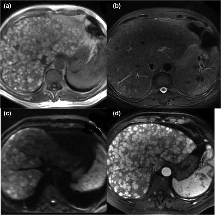
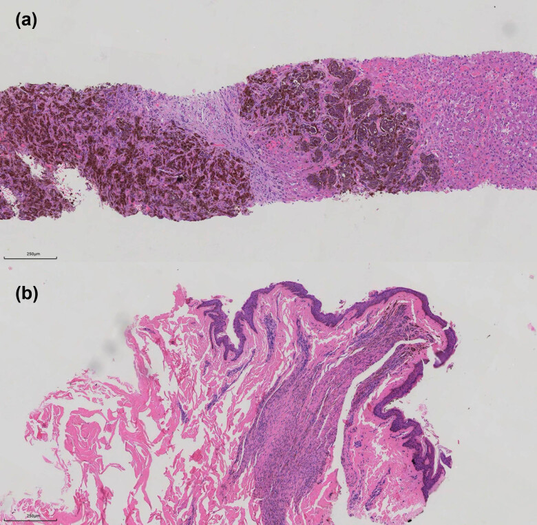
Malignant melanoma (MM) frequently occurs in the skin or mucosa, whereas malignant melanoma of unknown primary (MUP) is diagnosed in patients with lymph nodes or visceral organs as the site of origin, where it is challenging to detect the primary lesion by comprehensive examination. MUP is possibly related to the spontaneous regression of the primary lesion. In addition, primary hepatic melanoma (PHM) usually refers to the primary MM occurring in the liver, with no typical primary lesions and no manifestations of tumor metastasis. A 61-year-old male patient with liver as the site of origin was diagnosed with MM by Melan-A, HMB-45, and S-100 immunohistochemistry staining of liver biopsy tissue. Based on a comprehensive examination, no basis was found for melanoma in sites such as the skin, mucosa, five sense organs, brain, digestive tract, respiratory tract, or genitalia, and the patient was subsequently diagnosed with MUP. MMs require a comprehensive inspection, beginning with the liver, to search for the primary lesion; if the primary lesion is not found, the possibility of PHM or MUP should be considered.
Keywords: diagnosis; liver; malignant melanoma; unknown primary.
© 2023 the author(s), published by De Gruyter.
Conflict of interest statement
Conflict of interest: Authors state no conflict of interest.
Figures

References
-
- Skarbez K, Fanciullo L. Metastatic melanoma from unknown primary presenting as dorsal midbrain syndrome. Optom Vis Sci. 2012;89(12):e112–7. - PubMed
-
- Kamposioras K, Pentheroudakis G, Pectasides D, Pavlidis N. Malignant melanoma of unknown primary site. To make the long story short. A systematic review of the literature. Crit Rev Oncol Hematol. 2011;78(2):112–26. - PubMed
-
- Bae JM, Choi YY, Kim DS, Lee JH, Jang HS, Lee JH, et al. Metastatic melanomas of unknown primary show better prognosis than those of known primary: a systematic review and meta- analysis of observational studies. J Am Acad Dermatol. 2015;72(1):59–70. - PubMed
Publication types
LinkOut - more resources
Full Text Sources