

Generalized cystic lymphangiomatosis incidentally diagnosed in an asymptomatic adult: Imaging findings of a very rare case
- PMID: 37941984
- PMCID: PMC10628799
- DOI: 10.1016/j.radcr.2023.10.017
Generalized cystic lymphangiomatosis incidentally diagnosed in an asymptomatic adult: Imaging findings of a very rare case
Abstract
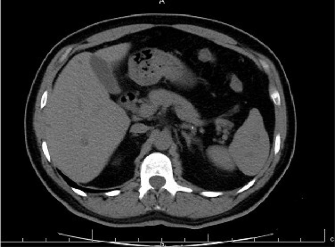
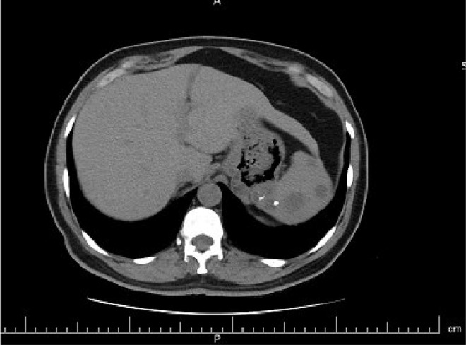
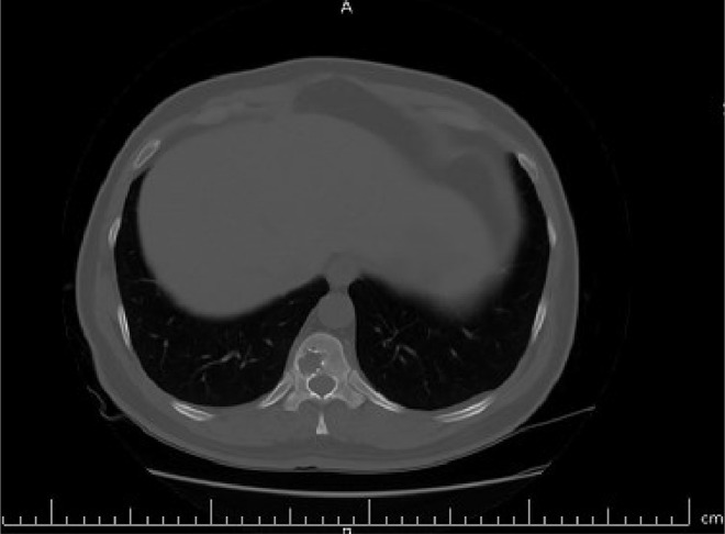
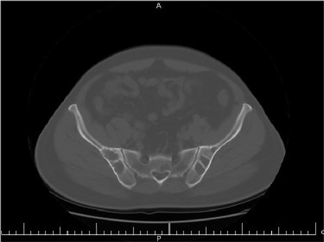
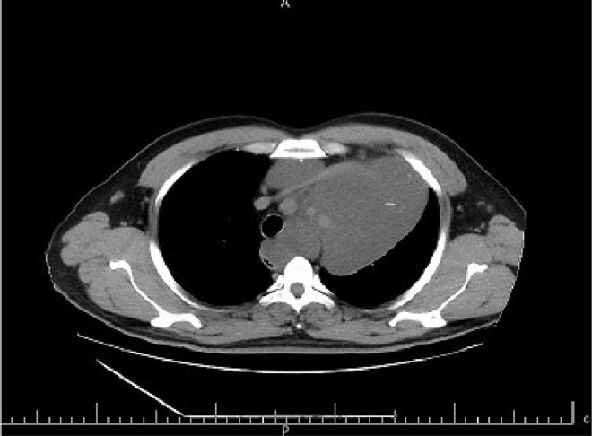
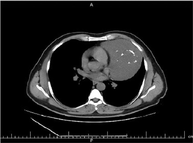
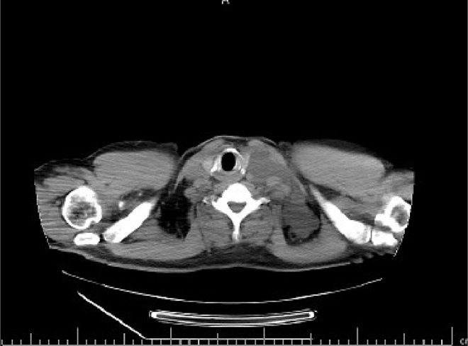
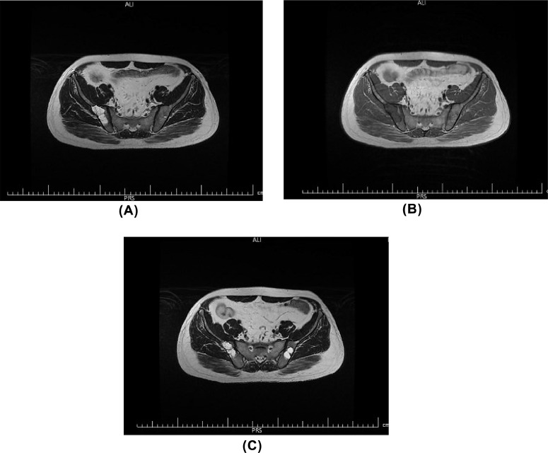
Lymphangiomas are benign lesions of vascular origin with lymphatic differentiation, most commonly found in the head and neck. Generalized lymphangiomatosis is a very rare condition in adults, which is characterized by a diffuse proliferation of lymphatic vessels. The lymphangioma is composed of lymphatic endothelium-lined cystic spaces. This condition can be histologically differentiated from other vascular disorders such as cavernous or capillary hemangioma. However, many cases of lymphangioma can be confused with other vascular disorders, because of overlapping histologic findings. radiologic examinations, such as CT scan and MR imaging, are useful for assessing the morphologic feature and also the extent of disease, it is important to know the radiologic findings of generalized lymphangiomatosis. In this paper, we report a case of generalized lymphangiomatosis in a 42-year-old male who presented with left flank pain and hematuria. The first differential diagnosis was renal colic; hence he underwent an abdominopelvic computed tomography scan (CT scan). In the performed CT scan multiple cystic lesions were seen in the liver and spleen. Also, lytic lesions were seen in bones. CT-guided biopsy was performed and the result was compatible with generalized lymphangiomatosis, confirmed by cytology. Generalized lymphangiomatosis is a rarely reported disease in children and young adults. Delayed diagnosis in older patients or misdiagnosis is common due to its rarity and nonspecific clinical presentation. Different imaging modalities can incidentally diagnose the disease in asymptomatic patients. So radiologists should be aware of the disease manifestations in imaging modalities to diagnose the disease sooner and help the clinician start the therapy if needed.
Keywords: Computed tomography scan; Generalized lymphangiomatosis; Lymphangioma; Lymphangiomatosis; Magnetic resonance imaging cytology.
© 2023 The Authors. Published by Elsevier Inc. on behalf of University of Washington.
Figures
References
-
- Francavilla ML, White CL, Oliveri B, Lee EY, Restrepo R. Intraabdominal lymphatic malformations: pearls and pitfalls of diagnosis and differential diagnoses in pediatric patients. Am J Roentgenol. 2017;208(3):637–649. - PubMed
Publication types
LinkOut - more resources
Full Text Sources
