. 2023 Oct 6;26(11):108149.
doi: 10.1016/j.isci.2023.108149. eCollection 2023 Nov 17.

The reproduction of gram-negative protoplasts and the influence of environmental conditions on this process

Affiliations

The reproduction of gram-negative protoplasts and the influence of environmental conditions on this process

Dheeraj Kanaparthi et al. iScience. .

Abstract

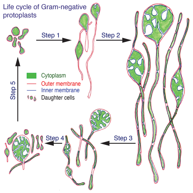
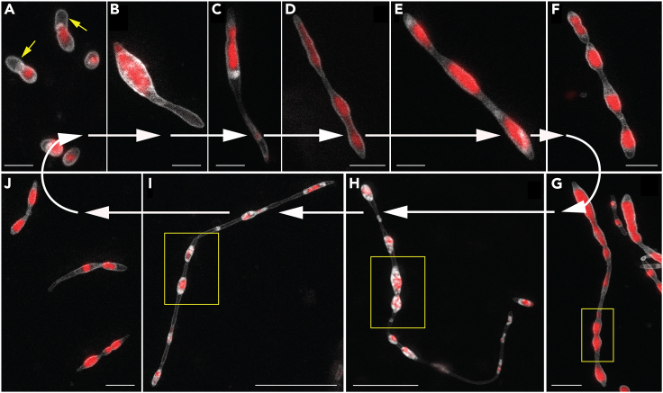
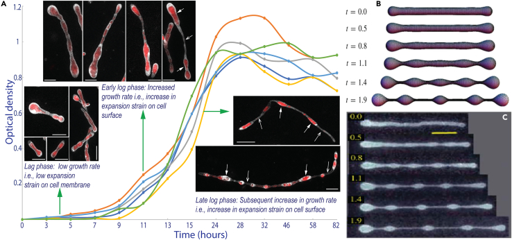
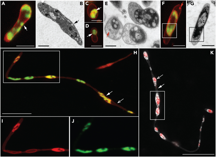
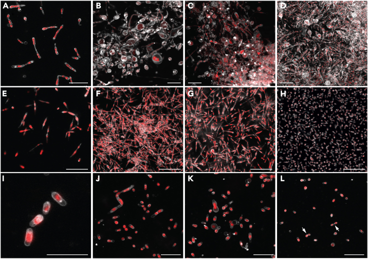
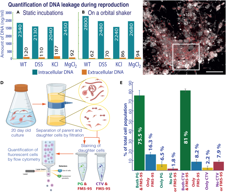
Bacterial protoplasts are known to reproduce independently of canonical molecular biological processes. Although their reproduction is thought to be influenced by environmental conditions, the growth of protoplasts in their natural habitat has never been empirically studied. Here, we studied the life cycle of protoplasts in their native environment. Contrary to the previous perception that protoplasts reproduce in an erratic manner, cells in our study reproduced in a defined sequence of steps, always leading to viable daughter cells. Their reproduction can be explained by an interplay between intracellular metabolism, the physicochemical properties of cell constituents, and the nature of cations in the growth media. The efficiency of reproduction is determined by the environmental conditions. Under favorable environmental conditions, protoplasts reproduce with nearly similar efficiency to cells that possess a cell wall. In short, here we demonstrate the simplest method of cellular reproduction and the influence of environmental conditions on this process.

Keywords: Cell biology; Evolutionary biology; Microbiology.


Conflict of interest statement

The authors declare that there are no conflicts of interest regarding the publication of this article.

Figures

None
Graphical abstract
Figure 1
Figure 1
The life cycle of RS-P All panels show STED microscopy images of cells stained with FM5-95 (membrane, white) and PicoGreen (DNA, red). White arrows guide the eye in following the life cycle progressing from (A) to (J). Arrows (yellow) in (A) highlight a cell with outer membrane extensions (are also seen in B and C). The steps involved in forming outer membrane extensions are shown in Figure 2. Boxed regions in (G)–(I) highlight cytoplasmic compartments within the filamentous cell undergoing binary fission (in sequence from G–I). (J) Individual daughter cells with membrane overhangs formed from the fragmentation of filamentous cells. Also, see Figure S2 for phase-contrast images. Scale bars: 2 μm (A–G and J) and 10 μm (H and I).
Figure 2
Figure 2
TEM images of early growth stage RS-P cells (A–C) Cells with folded excess membrane within the cell (highlighted regions). (D) RS-P cells with intracellular vesicles. The arrow in (D) points to the inner membrane undergoing budding into the periplasmic space. A cell with hollow vesicles in the periplasmic space was shown in (E). (F) The magnified periplasmic region of the cell in (E). (G–I) Cells with the elongated outer membrane. (J) The cytoplasmic compartment’s growth, filling in the void left by the outer membrane (arrow). Scale bars: 0.5 μm (A–J).
Figure 3
Figure 3
RS-P cells in different growth stages and comparison of their morphology with LVs (A) The growth curve and morphologies of RS-P in different growth stages (individual plots represent multiple repetitions, n = 6). Cells were stained, imaged, and color-coded, as in Figure 1. Closed and open arrows point to cytoplasmic compartments and hollow outer membrane connecting the cytoplasmic compartments, respectively (Figure 4). (B and C) Theoretically predicted and experimentally observed morphologies of vesicles subjected to elongational stress over time (t) (B and C were originally published by Narasimhan et al., 2015, reproduced here with permission from Cambridge University Press]. A similar comparison of RS-P cells with LVs undergoing expansion is shown in Figure S3. Scale bar: 1 μm (lag and early log phase), 10 μm (late log phase).
Figure 4
Figure 4
Membrane phase separation in RS-P Cells in (A), (C,) (D), (F), and (H), (I), and (J) are stained with FM5-95 (all membrane, red) and FAST DiI (Ld membrane, green). (A and B) Confocal and TEM images of early growth stage cells. Arrows in (A) point to regions of the cell with Ld membrane. Arrows in (B) point to similar intracellular vesicles. (C–G) Confocal and TEM images of later growth stage cells. Arrows in (B) and (C) point to the cytoplasmic compartment, exclusively enclosed in the Ld membrane. The arrow (red) in (E) points to the vesicles in periplasmic space. The arrow (red) in (F) shows similar periplasmic vesicles composed of Lo membrane. (H) Late log phase filamentous RS-P cells (see Figure S7 for multiple optical sections of this stack). (I and J) Magnified regions of the boxed region in (H) in individual Lo and Ld membrane channels. (K) RS-P cell similar in morphology to the cell shown in (I). The cell in this image is stained with membrane and DNA stain, as in Figure 1. A comparison of cells in (H) and (K) shows the inner cell membrane enriched in Ld membrane. Scale bars: 1 μm (A, C, D, and F), 500 nm (B, E, and G), and 10 μm (H and K).
Figure 5
Figure 5
Influence of salt composition on RS-P’s morphology (A–D) RS-P cells grown in 7% KCl-NB from early to late growth stages. (E–H) RS-P cells grown in 7% MgCl2-NB from early to late growth stages. (I–J) RS-P cells grown in 7% DSS-NB on an orbital shaker. Arrows in (L) point to cells undergoing binary fission. Cells were stained, imaged, and color-coded, as in Figure 1. Scale bars: 10 μm.
Figure 6
Figure 6
Reproductive efficiency and viability of RS-P daughter cells Graphs A and B show intracellular (blue bars) and extracellular (orange bars) DNA concentrations (n = 5 for each condition). (A) RS-P grown under static conditions. (B) RS-P grew on an orbital shaker. Annotations: WT (wild-type, R. sphaeroides with a cell wall grown in NB); DSS, KCl, and MgCl2RS-P grown in NB with respective salts. (C) RS-P cells grown in 7% DSS-NB in the late growth stage (scale bars: 10 μm). Cells in this image are stained, as described in Figure 1. (D) Schematic workflow of the experiment to quantify live daughter cells. Cells were stained with different combinations of FM5-95 (cell membrane), PicoGreen (PG, DNA), and CellTrace Violet (CTV, cell integrity) (n = 5, in the figure but multiple repetitions over the course of the work). (E) The percentage of RS-P daughter cells identified positive for different viability markers in flow cytometry. The original flow cytometry plots are shown in Figure S9.

Similar articles

Cited by

  • The reproduction process of Gram-positive protocells.
    Kanaparthi D, Lampe M, Krohn JH, Zhu B, Hildebrand F, Boesen T, Klingl A, Phapale P, Lueders T. Kanaparthi D, et al. Sci Rep. 2024 Mar 25;14(1):7075. doi: 10.1038/s41598-024-57369-4. Sci Rep. 2024. PMID: 38528088 Free PMC article.

References

    1. Vollmer W., Blanot D., De Pedro M.A. Peptidoglycan structure and architecture. FEMS Microbiol. Rev. 2008;32:149–167. doi: 10.1111/j.1574-6976.2007.00094.x. - DOI - PubMed
    1. Kern T., Giffard M., Hediger S., Amoroso A., Giustini C., Bui N.K., Joris B., Bougault C., Vollmer W., Simorre J.P. Dynamics characterization of fully hydrated bacterial cell walls by solid-state NMR: Evidence for cooperative binding of metal ions. J. Am. Chem. Soc. 2010;132:10911–10919. doi: 10.1021/ja104533w. - DOI - PubMed
    1. Westblade L.F., Errington J., Dörr T. Antibiotic tolerance. PLoS Pathog. 2020;16 doi: 10.1371/journal.ppat.1008892. - DOI - PMC - PubMed
    1. Kawai Y., Mercier R., Wu L.J., Domínguez-Cuevas P., Oshima T., Errington J. Cell growth of wall-free L-form bacteria is limited by oxidative damage. Curr. Biol. 2015;25:1613–1618. - PMC - PubMed
    1. Eisenberg A.D., Corner T.R. Osmotic behavior of bacterial protoplasts: temperature effects. J. Bacteriol. 1973;114:1177–1183. doi: 10.1128/jb.114.3.1177-1183.1973. - DOI - PMC - PubMed

LinkOut - more resources