Case Reports
. 2023 Oct 24:32:101949.
doi: 10.1016/j.ajoc.2023.101949. eCollection 2023 Dec.

A case of cilioretinal artery occlusion: Diagnostic procedures

Affiliations
Case Reports

A case of cilioretinal artery occlusion: Diagnostic procedures

Clara Rizzo et al. Am J Ophthalmol Case Rep. .

Abstract

Purpose: To evaluate characteristic imaging findings and functional outcomes of Cilioretinal Artery Occlusion (CLRAO) associated with giant cell arteritis (GCA).

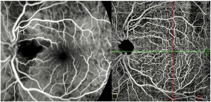
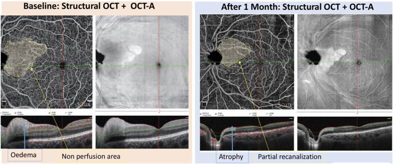
Observations: We report the case of a 70-year-old woman presenting with sudden vision loss caused by a GCA-associated-CLRAO in her left eye (LE). A thorough ophthalmologic examination together with optical coherence tomography (OCT), OCT-Angiography (OCT-A), fluorescein angiography and fundus autofluorescence were performed. At presentation, the best corrected visual acuity in the LE was 20/200 and funduscopic examination revealed optic disc edema associated with retinal whitening along the area perfused by the CLRA. After 1 month, OCT and OCT-A revealed an improvement of the retinal edema and a partial reduction of the non-perfused areas in the superficial and deep capillary plexuses, as well as in the outer retina and in the choriocapillaris. Fluorescein angiography showed a reduction in the perfusion of the affected area, a delayed perfusion of the temporal sector of the optic disc, as well as areas of choroidal hypoperfusion in the peripheral temporal retina. The patient's visual acuity did not change during the follow up.

Conclusion and importance: Despite a partial recanalization of the occluded vasculature being possible after GCA-associated-CLRAO, the patient's visual prognosis remains poor.

Keywords: Cilioretinal artery occlusion; Giant cell arteritis; Optical coherence tomography angiography.


Conflict of interest statement

The authors declare that they have no known competing financial interests or personal relationships that could have appeared to influence the work reported in this paper.

Figures

Fig. 1
Fig. 1
Funduscopy (left) and Autofluorescence (right) of the LE at baseline.
Fig. 2
Fig. 2
Fundoscopy (left images) and OCT (right images) at baseline (upper two images) and 1 month after treatment (lower two images).
Fig. 3
Fig. 3
FA of the posterior pole (early, intermediate and late phases, respectively from left to right) show absence of perfusion in the interpapillary macular region and hyperpermeability of the temporal sectors of the disc.
Fig. 4
Fig. 4
Magnified figures of the perimacular region on FA (early phase) and OCT-A (Superficial Plexus) after 1 month.
Fig. 5
Fig. 5
FA at 1 month follow up showing focal choroidal hypoperfusion areas in the temporal retinal sectors (red dotted area). (For interpretation of the references to color in this figure legend, the reader is referred to the Web version of this article.)
Fig. 6
Fig. 6
OCT-A of superficial and deep capillary plexus, outer retina and choricapillaries at baseline and after 1 month. At baseline, the superficial and deep capillary plexuses, show absence of perfusion due to the combination of ischemia and temporal iuxta-papillary edema. After 1 month, resolution of edema results in the reduction of non-perfusion area with persistence of ischemical area and correspective focal fiber atrophy (B-scan).
Fig. 7
Fig. 7
Details of OCT-A, enface and B-scan structural OCT (superficial capillary plexus) at baseline and after 1 month of treatment. The area of non-perfusion (yellow dotted) was reduced after 1 month due to a partial recanalization of the peripheral capillaries. (For interpretation of the references to color in this figure legend, the reader is referred to the Web version of this article.)

References

    1. Christodoulou P., Katsimpris I. Optical coherence tomography findings in a case of cilioretinal artery occlusion reversal, treated with mannitol and carbogen administration. Ann Eye Sci. 2019;4:13. doi: 10.21037/aes.2019.02.01. 13. - DOI
    1. Leavitt J.A., Larson T.A., Hodge D.O., Gullerud R.E. The incidence of central retinal artery occlusion in Olmsted County, Minnesota. Am J Ophthalmol. 2011;152(5):820–823. doi: 10.1016/j.ajo.2011.05.005. e2. - DOI - PMC - PubMed
    1. Brown G.C., Shields J.A. Cilioretinal arteries and retinal arterial occlusion. Arch Ophthalmol. 1979;97(1):84–92. doi: 10.1001/archopht.1979.01020010024006. - DOI - PubMed
    1. Brown G.C., Moffat K., Cruess A., Magargal L.E., Goldberg R.E. Cilioretinal Artery Obstruction: retina. 1983;3(3):182–187. doi: 10.1097/00006982-198300330-00007. - DOI - PubMed
    1. Hayreh S.S. Ocular vascular occlusive disorders: natural history of visual outcome. Prog Retin Eye Res. 2014;41:1–25. doi: 10.1016/j.preteyeres.2014.04.001. - DOI - PMC - PubMed

Publication types

LinkOut - more resources