. 2023 Sep 18;12(10):e1797-e1802.
doi: 10.1016/j.eats.2023.06.009. eCollection 2023 Oct.

Reproducible and Effective Biceps Tenodesis Method Utilizing In-Office Nano-Arthroscopy

Affiliations

Reproducible and Effective Biceps Tenodesis Method Utilizing In-Office Nano-Arthroscopy

Christopher A Colasanti et al. Arthrosc Tech. .

Abstract

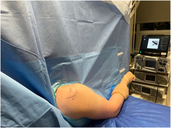
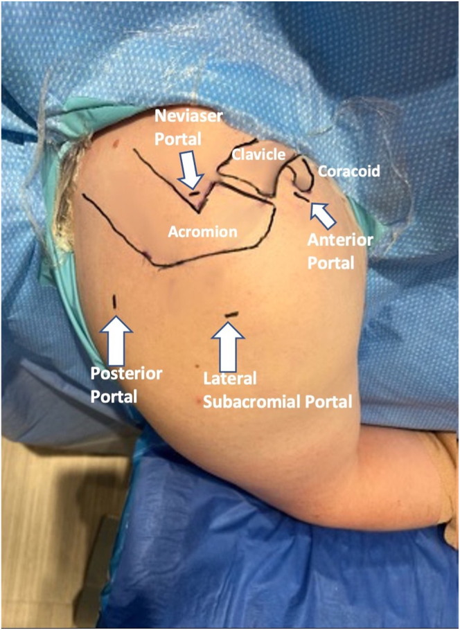
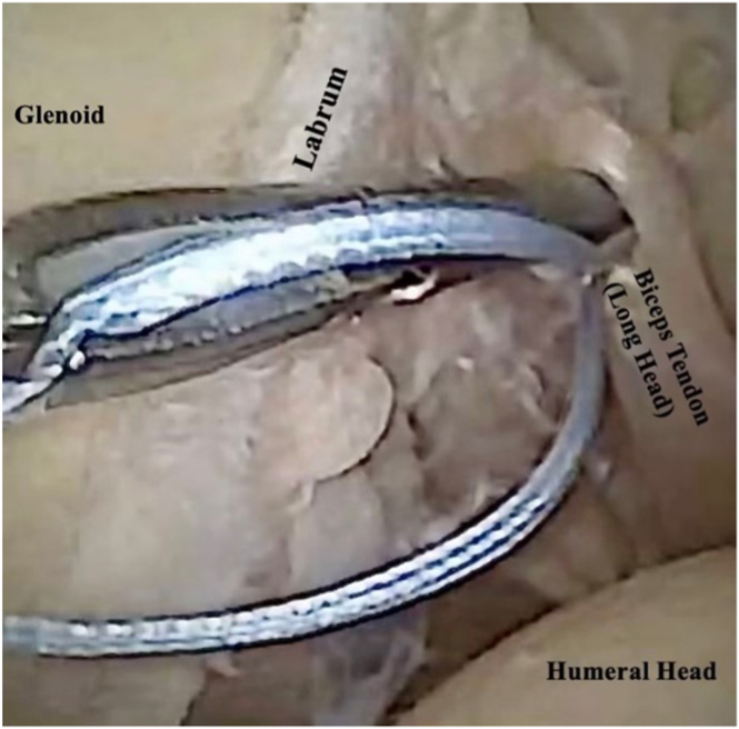
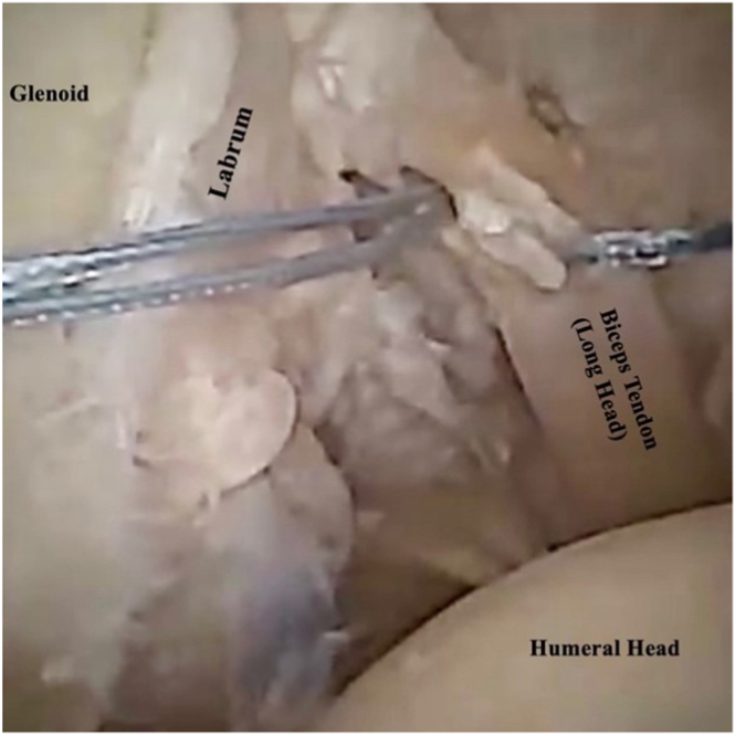
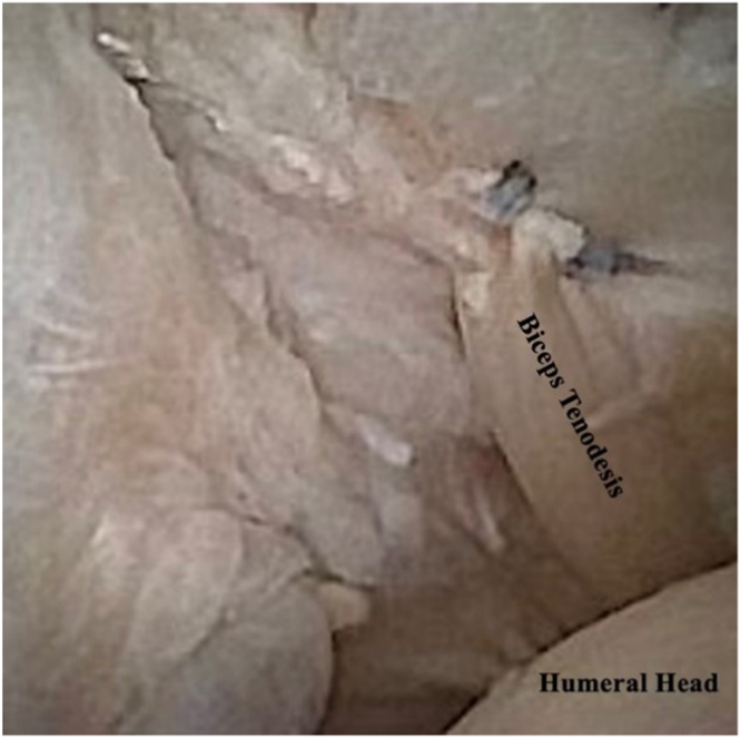
Biceps tendinopathy is a common cause of chronic anterior shoulder pain characterized by altered joint mechanics with considerable deficits in range of motion secondary to pain. The benefits of in-office nano-arthroscopy (IONA) include the ability to diagnosis and treat biceps tendinopathy, quicker patient recovery, reduced cost, and improved patient satisfaction. The purpose of this technical report is to describe the technique for performing IONA for biceps tendinopathy (biceps tenotomy/biceps tenodesis), with special consideration for obtaining adequate local anesthesia, proper indications, adequate visualization, and the advantages of performing these procedures in the office rather than the operating room.


Figures

Fig. 1
Fig. 1
An in-office nano-arthroscopy standard shoulder setup. With the back of the examination table positioned at approximately 70° to 80°, the patient is comfortably seated to mimic the beach-chair positioning. The ipsilateral shoulder (right) is positioned so that the posterior, lateral, and anterior aspects of the shoulder are unobstructed. The operative arm is rested on a well-padded mayo stand with arm in slight forward flexion and abduction. This positioning will help facilitate entry into the glenohumeral joint.
Fig. 2
Fig. 2
The patient is in an upright, beach-chair position, with the bed set at approximately 70° to 80°. Using sterile technique, the patient’s extremity (right shoulder) is prepped, using a mixture of chlorhexidine gluconate with isopropyl alcohol and draped. Standard portal sites are marked with respect to relevant surface anatomy markings, which include the acromion, clavicle, and coracoid. The posterior portal, which is the primary viewing portal, is made approximately 2 cm inferior and 2 cm medial to the posterolateral border of the acromion. The anterior, working portal is made 1 cm laterally to the coracoid process.
Fig. 3
Fig. 3
This is an arthroscopic view of the left shoulder through the posterior portal. Through the anterior portal, using a BirdBeak suture passer, the SutureTape is first passed approximately 1 to 1.5 cm from the bicep-labral anchor. Through the anterior portal, the BirdBeak suture passer is then once again passed approximately 1 cm lateral to the biceps tendon, through the capsule, to retrieve the suture tape. The 2 limbs are tied over the capsule at the level of the rotator interval using an arthroscopic knot pusher.
Fig. 4
Fig. 4
This is an arthroscopic view of the left shoulder through the posterior portal. Through the anterior portal, using a BirdBeak suture passer, SutureTape is passed a second time approximately 5 mm to 1 cm from the long head of biceps origin. Through the anterior portal, the BirdBeak suture passer is then once again passed approximately 1 cm lateral to the biceps tendon, through the capsule, to retrieve the suture tape. The 2 limbs are tied over the capsule at the level of the rotator interval using an arthroscopic knot pusher.
Fig. 5
Fig. 5
This is an arthroscopic view of the left shoulder through the posterior portal, demonstrating tenodesis of the biceps tendon to the rotator cuff interval, followed by tenotomy at the long head of biceps origin. The portals can be sealed primarily using adhesive wound closure strips or with simple nylon sutures if the surgeon feels they are necessary. A dry, sterile dressing is applied that facilitates early shoulder range of motion.

References

    1. Wilk K.E., Hooks T.R. The Painful long head of the biceps brachii: Nonoperative treatment approaches. Clin Sports Med. 2016;35:75–92. - PubMed
    1. Hsu A.R., Ghodadra N.S., Provencher M.T., Lewis P.B., Bach B.R. Biceps tenotomy versus tenodesis: A review of clinical outcomes and biomechanical results. J Shoulder Elbow Surg. 2011;20:326–332. - PubMed
    1. Castricini R., Familiari F., De Gori M., et al. Tenodesis is not superior to tenotomy in the treatment of the long head of biceps tendon lesions. Knee Surg Sports Traumatol Arthrosc. 2018;26:169–175. - PubMed
    1. Belk J.W., Kraeutler M.J., Houck D.A., Chrisman A.N., Scillia A.J., McCarty E.C. Biceps tenodesis versus tenotomy: A systematic review and meta-analysis of level I randomized controlled trials. J Shoulder Elbow Surg. 2021;30:951–960. - PubMed
    1. Golish S.R., Caldwell P.E., III, Miller M.D., et al. Interference screw versus suture anchor fixation for subpectoral tenodesis of the proximal biceps tendon: A cadaveric study. Arthroscopy. 2008;24:1103–1108. - PubMed

LinkOut - more resources