. 2023 Oct 27:23:100845.
doi: 10.1016/j.mtbio.2023.100845. eCollection 2023 Dec.

Extracellular vesicles hybrid plasmid-loaded lipid nanovesicles for synergistic cancer immunotherapy

Affiliations

Extracellular vesicles hybrid plasmid-loaded lipid nanovesicles for synergistic cancer immunotherapy

Qing Tong et al. Mater Today Bio. .

Abstract

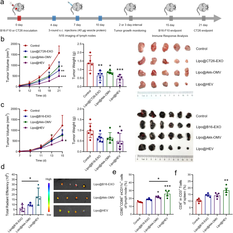
Combination immunotherapy of cancer vaccines with immune checkpoint inhibitors (ICIs) represents a promising therapeutic strategy for immunosuppressed and cold tumors. However, this strategy still faces challenges, including the limited therapeutic efficacy of cancer vaccines and immune-related adverse events associated with systematic delivery of ICIs. Herein, we demonstrate the antitumor immune response induced by outer membrane vesicle from Akkermansia muciniphila (Akk-OMV), which exhibites a favorable safety profile, highlighting the potential application as a natural and biocompatible self-adjuvanting vesicle. Utilizing tumor cell-derived exosome as an antigen source and Akk-OMV as a natural adjuvant, we construct a cancer vaccine formulation of extracellular vesicles hybrid lipid nanovesicles (Lipo@HEV) for enhanced prophylactic and therapeutic vaccination by promoting dendritic cell (DC) maturation in lymph node and activating cytotoxic T cell (CTL) response. The Lipo@HEV is further loaded with plasmid to enable gene therapy-mediated PD-L1 blockade upon peritumoral injection. Meanwhile, it penetrates into lymph node to initiate DC maturation and CTL activation, synergistically inhibiting the established tumor. The fabrication of extracellular vesicles hybrid plasmid-loaded lipid nanovesicles reveals a promising gene therapy-guided and vesicle-based hybrid system for therapeutic cancer vaccination and synergistic immunotherapy strategy.

Keywords: Akkermansia muciniphila; Cancer vaccine; Extracellular vesicles; Hybrid lipid nanovesicles; Immune checkpoint blockade; Liposomes.


Conflict of interest statement

The authors declare the following financial interests/personal relationships which may be considered as potential competing interests: Xiaoyuan Huang reports financial support was provided by 10.13039/501100001809National Natural Science Foundation of China and 10.13039/501100003819Natural Science Foundation of Hubei Province.

Figures

Image 1
Graphical abstract
Fig. 1
Fig. 1
Characterization of BEVs and antitumor vaccination efficacy of Akk-OMV. (a) TEM images of BEVs (scale bar: 100 nm). (b) Average particle sizes and zeta potentials of BEVs (A, Akk-OMV; B, Bifi-CMV; D, DH5α-OMV) measured by nanoparticle tracking analysis (n = 3). (c) SDS-PAGE analysis of protein components in BEVs (M, Marker; A, Akk-OMV; B, Bifi-CMV; D, DH5α-OMV). (d) Experimental design: BALB/c mice were subcutaneously injected at the tail base with PBS, Akk-OMV, Bifi-CMV or DH5α-OMV (10 μg) at 3 day-intervals for 5 times, and then inoculated with 2 × 105 4T1 tumor cells on day 0. Mice were euthanized on day 28 for RNA-sequencing analysis. (e) Tumor growth kinetics after vaccination of different BEVs (n = 6). (f) Transcriptome heatmap of DEGs in tumors and spleens of mice vaccinated with different BEVs and PBS (n = 5). GO pathway enrichment analysis in (g) tumors and (h) spleens of Akk-OMV versus the PBS control. (i) KEGG pathway enrichment analysis in spleens in Akk-OMV, Bifi-OMV and DH5α-OMV compared to the PBS control. Data are presented as mean ± SD. Statistical significance was calculated via one-way ANOVA with Tukey multiple comparisons test. **p < 0.01 versus corresponding control group.
Fig. 2
Fig. 2
Akk-OMV exhibited a favorable safety profile over E. Coli DH5α-OMV. (a) Experimental design for toxicity evaluation upon 3-round intraperitoneal injection. BALB/c mice were injected with Akk-OMV and DH5α-OMV (20 μg) on day 0, 3 and 6, and were euthanized on day 14. (b) Survival rate of each group at the end point (n = 6). (c) Spleen images and weights at the endpoint (n = 3 and 6). (d) Serum concentrations of hepatic and renal function indexes (n = 3 and 6). (e) Representative H&E staining images of spleens (Scale bar: upper images 1 mm, lower images 250 μm). (f) Experimental design for toxicity evaluation upon subcutaneous injection. C57BL/6 mice were injected at the tail base with Akk-OMV and DH5α-OMV (20 μg) on day 0, 3, 6, and were euthanized on day 9. (g) Skin damage images of different groups at endpoint (n = 7). The skin damage at the injection site was marked by red circle. (h) Representative H&E staining images of spleens (Scale bar: left images 1 mm, right images 250 μm). (i) Serum concentrations of hepatic and renal function indexes (n = 7). Data are presented as mean ± SD. Statistical significance was calculated via one-way ANOVA with Tukey multiple comparisons test. *p < 0.05, **p < 0.01, ***p < 0.001 versus corresponding control group. (For interpretation of the references to color in this figure legend, the reader is referred to the Web version of this article.)
Fig. 3
Fig. 3
Preparation and characterization of Lipo@HEV. (a) TEM images of liposome, 4T1 exosome, B16–F10 exosome, CT26 exosome, Akk-OMV and Lipo@HEV (scale bar: 100 nm). (b) Nano-flow cytometry analysis of vesicle fusion in Lipo@HEV prepared at weight ratios of 100:1:1, 50:1:1, 20:1:1, with PKH26-labeled OMV and PKH67-labeled exosome. (c) Representative images of vesicle fusion in Lipo@HEV and HEV via confocal laser scanning microscopy (CLSM) (scale bar: 10 μm, OMV labeled with PKH26, exosome labeled with PKH67). (d) Particle sizes and (e) zeta potentials of liposome, 4T1 exosome, B16–F10 exosome, Akk-OMV and Lipo@HEV measured by NTA (n = 3). (f) SDS-PAGE of protein components and Western blot of exosome markers (CD9 and TSG101) in tumor cell membrane (TCM), exosome, OMV, Lipo@HMV and Lipo@HEV. (g) Colocalization of PKH26/PKH67-labeled Lipo@HEV in B16–F10 cells and BMDCs via CLSM (scale bar: 50 μm). (h) Flow cytometry images and analysis of DC maturation markers in BMDCs (CD80+CD86+ in CD11c+) after incubation with exosome, OMV, HEV and different formulations of Lipo@HEV for 24 h (n = 3). Data are presented as mean ± SD. Statistical significance was calculated via one-way ANOVA with Tukey multiple comparisons test. *p < 0.05, **p < 0.01, ***p < 0.001 versus corresponding control group.
Fig. 4
Fig. 4
Prophylactic vaccination efficacy of Lipo@HEV. (a) Experimental design: C57BL/6 mice were subcutaneously injected at the tail base with PBS, Lipo@B16-EXO, Lipo@Akk-OMV, Lipo@B16-HEV and Lipo@4T1-HEV (40 μg vesicle protein) on day −13, −10 and −7, followed by inoculation with 2 × 105 B16–F10 cells on day 0. Mice were euthanized on day 18 for single-cell RNA-sequencing in tumors and TEM analysis in spleens. (b) Tumor growth kinetics after vaccination of different formulations (n = 6). Data are presented as mean ± SEM. (c) Cell markers profiling of defined cell subsets. (d) Uniform manifold approximation and projection (UMAP) plot of cells colored by cell types. (e) Percentages of different identified cell subsets (C0–C10, melanoma cell subsets; C11, fibroblasts; C12, endothelial cells; C13, macrophages; C14, erythrocytes; C15, granulocytes; C16, B cells). (f) Hallmark pathway enrichment analysis of DEGs in melanoma cell subsets. (g) Flow cytometry analysis of TEM (CD44+CD62L in CD3+CD8+) in spleens (n = 5). Data are presented as mean ± SD. Statistical significance was calculated via one-way ANOVA with Tukey multiple comparisons test. *p < 0.05, **p < 0.01, ***p < 0.001 versus corresponding control group.
Fig. 5
Fig. 5
Lipo@HEV promoted DC maturation in lymph node and activated CTL response against tumor growth. (a) Experimental design: C57BL/6 and BALB/c mice were inoculated with 2 × 105 B16–F10 and CT26 tumor cells on day 0, respectively, and were subcutaneously injected (40 μg vesicle protein) at the tail base with different vaccine formulations on day 4, 7 and 10. The DiR-labeled B16–F10 exosome and Akk-OMV were co-extruded with liposome to generate different DiR-labeled formulations, and C57BL/6 mice were subcutaneously injected at the tail base on day 4. The IVIS imaging was performed at 48 h post-injection. (b) Tumor growth kinetics, tumor weight and images after different treatments of control, Lipo@CT26-EXO, Lipo@Akk-OMV and Lipo@HEV (n = 5). (c) Tumor growth kinetics, tumor weight and images after different treatments of control, Lipo@B16-EXO, Lipo@Akk-OMV and Lipo@HEV (n = 6). (d) The fluorescence imaging and radiant efficiency of inguinal lymph nodes in different formulation groups (n = 4). (e) Flow cytometry analysis of DC maturation (CD80+CD86+ in CD11c+) of lymph nodes, (f) CD8+ in CD3+ T cells of spleens (n = 4). Data are presented as mean ± SD. Statistical significance was calculated via one-way ANOVA with Tukey multiple comparisons test. *p < 0.05, **p < 0.01, ***p < 0.001 versus corresponding control group.
Fig. 6
Fig. 6
Fabrication of Lipo-PD-L1@HEV for synergistic antitumor responses against the established tumor. (a) TEM image of Lipo-PD-L1@HEV (scale bar: 100 nm). (b) Average particle sizes and zeta potentials of Lipo-PD-L1@HEV and Lipo-PD-L1 analyzed by NTA (n = 3). (c) Fluorescence imaging and (d) total radiant efficiency of tumors, lymph nodes and other major organs at 48 h after peritumoral injection of DiR-labeled Lipo-PD-L1@HEV in 4T1 tumor-bearing mice (n = 5). (e) In vivo and ex vivo bioluminescence imaging and (f) average bioluminescence counts of tumors at 48 h after peritumoral injections of Lipo-luciferase@HEV and Lipo-luciferase in B16–F10 tumor-bearing mice (n = 4). (g) Experimental design for synergistic antitumor evaluation: C57BL/6 and BALB/c mice were inoculated with 2 × 105 B16–F10 and 4T1 tumor cells on day 0, respectively, followed by peritumoral injections of PBS, Lipo-PD-L1 and Lipo-PD-L1@HEV (20 μg plasmid) on day 9, 12 and 15. B16–F10 tumor-bearing mice were euthanized on day 17 for immune response analysis, and 4T1 tumor-bearing mice were utilized for tumor growth kinetics and survival rate evaluation. (h) Tumor growth kinetics after different treatments in B16–F10 tumor-bearing mice (n = 6). (i) Flow cytometry analysis of DC maturation (CD80+CD86+ in CD11c+) in lymph nodes, (j) CD8+ in CD3+ T cells in lymph nodes, (k) CD8+ in CD3+ T cells in tumors, and (l) Ratio of CD8+ T cells and Treg cells (CD25+FOXP3+ in CD4+) in tumors (n = 4). (m) Serum concentrations of TNF-α, IL-6 and IFN-γ analyzed by ELISA (n = 4). (n) Tumor growth kinetics, and (o) survival curves of 4T1 tumor-bearing mice received different treatments of control, Lipo-PD-L1 and Lipo-PD-L1@HEV in 4T1 tumor-bearing mice (n = 8). Data are presented as mean ± SD. Statistical significance was calculated via one-way ANOVA with Tukey multiple comparisons test, and survival rate analysis was calculated by log-rank (Mantel-Cox) test. *p < 0.05, **p < 0.01, ***p < 0.001 versus corresponding control group.
Fig. 7
Fig. 7
Schematic illustration of fabricated Lipo-PD-L1@HEV for synergistic cancer immunothearpy. After thin-film hydration of cationic liposome, incubation of cationic liposome with PD-L1 trap plasmid, and purification of tumor cell-derived exosome and Akk-OMV, the Lipo-PD-L1@HEV was fabricated by fusing extracellular vesicles with plasmid-loaded cationic liposome. Upon peritumoral injection, Lipo-PD-L1@HEV can localize in tumor for PD-L1 blockade, and penetrate into lymph node to efficiently initiate DC maturation, antigen presentation and CTL activation, synergistically inhibiting the established tumor.

References

    1. Saxena M., van der Burg S.H., Melief C.J.M., Bhardwaj N. Therapeutic cancer vaccines. Nat. Rev. Cancer. 2021;21:360–378. - PubMed
    1. Platten M., Bunse L., Wick A., Bunse T., Le Cornet L., Harting I., Sahm F., Sanghvi K., Tan C.L., Poschke I., et al. A vaccine targeting mutant IDH1 in newly diagnosed glioma. Nature. 2021;592:463–468. - PMC - PubMed
    1. Ott P.A., Hu Z., Keskin D.B., Shukla S.A., Sun J., Bozym D.J., Zhang W., Luoma A., Giobbie-Hurder A., Peter L., et al. An immunogenic personal neoantigen vaccine for patients with melanoma. Nature. 2017;547:217–221. - PMC - PubMed
    1. Lin M.J., Svensson-Arvelund J., Lubitz G.S., Marabelle A., Melero I., Brown B.D., Brody J.D. Cancer vaccines: the next immunotherapy frontier. Nat. Can. (Ott.) 2022;3:911–926. - PubMed
    1. Ye T., Li F., Ma G., Wei W. Enhancing therapeutic performance of personalized cancer vaccine via delivery vectors. Adv. Drug Deliv. Rev. 2021;177 - PubMed

LinkOut - more resources