

Voxel-based morphometry in single subjects without a scanner-specific normal database using a convolutional neural network
- PMID: 37943313
- PMCID: PMC11166757
- DOI: 10.1007/s00330-023-10356-1
Voxel-based morphometry in single subjects without a scanner-specific normal database using a convolutional neural network
Abstract
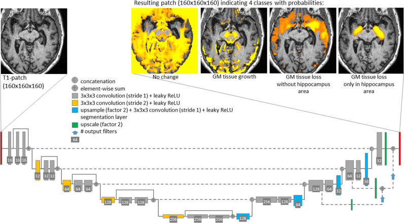
Objectives: Reliable detection of disease-specific atrophy in individual T1w-MRI by voxel-based morphometry (VBM) requires scanner-specific normal databases (NDB), which often are not available. The aim of this retrospective study was to design, train, and test a deep convolutional neural network (CNN) for single-subject VBM without the need for a NDB (CNN-VBM).
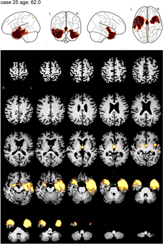
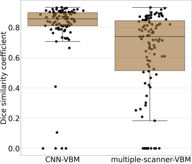
Materials and methods: The training dataset comprised 8945 T1w scans from 65 different scanners. The gold standard VBM maps were obtained by conventional VBM with a scanner-specific NDB for each of the 65 scanners. CNN-VBM was tested in an independent dataset comprising healthy controls (n = 37) and subjects with Alzheimer's disease (AD, n = 51) or frontotemporal lobar degeneration (FTLD, n = 30). A scanner-specific NDB for the generation of the gold standard VBM maps was available also for the test set. The technical performance of CNN-VBM was characterized by the Dice coefficient of CNN-VBM maps relative to VBM maps from scanner-specific VBM. For clinical testing, VBM maps were categorized visually according to the clinical diagnoses in the test set by two independent readers, separately for both VBM methods.
Results: The VBM maps from CNN-VBM were similar to the scanner-specific VBM maps (median Dice coefficient 0.85, interquartile range [0.81, 0.90]). Overall accuracy of the visual categorization of the VBM maps for the detection of AD or FTLD was 89.8% for CNN-VBM and 89.0% for scanner-specific VBM.
Conclusion: CNN-VBM without NDB provides a similar performance in the detection of AD- and FTLD-specific atrophy as conventional VBM.
Clinical relevance statement: A deep convolutional neural network for voxel-based morphometry eliminates the need of scanner-specific normal databases without relevant performance loss and, therefore, could pave the way for the widespread clinical use of voxel-based morphometry to support the diagnosis of neurodegenerative diseases.
Key points: • The need of normal databases is a barrier for widespread use of voxel-based brain morphometry. • A convolutional neural network achieved a similar performance for detection of atrophy than conventional voxel-based morphometry. • Convolutional neural networks can pave the way for widespread clinical use of voxel-based morphometry.
Keywords: Alzheimer disease; Brain mapping; Deep learning; Magnetic resonance imaging; Neural networks (computer).
© 2023. The Author(s).
Conflict of interest statement
The authors of this manuscript declare relationships with the following companies: Julia Krüger, Roland Opfer, and Lothar Spies are employees of jung diagnostics GmbH, Germany (
Figures
Comment in
-
Searching for the grail: may machine learning be a road to clinical use of brain MRI segmentation?Eur Radiol. 2024 Jun;34(6):3575-3577. doi: 10.1007/s00330-023-10438-0. Epub 2023 Nov 17. Eur Radiol. 2024. PMID: 37975922 No abstract available.
Similar articles
-
Clinical validation of artificial intelligence-based single-subject morphometry without normative reference database.J Alzheimers Dis. 2025 Jan;103(2):542-551. doi: 10.1177/13872877241304607. Epub 2025 Jan 12. J Alzheimers Dis. 2025. PMID: 39801073
-
Removing outliers from the normative database improves regional atrophy detection in single-subject voxel-based morphometry.Neuroradiology. 2024 Apr;66(4):507-519. doi: 10.1007/s00234-024-03304-3. Epub 2024 Feb 21. Neuroradiology. 2024. PMID: 38378906 Free PMC article.
-
Automatic segmentation of the thalamus using a massively trained 3D convolutional neural network: higher sensitivity for the detection of reduced thalamus volume by improved inter-scanner stability.Eur Radiol. 2023 Mar;33(3):1852-1861. doi: 10.1007/s00330-022-09170-y. Epub 2022 Oct 20. Eur Radiol. 2023. PMID: 36264314 Free PMC article.
-
Convolutional neural networks for classification of Alzheimer's disease: Overview and reproducible evaluation.Med Image Anal. 2020 Jul;63:101694. doi: 10.1016/j.media.2020.101694. Epub 2020 May 1. Med Image Anal. 2020. PMID: 32417716
-
Comparisons between Alzheimer disease, frontotemporal lobar degeneration, and normal aging with brain mapping.Top Magn Reson Imaging. 2005 Dec;16(6):409-25. doi: 10.1097/01.rmr.0000245457.98029.e1. Top Magn Reson Imaging. 2005. PMID: 17088691 Review.
Cited by
-
Searching for the grail: may machine learning be a road to clinical use of brain MRI segmentation?Eur Radiol. 2024 Jun;34(6):3575-3577. doi: 10.1007/s00330-023-10438-0. Epub 2023 Nov 17. Eur Radiol. 2024. PMID: 37975922 No abstract available.
References
-
- Mechelli A, Price CJ, Friston KJ, Ashburner J. Voxel-based morphometry of the human brain: methods and applications. Curr Med Imaging. 2005;1:105–113. doi: 10.2174/1573405054038726. - DOI
-
- Larvie M, Fischl B. Volumetric and fiber-tracing MRI methods for gray and white matter. Neuroimaging Pt I. 2016;135:39–60. - PubMed
MeSH terms
LinkOut - more resources
Full Text Sources
Medical
