. 2024 Jan 16;134(2):e170882.
doi: 10.1172/JCI170882.

Biallelic Cys141Tyr variant of SEL1L is associated with neurodevelopmental disorders, agammaglobulinemia, and premature death

Affiliations

Biallelic Cys141Tyr variant of SEL1L is associated with neurodevelopmental disorders, agammaglobulinemia, and premature death

Denisa Weis et al. J Clin Invest. .

Abstract

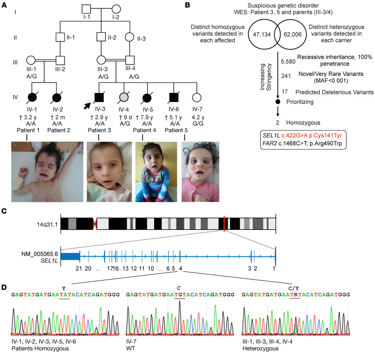
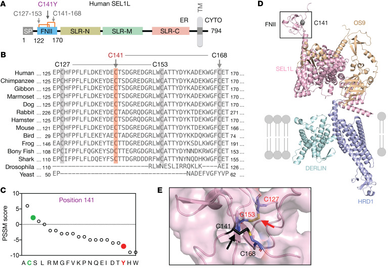
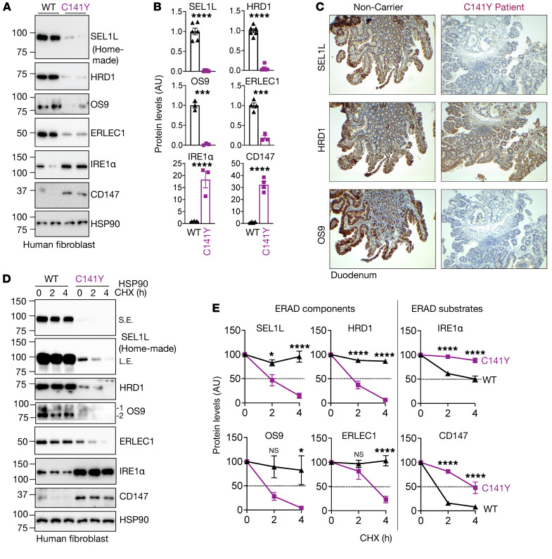
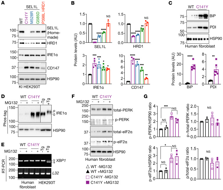
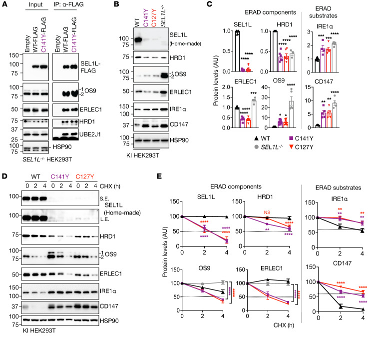
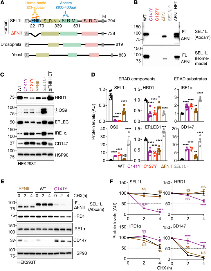
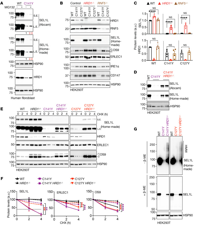
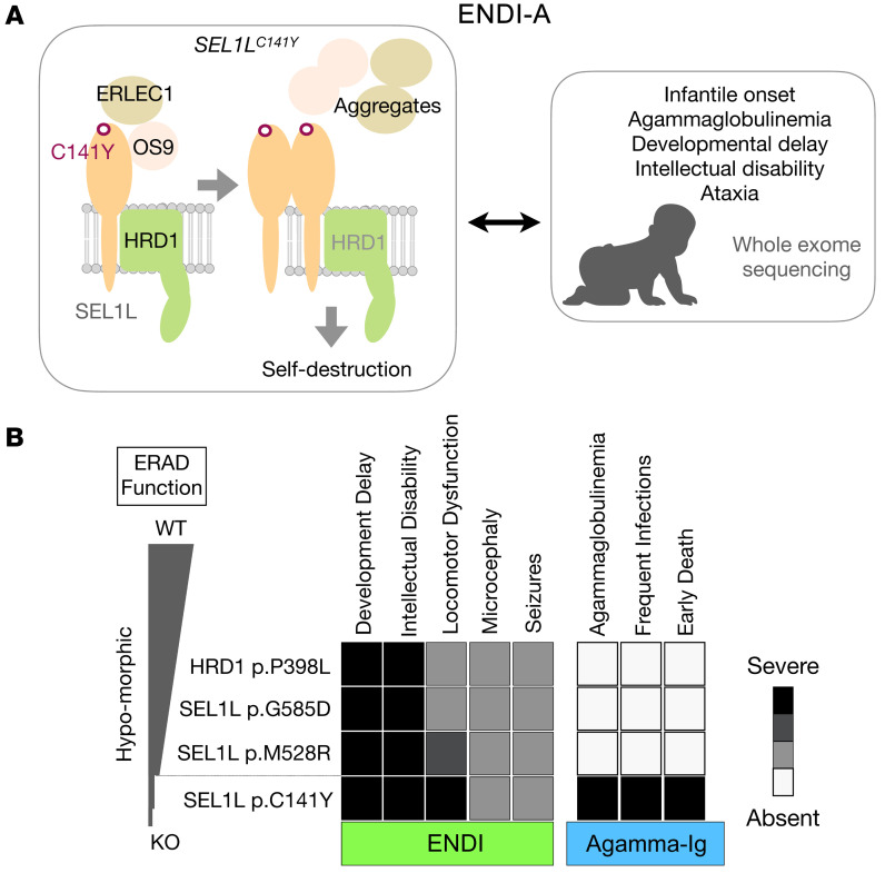
Suppressor of lin-12-like-HMG-CoA reductase degradation 1 (SEL1L-HRD1) ER-associated degradation (ERAD) plays a critical role in many physiological processes in mice, including immunity, water homeostasis, and energy metabolism; however, its relevance and importance in humans remain unclear, as no disease variant has been identified. Here, we report a biallelic SEL1L variant (p. Cys141Tyr) in 5 patients from a consanguineous Slovakian family. These patients presented with not only ERAD-associated neurodevelopmental disorders with onset in infancy (ENDI) syndromes, but infantile-onset agammaglobulinemia with no mature B cells, resulting in frequent infections and early death. This variant disrupted the formation of a disulfide bond in the luminal fibronectin II domain of SEL1L, largely abolishing the function of the SEL1L-HRD1 ERAD complex in part via proteasomal-mediated self destruction by HRD1. This study reports a disease entity termed ENDI-agammaglobulinemia (ENDI-A) syndrome and establishes an inverse correlation between SEL1L-HRD1 ERAD functionality and disease severity in humans.

Keywords: Adaptive immunity; Cell Biology; Genetic diseases; Protein misfolding.


Figures

Figure 1
Figure 1. Identification of SEL1LC141Y variant in humans using whole-exome sequencing.
(A) Family pedigrees for the kindreds from Slovakia (2 consanguineous pairs) for SEL1L p.C141Y, showing autosomal recessive inheritance. Arrows point to probands. Black shapes indicate affected patients and gray shapes show a newborn died from holoprosencephaly. The age indicated is as of 2022 or at time of death (cross). Photos of the patients are shown below. Photo of patient 1 was published in the book Pediatrics (51) to show marasmus and is republished here with permission. (B) Genetic analysis pipeline of whole-exome sequencing (WES) data for patients 3 and 5 (IV-3/6) and their parents, III-3/4. (C and D) Exonic and chromosomal locations of SEL1L variant (C), with Sanger sequencing in patients and other family members (D). R, heterozygosity; C, cysteine; Y, tyrosine.
Figure 2
Figure 2. Sequence and structural analyses of SEL1LC141Y variant.
(A) Schematic diagram of human SEL1L. SP, signal peptide; SLR-N/M/C, Sel1-like repeats at N-, middle-, and C-terminal; TM, transmembrane; CYTO, cytosol. Orange lines, 2 disulfide bonds (Cys127-Cys153, Cys141-Cys168). (B) The aa sequence alignment of SEL1L showing the conservation of SEL1L C141 residue (highlighted in orange) and neighboring cysteine residues (highlighted in gray) across species. (C) PSSM scores for position 141, with WT in green and variant in red. (D and E) Structural prediction of human SEL1L/OS9/HRD1/DERLIN ERAD complex using AlphaFold2 with close-up view of C141 residue and disulfide bonds (black arrows) (E).
Figure 3
Figure 3. SEL1LC141Y variant abolishes ERAD complex and function.
(A and B) Western blot analysis of ERAD proteins and endogenous ERAD substrates in WT and SEL1LC141Y patient fibroblasts with quantitation shown (B) (n = 3–6 per group). (C) Immunohistochemical staining of SEL1L (top), HRD1 (middle), and OS9 (bottom) in duodenal biopsies from patient 3 and noncarrier control. Original magnification, ×40. (D and E) Cycloheximide (CHX) chase analysis of ERAD proteins and endogenous ERAD substrates in WT and SEL1LC141Y patient fibroblasts with quantitation shown (E) (n = 3–6 per group). OS9 1 indicates isoform OS-9.1; OS9 2 indicates isoform OS-9.2. Both bands were quantitated together. n, individual cell samples. Data are represented as means ± SEM. *P < 0.05; ***P < 0.001; ****P < 0.0001, 2-tailed Student’s t test (B); 2-way ANOVA followed by Tukey’s multiple-comparisons test (E).
Figure 4
Figure 4. SEL1LC141Y causes severe ERAD dysfunction, but not an overt UPR.
(A and B) Western blot analysis of SEL1L, HRD1, and endogenous ERAD substrates in various KI HEK293T cells expressing different variants, with quantitation shown (B) (n = 3–4 per group). (C) Western blot analysis of ER chaperones in WT and SEL1LC141Y patient fibroblasts with quantitation shown below (n = 4–6 per group). (D) Western blot analysis of IRE1α phosphorylation using Phos-tag gel in WT and SEL1LC141Y patient fibroblasts treated with and without 10 μM MG132 for 2 hours (n = 3 per group). Tg, thapsigargin, ER stress inducer. (E) Reverse transcription PCR (RT-PCR) of XBP1 splicing levels in WT and SEL1LC141Y patient fibroblasts treated with and without 10 μM MG132 for 2 hours. Two independent repeats. u, unsplicing; s, splicing. (F and G) Western blot analysis of PERK and eIF2α phosphorylation in WT and SEL1LC141Y patient fibroblasts treated with and without 10 μM MG132 for 2 hours, with quantitation shown (G) (n = 3 per group). p, phosphorylation. n, individual cell samples. Data are represented as means ± SEM. *P < 0.05; **P < 0.01; ***P < 0.001; ****P < 0.0001, 2-tailed Student’s t test (B, CD147 protein level comparison between WT and P398L cells; C); 1-way ANOVA followed by Tukey’s post hoc test (B, comparison between WT and other variants; G).
Figure 5
Figure 5. Two disulfide bond pairs in the FNII domain of SEL1L are indispensable for ERAD complex stability and function.
(A) Immunoprecipitation of FLAG-agarose in SEL1L–/– HEK293T cells transfected with indicated SEL1L-FLAG constructs to examine their interactions with other ERAD components (n = 2 per group). (B and C) Western blot analysis of ERAD proteins and endogenous ERAD substrates in WT, the SEL1L variants KI or SEL1L–/– HEK293T cells, with quantitation shown (C). n = 5–9 (WT); n = 5–7 (C141Y); n = 5–9 (C127Y); n = 3–7 (SEL1L–/–). (D) Cycloheximide chase analysis of ERAD proteins and endogenous ERAD substrates in WT and SEL1L variants. (E) Quantitation of Figure 5D and Supplemental Figure 7. n = 4–10 (WT); n = 4–6 (C141Y); n = 4–6 (C127Y); n = 5–6 (SEL1L–/–). OS9 1 indicates isoform OS-9.1; OS9 2 indicates isoform OS-9.2. Both bands were quantitated together. n, individual cell samples. Data are represented as mean ± SEM. *P < 0.05; **P < 0.01; ***P < 0.001; ****P < 0.0001, 1-way ANOVA followed by Tukey’s post hoc test (C); 2-way ANOVA followed by Tukey’s multiple-comparisons test (E).
Figure 6
Figure 6. SEL1L FNII domain itself is dispensable for ERAD function.
(A) Schematic diagrams of human full-length and FNII truncated (ΔFNII, 115–170 aa) SEL1L and its homolog Hrd3 in drosophila and yeast with the epitopes recognized by either homemade or Abcam (ab78298) SEL1L antibodies indicated. SP, signal peptide; SLR-N/M/C, Sel1-like repeats at N-, middle-, and C-terminal; TM, transmembrane; Orange lines indicate 2 disulfide bonds. (BD) Western blot analysis of ERAD proteins and endogenous ERAD substrates in WT, SEL1L variants KI, ΔFNII, SEL1L–/–, and SEL1LWT/ΔFNII (ΔFNII HET) HEK293T cells, with quantitation shown (D). n = 5–11 (WT); n = 3–9 (SEL1LC141Y); n = 3–8 (SEL1LC127Y); n = 4–10 (ΔFNII); n = 3–6 (SEL1L–/–) independent samples. OS9 1 indicates isoform OS-9.1; OS9 2 indicates isoform OS-9.2. Both bands were quantitated together. (E and F) Cycloheximide chase analysis of ERAD proteins and endogenous ERAD substrates in various KI HEK293T cells, with quantitation shown (F). n = 4–9 (WT); n = 3–5 (ΔFNII); n = 3–5 (SEL1LC141Y). Data are represented as means ± SEM. *P < 0.05; **P < 0.01; ****P < 0.0001, 1-way ANOVA followed by Tukey’s post hoc test (D); 2-way ANOVA followed by Tukey’s multiple-comparisons test (F).
Figure 7
Figure 7. SEL1LC141Y variant causes proteasome-mediated self-destruction of SEL1L-HRD1 ERAD complex.
(A) Western blot analysis of SEL1L and HRD1 in WT and SEL1LC141Y patient fibroblasts treated with and without 10 μM MG132 for 2 hours, with quantitation shown in Supplemental Figure 9A. n = 6–8 (WT); n = 3–4 (C141Y). (B and C) Western blot analysis of ERAD proteins and endogenous ERAD substrates in WT or KI HEK293T cells with and without HRD1–/– or RNF5–/–, with quantitation shown in C and Supplemental Figure 9, C and E (n = 3–9 per group). (D) Western blot analysis of HRD1 in WT or KI HEK293T cells with and without HRD1–/– using 2 different SEL1L antibodies, with quantitation shown in Supplemental Figure 9B (n = 3–6 per group). (E and F) Cycloheximide chase analysis of ERAD proteins in WT and KI HEK293T cells with and without HRD1–/–, with quantitation shown (F) (n = 3–6 per group). OS9 1 indicates isoform OS-9.1; OS9 2 indicates isoform OS-9.2. Both bands were quantitated together. (G) Reducing and nonreducing SDS-PAGE and Western blot analysis of HMW aggregates of SEL1L in KI HEK293T cells with and without HRD1–/– (representative of 2 repeats). Data are represented as means ± SEM. **P < 0.01; ***P < 0.001; ****P < 0.0001, 1-way ANOVA followed by Tukey’s post hoc test (C); 2-way ANOVA followed by Tukey’s multiple-comparisons test (F).
Figure 8
Figure 8. Our models for disease causality of SEL1LC141Y and an inverse correlation between ERAD function and disease severity in humans.
(A) Human SEL1LC141Y variant causes a significant loss of SEL1L-HRD1 ERAD function due to aggregation and self-destruction, leading to ENDI-A. (B) In comparison with other variants, the SEL1LC141Y variant is much more severe in terms of ERAD dysfunction and disease severity.

Comment in

  • Hypomorphic human SEL1L and HRD1 variants uncouple multilayered ER-associated degradation machinery

Similar articles

Cited by

References

    1. Hampton RY, et al. Role of 26S proteasome and HRD genes in the degradation of 3-hydroxy-3-methylglutaryl-CoA reductase, an integral endoplasmic reticulum membrane protein. Mol Biol Cell. 1996;7(12):2029–2044. doi: 10.1091/mbc.7.12.2029. - DOI - PMC - PubMed
    1. Bordallo J, et al. Der3p/Hrd1p is required for endoplasmic reticulum-associated degradation of misfolded lumenal and integral membrane proteins. Mol Biol Cell. 1998;9(1):209–222. doi: 10.1091/mbc.9.1.209. - DOI - PMC - PubMed
    1. Kikkert M, et al. Human HRD1 is an E3 ubiquitin ligase involved in degradation of proteins from the endoplasmic reticulum. J Biol Chem. 2004;279(5):3525–3534. doi: 10.1074/jbc.M307453200. - DOI - PubMed
    1. Qi L, et al. New insights into the physiological role of endoplasmic reticulum-associated degradation. Trends Cell Biol. 2017;27(6):430–440. doi: 10.1016/j.tcb.2016.12.002. - DOI - PMC - PubMed
    1. Hwang J, Qi L. Quality control in the endoplasmic reticulum: crosstalk between ERAD and UPR pathways. Trends Biochem Sci. 2018;43(8):593–605. doi: 10.1016/j.tibs.2018.06.005. - DOI - PMC - PubMed