. 2023 Nov 9;18(11):e0293682.
doi: 10.1371/journal.pone.0293682. eCollection 2023.

Diacerein provokes apoptosis, improves redox balance, and downregulates PCNA and TNF-α in a rat model of testosterone-induced benign prostatic hyperplasia: A new non-invasive approach

Affiliations

Diacerein provokes apoptosis, improves redox balance, and downregulates PCNA and TNF-α in a rat model of testosterone-induced benign prostatic hyperplasia: A new non-invasive approach

Rabab Ahmed Rasheed et al. PLoS One. .

Abstract

One of the most prevalent chronic conditions affecting older men is benign prostatic hyperplasia (BPH), causing severe annoyance and embarrassment to patients. The pathogenesis of BPH has been connected to epithelial proliferation, inflammation, deranged redox balance, and apoptosis. Diacerein (DIA), the anthraquinone derivative, is a non-steroidal anti-inflammatory drug. This study intended to investigate the ameliorative effect of DIA on the prostatic histology in testosterone-induced BPH in rats. BPH was experimentally induced by daily subcutaneous injection of testosterone propionate for four weeks. The treated group received DIA daily for a further two weeks after induction of BPH. Rats' body and prostate weights, serum-free testosterone, dihydrotestosterone, and PSA were evaluated. Prostatic tissue was processed for measuring redox balance and histopathological examination. The BPH group had increased body and prostate weights, serum testosterone, dihydrotestosterone, PSA, and oxidative stress. Histologically, there were marked acinar epithelial and stromal hyperplasia, inflammatory infiltrates, and increased collagen deposition. An immunohistochemical study showed an increase in the inflammatory TNF-α and the proliferative PCNA markers. Treatment with DIA markedly decreased the prostate weight and plasma hormones, improved tissue redox balance, repaired the histological changes, and increased the proapoptotic caspase 3 expression besides the substantial reduction in TNF-α and PCNA expression. In conclusion, our study underscored DIA's potential to alleviate the prostatic hyperplastic and inflammatory changes in BPH through its antioxidant, anti-inflammatory, antiproliferative, and apoptosis-inducing effects, rendering it an effective, innovative treatment for BPH.


Conflict of interest statement

The authors have declared that no competing interests exist.

Figures

Fig 1
Fig 1. The design and the timeline of the study.
Fig 2
Fig 2. Statistical analysis of the effect of DIA on (a) rats’ body weight, (b) prostate weight, and (c) prostate index in the different research groups.
Data presented as mean ± SD (n = 10). One-way ANOVA and post hoc Tukey test. * significant to the control group, @ significant to the DIA group, # significant to the BPH group, p<0.0001. DIA: diacerein; BPH: benign prostatic hyperplasia.
Fig 3
Fig 3. Statistical analysis of the effect of DIA on serum markers and tissue redox balance.
(a) serum testosterone, (b) serum dihydrotestosterone (DHT), (c) serum PSA, (d) MDA enzyme content, (e) reduced GSH enzyme content, and (f) SOD enzyme content. Data presented as mean ± SD (n = 10). One-way ANOVA and post hoc Tukey test. * significant to the control group, @ significant to the DIA group, # significant to the BPH group, p<0.0001. DIA: diacerein; BPH: benign prostatic hyperplasia.
Fig 4
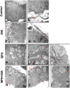
Fig 4. Photomicrographs of prostate sections stained with H&E and toluidine blue.
(a-c) The control group and (d-f) DIA groups showing normal histology of the prostate with variable-sized crammed regular acini (A) lined by one layer of cuboidal to columnar epithelium (arrowheads) with apical cytoplasmic vacuolation (V) and with basal rounded vesicular nuclei and distinct nucleoli (N). The epithelium is underlined by a clear basement membrane (B.M) and smooth muscle cells (SM). The acinar lumen shows few short papillary projections (P) and eosinophilic secretions (S). The intervening stroma is thin and fibromuscular, containing blood vessels (BV). (g-l) BPH group showing spaced-out pleomorphic acini (A), lined with hyperplastic epithelium (arrowheads) with frequent protruding papillary projections (P). The acinar lumen contains accumulated eosinophilic secretions (S) and sloughed epithelium (circle). The basement membrane is markedly thickened (BM) with focal disruption at some points (red arrows). The intervening stroma is thickened and edematous with markedly congested vasculature (BV) and inflammatory infiltrates (yellow arrows). (m-o) BPH+DIA group with improved prostatic histology showing packed regular acini (A) lined with one or few layers of cuboidal epithelium (arrowheads) resting on intact thickened basement membrane (BM). The acinar epithelial cells show vesicular nuclei with distinct nucleoli (N). The acinar lumen shows a few short projecting papillae (P) and eosinophilic secretions (S). (p) Statistical analysis of the acinar epithelial height in all study groups. Data presented as mean ± SD (n = 10). One-way ANOVA and post hoc Tukey test. * significant to the control group, @ significant to the DIA group, # significant to the BPH group, p<0.0001. DIA: diacerein; BPH: benign prostatic hyperplasia.
Fig 5
Fig 5. Photomicrographs of prostate sections stained with Masson’s trichrome (Magnification × 400).
(a, b) The control and DIA groups showing delicate collagen fibers in the stroma (arrows). (c, d) BPH group showing thick wavy collagen bundles between prostatic acini and around blood vessels (arrows). (e) BPH+DIA group displaying reduced deposited collagen (arrow). (f) Statistical analysis of the mean area % of deposited collagen in all study groups. Data presented as mean ± SD (n = 10). One-way ANOVA and post hoc Tukey test. * significant to the control group, @ significant to the DIA group, # significant to the BPH group, p<0.0001. DIA: diacerein; BPH: benign prostatic hyperplasia.
Fig 6
Fig 6. Photomicrographs of prostate sections immunostained with anti-caspase 3, anti-TNF-α, and anti-PCNA antibodies (Magnification × 400).
(a-c) the control group and (d-f) DIA group showing negative cytoplasmic caspase 3 and TNF-α reactions and minimal nuclear PCNA expression, respectively. (g-i) BPH group showing mild cytoplasmic caspase 3, strong TNF-α reactions, and intense nuclear PCNA expression, respectively (arrows). (j-l) BPH+DIA group exhibiting accentuated cytoplasmic caspase 3, attenuated TNF-α, and mild PCNA expressions, respectively (arrows). (m-o) Statistical analysis of the mean area% of caspase 3, TNF-α, and PCNA immunoreaction in all study groups, respectively. Data presented as mean ± SD (n = 10). One-way ANOVA and post hoc Tukey test. * significant to the control group, @ significant to the DIA group, # significant to the BPH group, p<0.0001. DIA: diacerein; BPH: benign prostatic hyperplasia.
Fig 7
Fig 7. TEM photomicrographs of ultrathin sections of prostate.
(a, b) control group and (c, d) DIA group showing acinar cells with basal euchromatic nuclei with islands of heterochromatin and regular nuclear membrane resting on regular basement membrane with smooth muscle cells in between. The cytoplasm contains rough endoplasmic reticulum, apical vacuoles, and secretory granules. Apical microvilli are seen protruding in the lumen. Delicate collagen fibers are spotted underneath the acinar cells and in the stroma. (e-g) BPH group showing multiple layers of acinar cells. Nuclei are euchromatic, with many mitotic figures and irregular nuclear membranes. The basement membrane is markedly thickened with focal disruption. The stroma shows thick collagen bundles arranged in various directions with hypertrophic active fibroblasts. (h-j) BPH+DIA group showing a single layer of acinar epithelial cells with normally looking nuclei. The apical parts show many secretory granules and heterogeneous vacuoles. The apical surface shows projecting microvilli. The basement membrane is regular and intact. Little collagen fibers are spotted above and underneath the basement membrane. N: nucleus, R: rER, V: vacuoles, S: smooth muscle, BM: basement membrane, F: fibroblasts, G: secretory granules, long arrow: collagen fibers, short arrow: microvilli. Scale bar: 2 μm.

References

    1. D’Amico R, Genovese T, Cordaro M, Siracusa R, Gugliandolo E, Peritore AF, et al.. Palmitoylethanolamide/Baicalein Regulates the Androgen Receptor Signaling and NF-κB/Nrf2 Pathways in Benign Prostatic Hyperplasia. Antioxidants (Basel). 2021;10. doi: 10.3390/antiox10071014 - DOI - PMC - PubMed
    1. Trujillo-Rojas L, Fernández-Novell JM, Blanco-Prieto O, Martí-Garcia B, Rigau T, Rivera Del Álamo MM, et al.. Rat age-related benign prostate hyperplasia is concomitant with an increase in the secretion of low ramified α-glycosydic polysaccharides. Theriogenology. 2022;189: 150–157. doi: 10.1016/j.theriogenology.2022.06.011 - DOI - PubMed
    1. El-Ashmawy NE, Khedr EG, El-Bahrawy HA, Helmy NN. Modulatory Effect of Silymarin on Apoptosis in Testosterone -Induced Benign Prostatic Hyperplasia in Rats. Pathol Oncol Res. 2020;26: 1947–1956. doi: 10.1007/s12253-019-00764-4 - DOI - PubMed
    1. Tusubira D, Aja PM, Munezero J, Ssedyabane F, Namale N, Ifie JE, et al.. Safety profile of colocasia esculenta tuber extracts in benign prostate hyperplasia. BMC Complement Med Ther. 2023;23: 187. doi: 10.1186/s12906-023-04018-4 - DOI - PMC - PubMed
    1. Devlin CM, Simms MS, Maitland NJ. Benign prostatic hyperplasia—what do we know? BJU Int. 2021;127: 389–399. doi: 10.1111/bju.15229 - DOI - PubMed

Publication types