. 2024 Feb 1;33(4):355-373.
doi: 10.1093/hmg/ddad188.

Clinical features, functional consequences, and rescue pharmacology of missense GRID1 and GRID2 human variants

Affiliations

Clinical features, functional consequences, and rescue pharmacology of missense GRID1 and GRID2 human variants

James P Allen et al. Hum Mol Genet. .

Abstract

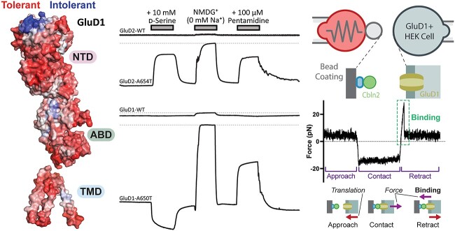
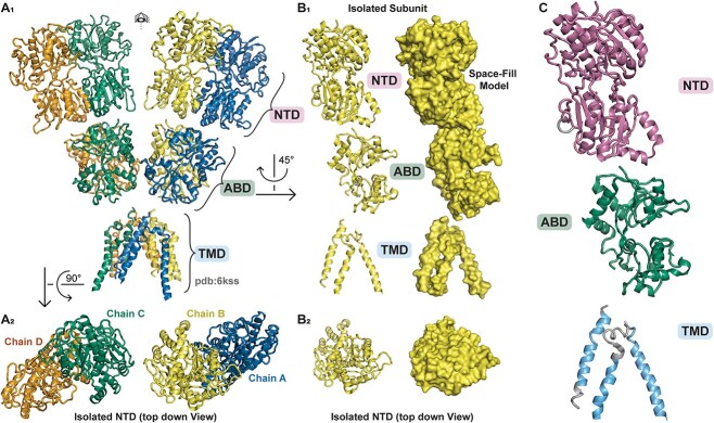
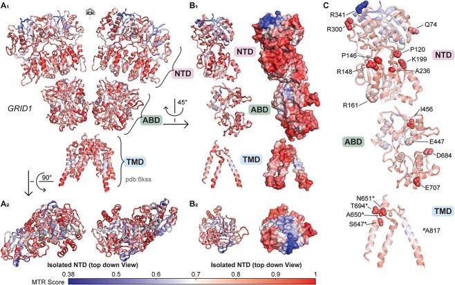
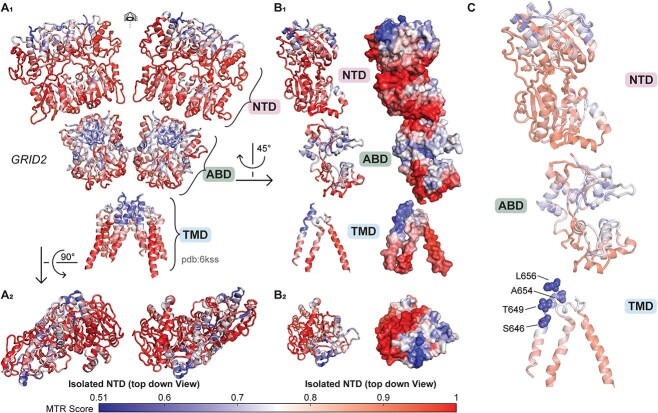
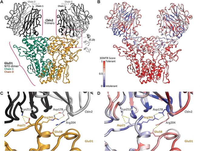
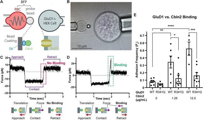
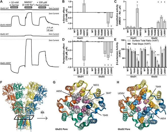
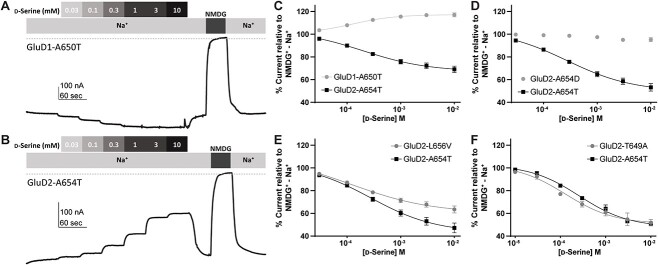
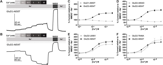
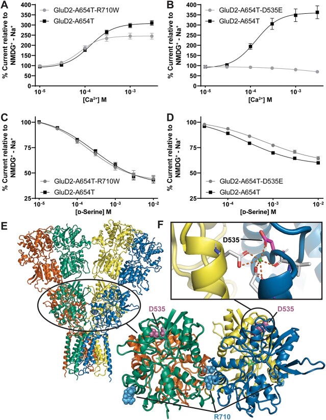
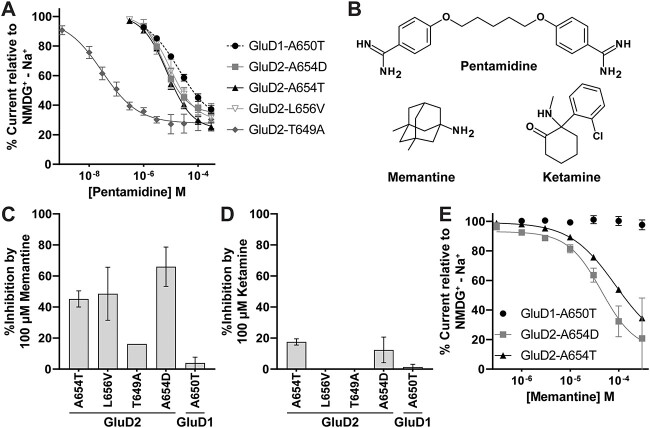
GRID1 and GRID2 encode the enigmatic GluD1 and GluD2 proteins, which form tetrameric receptors that play important roles in synapse organization and development of the central nervous system. Variation in these genes has been implicated in neurodevelopmental phenotypes. We evaluated GRID1 and GRID2 human variants from the literature, ClinVar, and clinical laboratories and found that many of these variants reside in intolerant domains, including the amino terminal domain of both GRID1 and GRID2. Other conserved regions, such as the M3 transmembrane domain, show different intolerance between GRID1 and GRID2. We introduced these variants into GluD1 and GluD2 cDNA and performed electrophysiological and biochemical assays to investigate the mechanisms of dysfunction of GRID1/2 variants. One variant in the GRID1 distal amino terminal domain resides at a position predicted to interact with Cbln2/Cbln4, and the variant disrupts complex formation between GluD1 and Cbln2, which could perturb its role in synapse organization. We also discovered that, like the lurcher mutation (GluD2-A654T), other rare variants in the GRID2 M3 domain create constitutively active receptors that share similar pathogenic phenotypes. We also found that the SCHEMA schizophrenia M3 variant GluD1-A650T produced constitutively active receptors. We tested a variety of compounds for their ability to inhibit constitutive currents of GluD receptor variants and found that pentamidine potently inhibited GluD2-T649A constitutive channels (IC50 50 nM). These results identify regions of intolerance to variation in the GRID genes, illustrate the functional consequences of GRID1 and GRID2 variants, and suggest how these receptors function normally and in disease.

Keywords: GRID1; GRID2; Cbln2; Cbln4; GluD1; GluD2; cerebellar ataxia; cerebellar atrophy; delta receptors; lurcher; schizophrenia.


Conflict of interest statement

S.F.T. is a member of the SAB for Eumentis Therapeutics, Sage Therapeutics, and Combined Brain, is a member of the Medical Advisory Board for the GRIN2B Foundation and the CureGRIN Foundation, is an advisor to GRIN Therapeutics and Neurocrine, is co-founder of NeurOp Inc. and Agrithera Inc., is a member of the Board of Directors of NeurOp Inc., and is co-inventor on IP owned by Emory University. H.Y. is PI of a grant to Emory University from Sage Therapeutics. S.J.M. is PI of a grant from GRIN Therapeutics to Emory University.

Figures

None
Graphical abstract
Figure 1
Figure 1
GluD1 protein structure described by Burada et al. (2020), pdb ID 6KSS (see  Supplementary Data 1). (A1) Full view of protein structure with four separate GluD1 protein subunits visible (chain A, blue; chain B, yellow; chain C, green; chain D, orange). (A2) Top down view of the amino-terminal domain (NTD) of the GluD1 tetramer. (B1-2) Isolated GluD1 subunit shown with ribbon and space fill model. (C) GluD1 protein NTD in pink, agonist-binding domain [56] in green, and transmembrane domain (TMD) in cyan.
Figure 2
Figure 2
Genetic regional intolerance to variation of GRID1. The 3D MTR analysis for GRID1 was developed from the GluD1 structure described by Burada et al. (2020) with 3D MTR residue window of 31 (see Supplementary Data 1 and 2). Low 3D MTR scores (blue) indicate residues less tolerant to variation, while high scores indicate residues that are more tolerant to variation [81]. (A1) Full view of GRID1 3D MTR scores on GluD1 protein structure. (A2) Top down view of the amino-terminal domain (NTD) of GRID1 3D MTR scores on GluD1 protein structure. (B1-2) Isolated subunit GRID1 3D MTR scores shown with ribbon and space fill model. (C) GRID1 variants clinically relevant or tested here that are not present in gnomAD 2.1.1 from Supplementary Tables S2 and S4 shown in context of 3D MTR scores. # denotes position of missense variants residing at listed positions that are not represented in the displayed figure due to lack of electron density. * denotes that variants were evaluated in this study. ^ denotes that variants at listed positions are found in gnomAD (v2.1.1) evaluated because they were in SCHEMA patients.
Figure 3
Figure 3
Genetic regional intolerance to variation of GRID2. The 3D MTR analysis for GRID2 was developed from the GluD1 structure described by Burada et al. (2020) with 3D MTR residue window of 31 (see Supplementary Data 1 and 2). Low 3D MTR scores (blue) indicate residues less tolerant to variation, while high scores [81] indicate residues that are more tolerant to variation. (A1) Full view of GRID2 3D MTR scores mapped onto the GluD1 protein structure, assuming that GluD2 has a similar architecture to GluD1. (A2) Top down view of the amino-terminal domain (NTD) of GRID2 3D MTR scores on the GluD1 protein structure. (B1-2) Isolated subunit GRID2 3D MTR scores shown with ribbon and space fill model. (C) GRID2 variants clinically relevant or tested here that are not present in gnomAD v2.1.1 from Supplementary Tables S3 and S5 shown in context of GRID2 3D MTR scores.
Figure 4
Figure 4
GluD1-Cbln protein-protein interactions. (A) Side view of a homology model (see  Supplementary Data 1) of human GRID1 in complex with human CBLN2. This structure was one frame from the MD simulation modeling the interactions between these proteins (Supplementary Fig. S2). (B) 3D MTR analysis of CBLN2 and GRID1 using the closest 21 residues in the analysis (see  Supplementary Data 2). Low MTR scores (blue) indicate residues intolerant to variation, while high scores indicate residues tolerant to variation [81]. (C) Interacting residues between the GluD1-Cbln2 complex. Labeled residues are predicted to form both hydrogen bonding and salt bridge interactions between GluD1 (Asp21, Arg341, Glu58) and Cbln2 (Lys212, Asp178, Arg204). R341-D178, E58-R204, and D21-K212 salt bridges are predicted to occur during 99.5%, 99.5%, and 100% of analyzed frames. View shown is depicted in panel A by the eye cartoon. On the chain not shown here, R341 interacts with a D176. (D) 3D MTR of the site of GluD1-Cbln2 interaction (same view as in C).
Figure 5
Figure 5
GluD1-R341Q reduces binding between GluD1-NTD and Cbln2. (A) Schematic of the biomembrane force probe (BFP) experiment. (B) Photomicrograph of the BFP experiment. (C) Force trace of a single BFP trial where there was no GluD1/Cbln2 interaction, as demonstrated by the lack of a tension force (+) upon probe retraction. (D) Force trace of a single BFP trial where there was a GluD1/Cbln2 interaction as evidence by the positive force reading upon probe retraction. (E) Adhesion frequencies between HEK293T cells expressing similar levels of GluD1 WT or R341Q (Supplementary Fig. S5) and BFP beads coated with indicated concentrations of Cbln2. Each point represents an adhesion frequency evaluated from the observed number of binding events divided by the total number of repeated contacts (50-100) between a single pair of cell and bead. Also shown are mean ± SEM for each condition. Abbreviations: RBC, red blood cell; SA, streptavidin. Means were compared with an unpaired ANOVA with post hoc Tukey test, where **** indicates P < 0.0001, ***P < 0.001, **P < 0.01, and *P < 0.05. Only pertinent significant comparisons are shown.
Figure 6
Figure 6
Screen for constitutive channel activity of GRID1 and GRID2 human variants. (A) Two electrode voltage clamp assay in Xenopus laevis oocytes expressing cRNA encoding GRID1 and GRID2 human variants. The zero current line is shown (dashed line). (B) Changes in current upon application of 10 mM d-serine. Positive values represent potentiation of constitutive current, while negative values are inhibition of constitutive current. (C) Constitutive current determined by ionic substitution of Na+ for NMDG+. (D) Application of 100 μM pentamidine inhibits constitutively active GluD1 and GluD2 variant receptors. (E) β-Lactamase-GluD1 and -GluD2 protein fusion constructs were assayed to determine surface expression of GluD1 and GluD2 variant receptors (see Methods). Values are normalized to wild-type surface/total ratios, and wild-type total protein expression. (F) GluD protein structure (GluD2 homology model) indicating the view in (G) and (H). (G) GluD1 ion channel pore (homology model) and (H) GluD2 ion channel pore (homology model) showing the localization of constitutively active variants in the channel pore. *P < 0.05 ANOVA with Dunnett’s multiple comparisons test.
Figure 7
Figure 7
Modulation of constitutively active GluD1 and GluD2 M3 variants by d-serine. Representative concentration-response curves showing d-serine modulation of the constitutive current observed for (A) GluD1-A650T and (B) GluD2-A654T. The differences between the baseline current and the current in NMDG+ are used to establish theoretical maximum inhibition for each recording. The dotted line represents zero current. (C–F) D-Serine concentration-response curve for d-serine modulation of constitutively active M3 variants GluD1-A650T (C), GluD2-A654D (D), GluD2-L656V (E) and GluD2-T649A. For each experiment, same day control curves recorded for the lurcher variant GluD2-A654T are shown. Unlike GluD2-A654T, GluD2-L656V, GluD2-T649A, we observed that GluD1-A650T is modestly potentiated by d-serine rather than inhibited. A654D shows a near complete loss of d-serine potency.
Figure 8
Figure 8
Modulation of constitutively active GluD1 and GluD2 M3 variants by extracellular Ca2+. Representative Ca2+ concentration-response curves for (A) GluD1-A650T and (B) GluD2-A654T. The dotted line represents zero current. (C–F) Ca2+ concentration-response of GluD1-A650T, GluD2-A654D, GluD2-L656V, GluD2-T649A, and GluD2-A654T. For each experiment, same day control curves recorded for the lurcher variant GluD2-A654T are shown. All recordings normalized to NMDG+ − Na+ difference current. Unlike GluD2-A654T, GluD1-A650T and GluD2-A654D were modestly inhibited by Ca2+.
Figure 9
Figure 9
Non-constitutively active GRID2 agonist binding domain variants alter d-serine and Ca2+ potency. For each experiment, same day control curves recorded for the lurcher variant GluD2-A654T are shown. (A) Ca2+ concentration-response curve for inhibition of GluD2-A654T-R710W and -A654T (B) Ca2+ concentration-response curve for inhibition of GluD2-A654T, D535E and GluD2-A654T. Unlike GluD2-A654T, GluD2-A654T,D535E is weakly inhibited by Ca2+. (C) d-Serine concentration-response of GluD2-A654T, R710W and GluD2-A654T. (D) d-Serine concentration-response of GluD2-A654T,D535E and GluD2-A654T. (E) GluD2 protein structure (GluD2 homology model) with a closer view of the GluD2 agonist binding domain shows the location of GluD2-R710W and GluD2-D535E. (F) Variant GluD2-D535E is at a site that is important for Ca2+ binding (PBD: 2V3T).
Figure 10
Figure 10
Rescue pharmacology of GluD1 and GluD2 constitutively active variants using FDA-approved channel blockers in Xenopus oocytes. (A) Pentamidine composite concentration-response curves for GluD2 variants show that pentamidine has an IC50 value that is > 200 fold lower (i.e. more potent) for GluD2-T649A (IC50 37 nM) than for GluD2-A654T. Recordings were performed in 1 mM Ca2+. (B) Structures of compounds tested in this study are shown. (C and D) Results are shown for a single concentration screen for memantine (C) and ketamine (D) inhibition of GluD receptor channel constitutive activity. (E) Memantine concentration-response curves are shown for GluD2-A654D and GluD2-A654T.

References

    1. Nakamoto C, Konno K, Miyazaki T. et al. Expression mapping, quantification, and complex formation of GluD1 and GluD2 glutamate receptors in adult mouse brain. J Comp Neurol 2020;528:1003–27. - PubMed
    1. Lomeli H, Sprengel R, Laurie DJ. et al. The rat delta-1 and delta-2 subunits extend the excitatory amino acid receptor family. FEBS Lett 1993;315:318–22. - PubMed
    1. Landsend AS, Amiry-Moghaddam M, Matsubara A. et al. Differential localization of delta glutamate receptors in the rat cerebellum: coexpression with AMPA receptors in parallel fiber-spine synapses and absence from climbing fiber-spine synapses. J Neurosci 1997;17:834–42. - PMC - PubMed
    1. Mayat E, Petralia RS, Wang YX. et al. Immunoprecipitation, immunoblotting, and immunocytochemistry studies suggest that glutamate receptor delta subunits form novel postsynaptic receptor complexes. J Neurosci 1995;15:2533–46. - PMC - PubMed
    1. Naur P, Hansen KB, Kristensen AS. et al. Ionotropic glutamate-like receptor delta2 binds D-serine and glycine. Proc Natl Acad Sci U S A 2007;104:14116–21. - PMC - PubMed

Publication types