. 2023 Nov 10;9(1):56.
doi: 10.1038/s41540-023-00320-6.

An integrated study to decipher immunosuppressive cellular communication in the PDAC environment

Affiliations

An integrated study to decipher immunosuppressive cellular communication in the PDAC environment

Gülben Avşar et al. NPJ Syst Biol Appl. .

Abstract

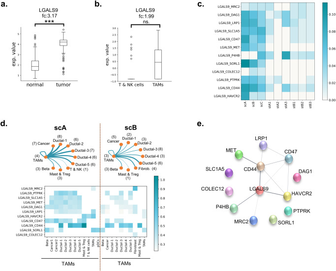
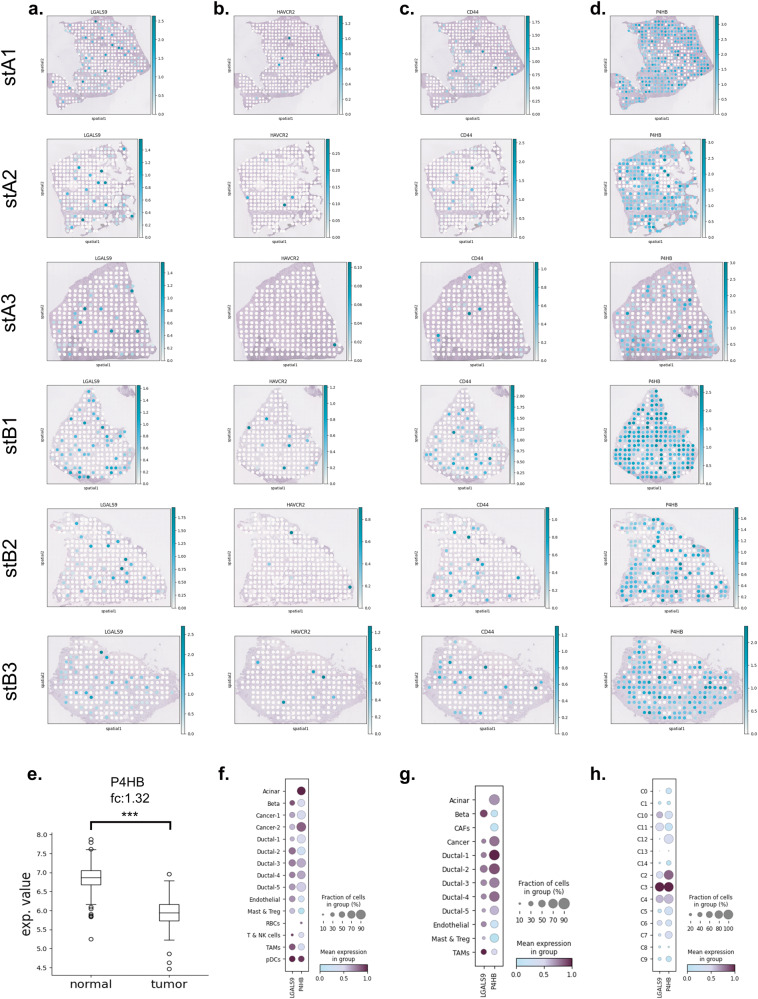
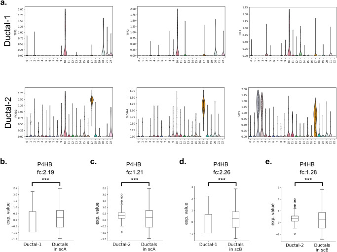
Pancreatic ductal adenocarcinoma (PDAC) is one the most aggressive cancers and characterized by a highly rigid and immunosuppressive tumor microenvironment (TME). The extensive cellular interactions are known to play key roles in the immune evasion, chemoresistance, and poor prognosis. Here, we used the spatial transcriptomics, scRNA-seq, and bulk RNA-seq datasets to enhance the insights obtained from each to decipher the cellular communication in the TME. The complex crosstalk in PDAC samples was revealed by the single-cell and spatial transcriptomics profiles of the samples. We show that tumor-associated macrophages (TAMs) are the central cell types in the regulation of microenvironment in PDAC. They colocalize with the cancer cells and tumor-suppressor immune cells and take roles to provide an immunosuppressive environment. LGALS9 gene which is upregulated in PDAC tumor samples in comparison to healthy samples was also found to be upregulated in TAMs compared to tumor-suppressor immune cells in cancer samples. Additionally, LGALS9 was found to be the primary component in the crosstalk between TAMs and the other cells. The widespread expression of P4HB gene and its interaction with LGALS9 was also notable. Our findings point to a profound role of TAMs via LGALS9 and its interaction with P4HB that should be considered for further elucidation as target in the combinatory immunotherapies for PDAC.


Conflict of interest statement

The authors declare no competing interests.

Figures

Fig. 1
Fig. 1. Cellular heterogeneity in PDAC tissues.
The cell clusters and cell type specific marker genes in a scA and b scB. The domains and cell type specific genes in c stA and d stB.
Fig. 2
Fig. 2. Communication profiles in the datasets.
The number of interactions between and within the cell types in a scA and b scB, and the domains in c stA and d stB (The color intensity in the heatmaps shows the number of observed pairs in the corresponding dataset). e The most frequent 15 L-R pairs (the x-axis refers to the percentage of the corresponding L-R pair in the complete list of interactions), f The enriched GO terms for the proteins with interactions between ductal cells and TAMs.
Fig. 3
Fig. 3. The observed cellular interactions.
a The top 15 L-R pairs in ST datasets (the color intensity shows the score of the interaction in the corresponding dataset), b the associated processes of top 15 L-R pair elements in ST datasets, c the communication between cancer cells, tumor-promoting immune cells and tumor-suppressor immune cells, d the top 25 L-R pairs in scA.
Fig. 4
Fig. 4. LGALS9 in TME.
a LAGLS9 overexpression in TCGA-PAAD compared to GTEX datasets. b LGALS9 overexpression in PDAC patients with the comparison of TAMs and T & NK cells. c the LGALS9-participated interactions in SC and ST datasets. d The distribution of LGALS9 interactions within TAMs and the other cell types (the number between the parentheses indicates the number of interactions with TAMs). e PPI network of LGALS9 and its receptors which were found in SC and ST datasets. (The color intensity in the heatmaps shows the score of the pair in the corresponding dataset).
Fig. 5
Fig. 5. Spatial distribution of the genes.
The expression profiles of a LGALS9, b HAVCR2, c CD44, and d P4HB gene on tissue samples (The color intensity refers to the expression value of the corresponding gene in the dataset.). e The regulation of P4HB in bulk datasets. The expression profiles of LGALS9 and P4HB in f scA, g scB, and h scC.
Fig. 6
Fig. 6. P4HB profile in SC datasets.
a The ductal cell clusters in adj dataset. The regulation of P4HB in comparison of b, c adj with scA, and d, e adj with scB.
Fig. 7
Fig. 7. The overview of the study.
The observed landscape for the immunosuppressive PDAC TME.

References

    1. Garcia PE, et al. Differential contribution of pancreatic fibroblast subsets to the pancreatic cancer stroma. Cell Mol. Gastroenterol. Hepatol. 2020;10:581–599. - PMC - PubMed
    1. Chen K, et al. Development and validation of prognostic and diagnostic model for pancreatic ductal adenocarcinoma based on scRNA-seq and bulk-seq datasets. Hum. Mol. Genet. 2022;31:1705–1719. - PMC - PubMed
    1. Seifert AM, et al. Detection of pancreatic ductal adenocarcinoma with galectin-9 serum levels. Oncogene. 2020;39:3102–3113. - PMC - PubMed
    1. Nikšić M, et al. Pancreatic cancer survival trends in the US from 2001 to 2014: a CONCORD-3 study. Cancer Commun. 2023;43:87–99. - PMC - PubMed
    1. Liu X, et al. The crosstalk between malignant cells and tumor-promoting immune cells relevant to immunotherapy in pancreatic ductal adenocarcinoma. Front Cell Dev. Biol. 2022;9:3857. - PMC - PubMed

MeSH terms