. 2023 Dec;30(12):1867-1877.
doi: 10.1038/s41594-023-01128-y. Epub 2023 Nov 9.

Cryo-EM reveals how Hsp90 and FKBP immunophilins co-regulate the glucocorticoid receptor

Affiliations

Cryo-EM reveals how Hsp90 and FKBP immunophilins co-regulate the glucocorticoid receptor

Chari M Noddings et al. Nat Struct Mol Biol. 2023 Dec.

Abstract

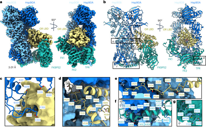
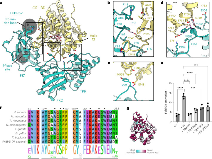
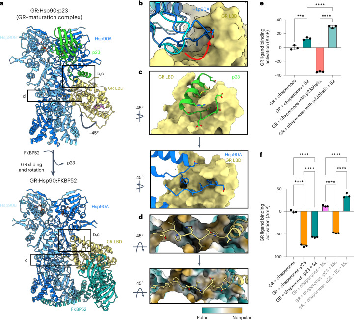
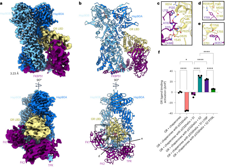
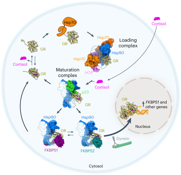
Hsp90 is an essential molecular chaperone responsible for the folding and activation of hundreds of 'client' proteins, including the glucocorticoid receptor (GR). Previously, we revealed that Hsp70 and Hsp90 remodel the conformation of GR to regulate ligand binding, aided by co-chaperones. In vivo, the co-chaperones FKBP51 and FKBP52 antagonistically regulate GR activity, but a molecular understanding is lacking. Here we present a 3.01 Å cryogenic electron microscopy structure of the human GR:Hsp90:FKBP52 complex, revealing how FKBP52 integrates into the GR chaperone cycle and directly binds to the active client, potentiating GR activity in vitro and in vivo. We also present a 3.23 Å cryogenic electron microscopy structure of the human GR:Hsp90:FKBP51 complex, revealing how FKBP51 competes with FKBP52 for GR:Hsp90 binding and demonstrating how FKBP51 can act as a potent antagonist to FKBP52. Altogether, we demonstrate how FKBP51 and FKBP52 integrate into the GR chaperone cycle to advance GR to the next stage of maturation.


Conflict of interest statement

The authors declare no competing interests.

Figures

Fig. 1
Fig. 1. Architecture of the GR:Hsp90:FKBP52 complex.
a, Composite cryo-EM map of the GR:Hsp90:FKBP52 complex. Hsp90A, dark blue; Hsp90B, light blue; GR, yellow; FKBP52, teal. Color scheme is maintained throughout. b, Atomic model in cartoon representation with boxes corresponding to the interfaces shown in detail in cg. c, Interface 1 of the Hsp90:GR interaction, depicting the Hsp90A Src loop (Hsp90A345–360) interacting with the GR hydrophobic patch. GR is in surface representation. d, Interface 2 of the Hsp90:GR interaction, depicting GR Helix 1 (GR532–539) packing against the entrance to the Hsp90 lumen. Hsp90A/Hsp90B are in surface representation. e, Interface 3 of the Hsp90:GR interaction, depicting GR pre-Helix 1 (GR519–531) threading through the Hsp90 lumen. Hsp90A/Hsp90B are in surface representation. f, Interface 1 of the Hsp90:FKBP52 interaction, depicting FKBP52 TPR H7e (FKBP52387–424) interacting with the Hsp90A/Hsp90B CTD dimer interface. Hsp90A/Hsp90B are in surface representation. g, Interface 2 of the Hsp90:FKBP52 interaction, depicting the Hsp90B MEEVD motif (Hsp90B700–706) binding in the helical bundle of the FKBP52 TPR domain. FKBP52 is in surface representation.
Fig. 2
Fig. 2. The GR:FKBP52 interaction and functional significance.
a, Atomic model depicting the three interfaces between GR (yellow) and FKBP52 (teal) in the GR:Hsp90:FKBP52 complex. The FKBP52 proline-rich loop and PPIase catalytic site are highlighted in gray. Dexamethasone is colored in pink. b, Interface 1 between GR (yellow) and the FKBP52 FK1 domain (teal), showing interacting side chains and hydrogen bonds (dashed pink lines). c, Interface 2 between GR (yellow) and the FKBP52 FK2 domain (teal), showing interacting side chains and hydrogen bonds (dashed pink lines). d, Interface 3 between GR (yellow) and the FKBP52 FK2–TPR linker (teal), showing interacting side chains and hydrogen bonds (dashed pink lines). e, GR activation assay in wild-type yeast strain JJ762 expressing FKBP52 (‘52’) or FKBP52 mutants. The fold increase in GR activities compared to the empty vector (e.v.) control are shown (mean ± s.d.). n = 3 biologically independent samples per condition. Significance was evaluated using a one-way analysis of variance (F(5,12) = 26.10; P < 0.0001) with post-hoc Dunnett’s multiple comparisons test (n.s. P > 0.05; *P ≤ 0.05; **P ≤ 0.01; ***P ≤ 0.001). P values: P(e.v. versus 52) <0.0001, P(52 versus 52ΔFK1) <0.0001, P(52 versus 52 S118A) <0.0001, P(52 versus 52 Y161D) 0.0003, P(52 versus 52 W259D) 0.0005. f, Sequence alignment of eukaryotic FKBP52 showing conserved residues involved in the GR:FKBP52 interaction (denoted by a black asterisk). The bottom aligned sequence is human FKBP51. The alignment is colored according to the ClustalW convention. g, GR protein sequence conservation mapped onto the GR atomic model from the GR:Hsp90:FKBP52 complex. Residue conservation is depicted from most variable (cyan) to most conserved residues (maroon). GR residues that interact with FKBP52 are shown as spheres. Source data
Fig. 3
Fig. 3. FKBP52 competes with p23 to bind GR:Hsp90.
a, Atomic model of the GR–maturation complex (top) and the GR:Hsp90:FKBP52 complex (bottom) with boxes corresponding to the interfaces shown in detail in bd. Hsp90A, dark blue; Hsp90B, light blue; GR, yellow; p23, green; FKBP52, teal. b, Position of the Hsp90A Src loop in the GR–maturation complex (Hsp90A, cyan) versus the GR:Hsp90:FKBP52 complex (Hsp90A, dark blue). GR (yellow, surface representation). Hsp90A Src loop residues interacting with the GR hydrophobic patch are shown. c, Interface between the p23 tail-helix (green) and the GR hydrophobic patch (yellow, surface representation) in the GR–maturation complex (top). The p23 tail-helix is replaced by the Hsp90A Src loop (dark blue) in the GR:Hsp90:FKBP52 complex (bottom), which flips up to interact with the GR hydrophobic patch (yellow, surface representation). Interacting side chains are shown. d, Interaction between GR pre-Helix 1 (GR523–531) in the Hsp90 lumen in the GR–maturation complex (top) versus the GR:Hsp90:FKBP52 complex (bottom). Hsp90A/Hsp90B are in surface representation colored by hydrophobicity. GR pre-Helix 1 translocates through the Hsp90 lumen by two residues in the transition from the GR–maturation complex to the GR:Hsp90:FKBP52 complex. e, Equilibrium binding of 10 nM F-dex to 100 nM GR DBD–LBD with chaperones and 15 μM FKBP52 (‘52’) (mean ± s.d.). n = 3 biologically independent samples per condition. ‘Chaperones’: 15 μM Hsp70, Hsp90, Hop, and p23 or p23Δhelix; 2 μM Ydj1 and Bag-1. Significance was evaluated using a one-way analysis of variance (ANOVA) (F(3,8) = 541.2; P < 0.0001) with post-hoc Šídák’s test (n.s. P > 0.05; *P ≤ 0.05; **P ≤ 0.01; ***P ≤ 0.001; ****P ≤ 0.0001). P values: P(chaperones versus chaperones + 52) 0.0002, P(chaperones + 52 versus chaperones with p23Δhelix + 52) <0.0001, P(chaperones with p23Δhelix versus chaperones with p23Δhelix + 52) <0.0001. f, Equilibrium binding of 10 nM F-dex to 100 nM GR DBD–LBD with chaperones, 15 μM FKBP52 (‘52’) and 20 mM sodium molybdate (‘Mo.’) (mean ± s.d.). n = 3 biologically independent samples per condition. ‘Chaperones’: 15 μM Hsp70, Hsp90, Hop and p23; 2 μM Ydj1 and Bag-1. ‘-p23’ indicates p23 was left out of the chaperone mixture. Significance was evaluated using a one-way ANOVA (F(5,12) = 761.5; P < 0.0001) with post-hoc Šídák’s test (n.s. P > 0.05; *P ≤ 0.05; **P ≤ 0.01; ***P ≤ 0.001; ****P ≤ 0.0001). P value <0.0001 for each comparison. Source data
Fig. 4
Fig. 4. Architecture of the GR:Hsp90:FKBP51 complex.
a, Composite cryo-EM map of the GR:Hsp90:FKBP51 complex. Hsp90A, dark blue; Hsp90B, light blue; GR, yellow; FKBP51, purple. Color scheme is maintained throughout. b, Atomic model in cartoon representation with boxes corresponding to the interfaces shown in detail in ce. c, Interface 1 between GR (yellow) and the FKBP51 FK1 domain (purple), showing interacting side chains and hydrogen bonds (dashed pink lines). d, Interface 2 between GR (yellow) and the FKBP51 FK2 domain (purple), showing interacting side chains and hydrogen bonds (dashed pink lines). e, Interface 3 between GR (yellow) and the FKBP51 FK2–TPR linker (yellow), showing interacting side chains and hydrogen bonds (dashed pink lines). f, Equilibrium binding of 10 nM F-dex to 100 nM GR DBD–LBD with chaperones, 15 μM FKBP51 (‘51’), 15 μM FKBP52 (‘52’) or mutants (mean ± s.d.). n = 3 biologically independent samples per condition. ‘Chaperones’: 15 μM Hsp70, Hsp90, Hop, and p23 or p23Δhelix; 2 μM Ydj1 and Bag-1. Significance was evaluated using a one-way analysis of variance (F(5,12) = 404.1; P < 0.0001) with post-hoc Šídák’s test (n.s. P > 0.05; *P ≤ 0.05; **P ≤ 0.01; ***P ≤ 0.001; ****P ≤ 0.0001). P values: P(chaperones versus chaperones with p23Δhelix) <0.0001, P(chaperones versus chaperones with p23Δhelix + 51) 0.0343, P(chaperones with p23Δhelix + 51 versus chaperones with p23Δhelix + 51 L119P) <0.0001, P(chaperones with p23Δhelix + 51 versus chaperones with p23Δhelix + 52) <0.0001, P(chaperones with p23Δhelix + 52 versus chaperones with p23Δhelix + 52 P119L) <0.0001, P(chaperones with p23Δhelix + 51 P119L versus chaperones with p23Δhelix + 52 P119L) <0.0001. Source data
Fig. 5
Fig. 5. Mechanism of GR regulation by FKBP51 and FKBP52 during the GR chaperone cycle.
Schematic depicting how the FKBP co-chaperones integrate with the GR chaperone cycle and how this cycle may take place within a cellular context. Starting on the top left, GR (yellow, cartoon representation) is in dynamic equilibrium between cortisol-bound and unbound (apo) states. Hsp70 (orange) binds GR and locally unfolds GR to inhibit cortisol binding, stabilizing GR in a partially unfolded, apo state. Hsp70 transfers the partially unfolded GR to Hsp90 (light and dark blue):Hop (pink) to form the GR–loading complex, in which GR is stabilized in a partially unfolded, apo state. Cortisol (pink), which enters the cell through diffusion, binds to GR during the transition from the GR–loading complex to the GR–maturation complex when Hsp90 refolds the GR to a native conformation. In the GR–maturation complex, the cortisol-bound, folded GR is stabilized by Hsp90 and p23 (green) and is protected from Hsp70 re-binding. Depending on the relative concentrations of the FKBPs, either FKBP51 (purple) or FKBP52 (teal) can bind the GR:Hsp90:p23 complex, competing with p23, and stabilizing the rotated position of GR. FKBP51 sequesters GR:Hsp90 in the cytosol until ATP hydrolysis on Hsp90 allows release of GR back to the chaperone cycle. In contrast, FKBP52 promotes rapid nuclear translocation of GR:Hsp90 (refs. ,,,). Once in the nucleus, the cortisol-bound GR can dimerize, nucleate the assembly of transcriptional regulatory complexes, and regulate transcription, including activating expression of FKBP51, leading to a negative feedback loop that regulates GR activity in the cell,–.
Extended Data Fig. 1
Extended Data Fig. 1. Sample Preparation.
a, The in vitro reconstituted GR chaperone cycle. On the left, GR is active and able to bind ligand. Hsp70, aided by the co-chaperone Hsp40, engages GR and inhibits ligand binding. Hsp70 loads apo GR onto Hsp90 and Hop, which forms the ‘GR-loading complex’ (PDB ID 7KW7). Hsp70 and Hop are released, Hsp90 hydrolyzes ATP to fully close, and the co-chaperone p23 binds, forming the ‘GR-maturation complex’ (PDB ID 7KRJ). GR binds ligand in the transition from the GR-loading complex to the GR-maturation complex. In the maturation complex, GR is in a fully folded, native conformation and bound to ligand. Upon Hsp90 re-opening, GR is released from the complex to return to the cycle. b, Domain organization of the proteins in the GR:Hsp90:FKBP complexes and p23. c, Structural motifs of Hsp90, GR, and FKBP51/52. d, Coomassie-stained SDS-PAGE (4-12% acrylamide gel) with MBP-GR pulldown elutions from the in vitro reconstituted GR chaperone cycle. Lane 1- elution from MBP-GR pulldown for FKBP51-containing reaction; Lane 2- sample from Lane 1 after size exclusion chromatography (SEC) (e) and chemical crosslinking with 0.02% glutaraldehyde; Lane 3- elution from MBP-GR pulldown for FKBP52-containing reaction; Lane 4- sample from Lane 3 after SEC (e) and chemical crosslinking with 0.02% glutaraldehyde. This experiment was repeated 7 independent times with similar results. e, Size exclusion chromatography (SEC) profile of the elution from the MBP-GR pulldown. The green trace represents the SEC profile from the reconstituted GR chaperone cycle with FKBP51, while the blue trace represents the SEC profile from the reconstituted GR chaperone cycle with FKBP52. mAU = milli-absorbance units. Coomassie-stained SDS-PAGE (4-12% acrylamide gel) of the fractions from SEC corresponding to the GR:Hsp90:FKBP52 sample (top) or the GR:Hsp90:FKBP51 sample (bottom). Colors indicate which gel lanes correspond to specific regions of the SEC profile. Sample fractions from the region highlighted in purple were collected and used for cryo-EM data collection. This experiment was repeated 11 independent times with similar results. f, Representative electron micrograph for the cryo-EM dataset of the GR:Hsp90:FKBP52 complex (left) (−1.48 μm defocus) and GR:Hsp90:FKBP51 complex (right) (−2.23 μm defocus). A total of 11,162 and 26,413 micrographs were obtained, respectively. Source data
Extended Data Fig. 2
Extended Data Fig. 2. Cryo-EM Data Analysis for the GR:Hsp90:FKBP52 Complex.
a, Cryo-EM data processing procedure for the GR:Hsp90:FKBP52 complex performed in RELION and CryoSparc. Gold-standard Fourier shell correlation (GSFSC) curves of the final 3D reconstructions, including the focused maps and the consensus map, are shown (bottom). The blue lines intercept the y-axis at an FSC value of 0.143. Angular distribution of particles and local resolution are shown for the consensus map (bottom, middle). b, Map-to-model FSC curves between the GR:Hsp90:FKBP52 atomic model and the consensus GR:Hsp90:FKBP52 map, along with different views of the model within the map. The black dotted line intercepts the y-axis at an FSC value of 0.5.
Extended Data Fig. 3
Extended Data Fig. 3. Hsp90:GR Interfaces in the GR:Hsp90:FKBP52 Complex.
a, Hsp90:GR:FKBP52 complex map density with atomic model showing ATP-magnesium density in both Hsp90 protomers (Hsp90A/B). Bottom images show increased contour level on the map density to indicate that the ATP γ-phosphate position has much stronger density relative to the α and β-phosphates, likely corresponding to molybdate, which may act as a γ-phosphate analog (see Methods). b, Atomic model of GR from the GR:Hsp90:FKBP52 complex (yellow) aligned with GR from the crystal structure (PDB ID 1M2Z) (light pink) with co-activator peptide NCoA2 (purple) and GR from the GR-maturation complex structure (PDB ID 7KRJ). GR Helix 12 is indicated. c-f, Atomic model of GR:Hsp90:FKBP52 complex with Hsp90A (dark blue), Hsp90B (light blue), GR (yellow). Side chains in contact between GR and Hsp90 are shown, along with hydrogen bonds (dashed pink lines).c, Interface 1 of the GR:Hsp90 interaction depicting the GR hydrophobic patch (GR Helices 9 and 10) interacting with the Hsp90A Src loop (Hsp90345–360), Hsp90AW320, and Hsp90A NTD/MD helices. d, Interface 2 of the GR:Hsp90 interaction depicting the GR pre-Helix 1 strand and Helix 1 packing up against the Hsp90B amphipathic α-helices. e, Interface 2 of the GR:Hsp90 interaction depicting GR in surface representation colored by hydrophobicity (green = polar, brown = nonpolar) with Hsp90BY627 sticking into the BF3 druggable hydrophobic pocket. f, Interface 3 of the GR:Hsp90 interaction depicting the GR pre-Helix 1 strand threading through the Hsp90 lumen between Hsp90A and Hsp90B.
Extended Data Fig. 4
Extended Data Fig. 4. Hsp90:FKBP52 Interfaces in the GR:Hsp90:FKBP52 Complex.
Atomic model of the GR:Hsp90:FKBP52 complex with Hsp90A (dark blue), Hsp90B (light blue), GR (yellow), and FKBP52 (teal). Side chains in contact between Hsp90 and FKBP52 are shown, along with hydrogen bonds (dashed pink lines). a, Interface 1 of the Hsp90:FKBP52 interaction depicting the FKBP52 TPR H7e binding to the Hsp90A/B CTD dimer interface. The helix of FKBP52 H7e breaks to fit into the cleft formed by the Hsp90 CTDs. b, Interface 2 of the Hsp90:FKBP52 interaction depicting the Hsp90B MEEVD motif binding the FKBP52 TPR helical bundle. c, Interface 3 of the Hsp90:FKBP52 interaction depicting the FKBP52 TPR Helices 5 and 6 binding to the Hsp90B CTD. d, FKBP52 (teal) from the GR:Hsp90:FKBP52 atomic model aligned with the cryo-EM structure of FKBP51 (light blue) (PDB ID 7L7I) (top) and crystal structures of FKBP52 (PDB ID 1P5Q, 1Q1C) (bottom) showing the difference in interdomain angles. 1P5Q contains the FKBP52 FK1 and FK2 domain, while 1Q1C contains the FKBP52 FK2 and TPR domains. e, The GR:Hsp90:FKBP52 atomic model with FKBP52 (teal), GR (yellow), and dexamethasone (pink) with proline-isomerase inhibitors, rapamycin (brown) or FK506 (orange), docked into the atomic model to indicate the steric clash with GR. Rapamycin was docked in based on the FKBP52:rapamycin crystal structure (PDB ID 4DRJ) and FK506 was docked in based on the FKBP52:FK506:FRB crystal structure (PDB ID 4LAX).
Extended Data Fig. 5
Extended Data Fig. 5. Analysis of the GR:Hsp90:FKBP52 Structure.
a, Expression of human FKBP52 or FKBP52 mutants in wild-type yeast strain JJ762 assayed by immunoblot with a monoclonal antibody specific for FKBP52. A monoclonal antibody against PGK1 was used as a loading control. The asterisk marks an unknown protein that cross reacts with the anti-FKBP52 antibody. This experiment was performed one time. b, Atomic model of the GR:Hsp90:FKBP52 complex shown in surface representation. FKBP52 (teal), GR (yellow). The NCoA2 (nuclear coactivator 2) co-activator peptide is docked in based on the GR:NCoA2 crystal structure (PDB ID 1M2Z). While most of the coactivator peptide binding is sterically permitted, the N-terminus of NCoA2 clashes with the FKBP52 TPR domain (red circle). c, Atomic models of the GR-maturation complex (GR:Hsp90:p23) (left) and the GR:Hsp90:FKBP52 complex without FKBP52 (right) depicting that GR LBD dimerization is permitted once FKBP52 is released. Hsp90A (dark blue, surface representation), Hsp90B (light blue, surface representation), GR (yellow, surface representation), p23 (green, surface representation). In both complexes, the GR LBD dimerization site is accessible, however; binding of the second GR LBD (light pink) to the GR-maturation complex clashes with the Hsp90B CTD, shown with a red circle (left). Binding of the second GR LBD (light pink) to the GR:Hsp90:FKBP52 complex (right). Docking of the dimerized GR LBD is based on the GR LBD dimer crystal structure (PDB ID 1M2Z). d, Equilibrium binding of 10 nM fluorescent dexamethasone to 100 nM GR DBD-LBD with addition of 15 μM FKBP51 (‘51’) or FKBP52 (‘52’) measured by fluorescence polarization (mean ± SD). n = 3 biologically independent samples per condition. Fluorescence polarization values are baseline subtracted in accordance with the measured fluorescent dexamethasone baseline polarization value. Statistical significance was evaluated by an ordinary one-way ANOVA (F(2,6) = 1414, p < 0.0001) with post-hoc Tukey’s multiple comparisons test. P-values: p(GR vs. GR + 51) < 0.0001, p(GR vs. GR + 52) < 0.0001, p(GR + 51 vs. GR + 52) = 0.004. (n.s. P > 0.05; * P ≤ 0.05; ** P ≤ 0.01; *** P ≤ 0.001; **** P ≤ 0.0001). Source data
Extended Data Fig. 6
Extended Data Fig. 6. Cryo-EM Data Analysis for the GR:Hsp90:FKBP51 Complex.
a, Cryo-EM data processing procedure for the GR:Hsp90:FKBP51 complex performed in RELION and CryoSparc. Gold-standard Fourier shell correlation (GSFSC) curves of the final 3D reconstructions, including the focused maps and the consensus map, are shown (bottom). The blue lines intercept the y-axis at an FSC value of 0.143. Angular distribution of particles and local resolution are shown for the consensus map (bottom, middle). b, Map-to-model FSC curves between the GR:Hsp90:FKBP51 atomic model and the consensus GR:Hsp90:FKBP51 map, along with different views of the model within the map. The black dotted line intercepts the y-axis at an FSC value of 0.5.
Extended Data Fig. 7
Extended Data Fig. 7. Interfaces in the GR:Hsp90:FKBP51 Complex.
Atomic model of the GR:Hsp90:FKBP51 complex with Hsp90A (dark blue), Hsp90B (light blue), GR (yellow), and FKBP51 (purple). Side chains in contact between Hsp90 and FKBP51 or Hsp90 and GR are shown, along with hydrogen bonds (dashed pink lines). a, Hsp90:GR:FKBP51 complex map density with atomic model showing ATP-magnesium density in both Hsp90 protomers (Hsp90A/B). Bottom images show increased contour level on the map density to indicate that the ATP γ-phosphate position has much stronger density relative to the α and β-phosphates, likely corresponding to molybdate, which may act as a γ-phosphate analog (see Methods). b, Interface 1 of the Hsp90:FKBP51 interaction depicting the FKBP51 TPR H7e binding to the Hsp90A/B CTD dimer interface. The helix of FKBP51 H7e breaks to fit into the cleft formed by the Hsp90 CTDs. c, Interface 2 of the Hsp90:FKBP51 interaction depicting the Hsp90B MEEVD motif binding the FKBP51 TPR helical bundle. d, Interface 3 of the Hsp90:FKBP51 interaction depicting the FKBP51 TPR Helices 5 and 6 binding to the Hsp90B CTD. e, Interface 1 of the GR:Hsp90 interaction depicting the GR hydrophobic patch (GR Helices 9 and 10) interacting with the Hsp90A Src loop (Hsp90345–360), Hsp90AW320, and Hsp90A NTD/MD helices. f, Interface 2 of the GR:Hsp90 interaction depicting GR pre-Helix 1 strand and Helix 1 packing up against the Hsp90B amphipathic α-helices. g, Interface 3 of the GR:Hsp90 interaction depicting the GR pre-Helix 1 strand threading through the Hsp90 lumen between Hsp90A and Hsp90B. h, The GR:Hsp90:FKBP51 atomic model with FKBP51 (purple), GR (yellow), and dexamethasone (pink) with proline-isomerase inhibitors, rapamycin (brown) or FK506 (orange), docked into the atomic model to indicate the steric clash with GR. Rapamycin was docked in based on the FKBP51:rapamycin crystal structure (PDB ID 4DRI) and FK506 was docked in based on the FKBP51:FK506:FRB crystal structure (PDB ID 3O5R). The FKBP51-specific inhibitor SAFit2 was docked into the atomic model to indicate there is no steric clash with GR at the backbone level (although some side chains clash). SAFit2 was docked in based on the FKBP51:SAFit2 crystal structure (PDB ID 6TXX).
Extended Data Fig. 8
Extended Data Fig. 8. Effect of FKBP51 on GR Ligand Binding in vitro.
a, Equilibrium binding of 10 nM fluorescent dexamethasone to 100 nM GR DBD-LBD with chaperones and 15 μM FKBP51 (mean ± SD). n = 3 biologically independent samples per condition. ‘Chaperones’ = 15 μM Hsp70, Hsp90, Hop, and p23; 2 μM Ydj1 and Bag-1. Statistical significance was evaluated by an unpaired two-tailed t-test, p-value = 0.5737. (n.s. P > 0.05; * P ≤ 0.05; ** P ≤ 0.01; *** P ≤ 0.001; **** P ≤ 0.0001). b, Equilibrium binding of 10 nM fluorescent dexamethasone to 100 nM GR DBD-LBD with chaperones, 15 μM FKBP51, and 20 mM sodium molybdate (‘Mo’) (mean ± SD). n = 3 biologically independent samples per condition. ‘Chaperones’ = 15 μM Hsp70, Hsp90, Hop, and p23; 2 μM Ydj1 and Bag-1. Statistical significance was evaluated by an ordinary one-way ANOVA (F(5,12) = 647.1, p < 0.0001) with post-hoc Šídák’s multiple comparisons test. P-values: p(Chaperones vs. Chaperones -p23) < 0.0001, p(Chaperones vs. Chaperones -p23 + 51) < 0.0001, p(Chaperones -p23 vs. Chaperones -p23 + 51) = 0.0123, p(Chaperones + Mo. vs. Chaperones -p23 + Mo.) < 0.0001, p(Chaperones + Mo. vs. Chaperones -p23 + 51 + Mo.) = 0.1640, p(Chaperones -p23 + Mo. vs. Chaperones -p23 + 51 + Mo.) < 0.0001. (n.s. P > 0.05; * P ≤ 0.05; ** P ≤ 0.01; *** P ≤ 0.001; **** P ≤ 0.0001). Source data
Extended Data Fig. 9
Extended Data Fig. 9. Modeling FKBP Binding to All Five Steroid Hormone Receptors (SHRs).
a, FKBP52 bound to the glucocorticoid receptor (GR, yellow), progesterone receptor (PR, tan), mineralocorticoid receptor (MR, orange), estrogen receptor (ERα, pink), or androgen receptor (AR, green) based on the structure of the GR:Hsp90:FKBP52 complex. Due to the structural conservation of the LBDs across the five SHRs, all SHRs fit well with FKBP52 using the GR:Hsp90:FKBP52 atomic model, with no backbone clashes between the SHRs and FKBP52. The PDB IDs used to dock in the SHRs are as follows: PR (1A28), MR (2AA7), ERα (1ERE), AR (1T7R). b, Sequence conservation across human steroid hormone receptors (GR, MR, ERα, ERβ, AR) plotted onto the GR structure from the GR:Hsp90:FKBP52 atomic model. Residues are colored from most variable (cyan) to most conserved (maroon). Residues that interact with FKBP51/52 are shown as spheres.

Update of

References

    1. Zhao R, et al. Navigating the chaperone network: an integrative map of physical and genetic interactions mediated by the hsp90 chaperone. Cell. 2005;120:715–727. doi: 10.1016/j.cell.2004.12.024. - DOI - PubMed
    1. Taipale M, Jarosz DF, Lindquist S. HSP90 at the hub of protein homeostasis: emerging mechanistic insights. Nat. Rev. Mol. Cell Biol. 2010;11:515–528. doi: 10.1038/nrm2918. - DOI - PubMed
    1. Taipale M, et al. Quantitative analysis of HSP90–client interactions reveals principles of substrate recognition. Cell. 2012;150:987–1001. doi: 10.1016/j.cell.2012.06.047. - DOI - PMC - PubMed
    1. Schopf FH, Biebl MM, Buchner J. The HSP90 chaperone machinery. Nat. Rev. Mol. Cell Biol. 2017;18:345–360. doi: 10.1038/nrm.2017.20. - DOI - PubMed
    1. Picard D, et al. Reduced levels of hsp90 compromise steroid receptor action in vivo. Nature. 1990;348:166–168. doi: 10.1038/348166a0. - DOI - PubMed

MeSH terms

Substances