. 2023 Dec;20(12):1920-1929.
doi: 10.1038/s41592-023-02065-w. Epub 2023 Nov 9.

Adaptable, turn-on maturation (ATOM) fluorescent biosensors for multiplexed detection in cells

Affiliations

Adaptable, turn-on maturation (ATOM) fluorescent biosensors for multiplexed detection in cells

Harsimranjit Sekhon et al. Nat Methods. 2023 Dec.

Abstract

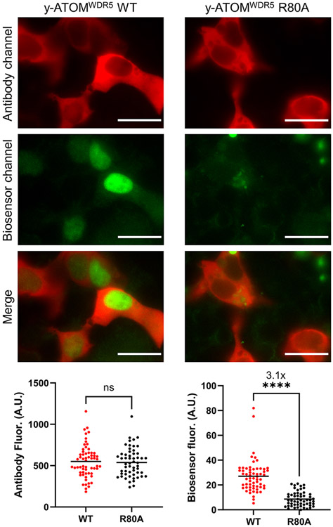
A grand challenge in biosensor design is to develop a single-molecule, fluorescent protein-based platform that can be easily adapted to recognize targets of choice. Here, we created a family of adaptable, turn-on maturation (ATOM) biosensors consisting of a monobody (circularly permuted at one of two positions) or a nanobody (circularly permuted at one of three positions) inserted into a fluorescent protein at one of three surface loops. Multiplexed imaging of live human cells coexpressing cyan, yellow and red ATOM sensors detected biosensor targets that were specifically localized to various subcellular compartments. Fluorescence activation involved ligand-dependent chromophore maturation with turn-on ratios of up to 62-fold in cells and 100-fold in vitro. Endoplasmic reticulum- and mitochondria-localized ATOM sensors detected ligands that were targeted to those organelles. The ATOM design was validated with three monobodies and one nanobody inserted into distinct fluorescent proteins, suggesting that customized ATOM sensors can be generated quickly.


Figures

Extended Data Fig. 1 |
Extended Data Fig. 1 |. Ligand-dependent ATOM turn-on in cells is not due to increased biosensor levels.
HEK 293T cells were co-transfected with one plasmid encoding y-ATOMWDR5, y-ATOMSH2, y-ATOMRAS, or y-ATOMmCh and a second plasmid expressing WDR5, SH2, hRAS, or mCh. After 48 h, cells were fixed, stained with an anti-GFP antibody conjugated with Alexa594, and imaged in the red channel (antibody; data depicted as gray points) and yellow channel (biosensor; data depicted in indicated colors). Each data point represents a cell (50–100 per experiment). Scale bars are 200 μm. The results are representative of three BR. Number of cells, means, standard deviations, and P values obtained by a two-tailed t-test for all valid comparisons are included in Extended Data Table 1.
Extended Data Fig. 2 |
Extended Data Fig. 2 |. r-ATOMWDR5, y-ATOMSH2, and c-ATOMRAS, co-expressed in the same cells, exhibit high turn-on values in the presence of their target ligands but not in the presence of noncognate ligands.
To match Fig. 4, the cyan channel (hRAS) is pseudocolored red, the yellow channel (SH2) is pseudocolored green, and the red channel (WDR5) is pseudocolored blue. HEK 293T cells were co-transfected with an equimolar mixture of three biosensor plasmids and a fourth plasmid encoding WDR5 (first row), SH2 (second row), and hRAS (third row). The fourth row shows the same experiment, but the fourth plasmid expressed all three ligands as described in the text. Scale bars are 200 μm. Turn-on ratios in each color channel (bottom plots) were calculated by dividing the average intensity of cells expressing the biosensor and its cognate ligand by the average of the intensities of cells expressing the same biosensor and the two negative control ligands. Each data point represents a cell (40–60 per experiment). P < 10−4 for all comparisons shown. The results are representative of three BR. Number of cells, means, standard deviations, and P values obtained by a two-tailed t-test for all valid comparisons are included in Extended Data Table 1.
Extended Data Fig. 3 |
Extended Data Fig. 3 |. y-ATOMWDR5 detects endogenous WDR5 and identifies nuclear localization in HEK 293T cells.
Staining with anti-GFP antibody (red) indicated that WT and R80A mutant y-ATOMWDR5 were both expressed in cytoplasm and nucleus, with highest levels in the cytoplasm. Imaging in the yellow channel (pseudocolored green) showed the WT biosensor was activated in the nucleus but in the OFF state in the cytoplasm. R80A y-ATOMWDR5 was dark in both cytoplasm and nucleus. Scale bars are 20 μm. Each data point represents one cell (61 cells per experiment). ****, P < 10−4; ns, not significant. Results are representative of 3 BR. Number of cells, means, standard deviations, and P values obtained by a two-tailed t-test for all valid comparisons are included in Extended Data Table 1.
Extended Data Fig. 4 |
Extended Data Fig. 4 |. Performance of NIR-FbGFP localized to cytoplasm, mitochondria, and ER of HEK 293T cells.
Cells were co-transfected with plasmids encoding NIR-FbGFP and Clover (tagged with indicated localization sequences) and imaged in the far-red channel to quantify compartment-specific turn-on. (b) The same cells were imaged in the green channel to determine relative Clover expression. Scale bars are 200 μm. Results are representative of 3 BR.
Fig. 1 |
Fig. 1 |. Biosensor design and screening y-ATOM sensors in HEK 293T cells.
a, X-ray structures of MBWDR5 (blue structure at left) and NBmCh (blue structure to the right) are shown bound to WDR5 and mCh (gray structures), respectively. MB and NB domains were circularly permuted at loops MB1–MB2 and NB1–NB4 (red), respectively (Protein Data Bank 6BYN and 8IM0). b, MB and NB permutants were inserted into YFP (pink) at one of the three loops depicted in blue (FP1–FP3). The YFP chromophore is shown in green spheres (Protein Data Bank 5WJ2). c, In the proposed turn-on mechanism, the MB or NB domain (blue) is partially unfolded in the absence of ligand (black), which disrupts the conformation of the FP domain (cylinder), leaving the chromophore in an unmatured state. Ligand binding induces the MB–NB domain to fold, restoring the native conformation of the FP and allowing the chromophore to mature. d,e, Fluorescence turn-on of MB-based y-ATOM sensors (d) and NB-based y-ATOM sensors (e) was determined in HEK 293T cells, with the best-performing combination of MB/NB and FP domains indicated by the colored boxes. Cognate and noncognate ligands for each sensor are designated by colored and gray symbols, respectively. Ab on the y axes denotes fluorescence intensity of the anti-GFP antibody used to measure total biosensor expression. Each data point represents a cell. ****P < 10−4, ***P < 10−3, **P < 10−2, *P < 0.05; NS, not significant. Number of cells, means, standard deviations (s.d.) and P values obtained by a two-tailed t-test for all valid comparisons are included in Extended Data Table 1. The results are representative of at least three BRs. See Supplementary Figs. 1-3 and 8 for cell images.
Fig. 2 |
Fig. 2 |. y-ATOM sensors show high selectivity and fluorescence turn-on in human cells.
Images on the diagonal represent cells transfected with plasmids expressing biosensor and cognate ligand. The off-diagonal images are cells transfected with plasmids expressing biosensor and decoy ligands. Higher contrast settings are shown in the insets to demonstrate that biosensors were expressed (but dim) in the off-diagonal cells. Fluorescence turn-on is quantified at the bottom. Each data point represents a cell. P < 10−4 for all comparisons shown. Scale bars, 200 μm. The results are representative of four BR. Number of cells, means, s.d. and P values obtained by a two-tailed t-test for all valid comparisons are included in Extended Data Table 1. A.U., arbitrary units.
Fig. 3 |
Fig. 3 |. Characterization of purified y-ATOM sensors at 37 °C.
Sensors and ligands are colored cyan (hRAS), orange (SH2), red (WDR5) and purple (mCh). a, Fluorescence turn-on resulted from ligand-induced chromophore maturation, as evidenced by the increase in absorbance at 514 nm (dashed lines) and fluorescence at 530 nm (solid lines). Samples contained 10 μM ligand. Data were recorded at time zero (black lines) and 12 h (colored lines). b, Fluorescence was activated in a ligand-specific manner with half-times of 1.7–7.4 h. Lines are best fits of the data to a single exponential function. Filled and empty circles represent cognate and decoy ligands, respectively. c, y-ATOM sensors bound their cognate targets with low nM to low μM Kd. Lines are best fits of the data to the one-site binding equation. Data are plotted as mean ± s.d. (n = 3 technical repeats, defined as data obtained from the same stock of purified protein; data are representative of two to three BRs). A.U., arbitrary units. Fitted values are reported in Table 1.
Fig. 4 |
Fig. 4 |. Multiplexed three-color imaging shows ATOM sensors detect subcellular localization of protein targets.
For visual clarity, the cyan channel (hRAS) is pseudocolored red, the yellow channel (SH2) is pseudocolored green and the red channel (WDR5) is pseudocolored blue. a, Cells cotransfected with a mixture of c-ATOMRAS, y-ATOMSH2 and r-ATOMWDR5 expressing plasmids, and a fourth plasmid encoding all three ligands showed fluorescence turn-on specific to the presence of ligands (top row and middle row). The higher contrast settings in the insets demonstrated that biosensors expressed but were dim. Focusing on a single cell (bottom row) revealed distinct biosensor fluorescence localized to the plasma membrane (c-ATOMRAS), cytoplasm (y-ATOMSH2) and (r-ATOMWDR5). Scale bars are 100 μm in top and middle rows, and 10 μm in the bottom row. b, Sensor turn-on was calculated by dividing the average intensity of the cells in the top row of a (red circles) by that of the cells in the middle row of a (gray circles). Each data point represents a cell (42 per experiment). P < 10−4 for all comparisons shown. The results are representative of four BRs. c, A line trace through the cell shown in the bottom row of a confirmed the expected locations of hRAS, SH2 and WDR5 in the plasma membrane (red trace), cytoplasm (green trace) and nucleus (blue trace), respectively. Number of cells, means, s.d. and P values obtained by a two-tailed t-test for all valid comparisons are included in Extended Data Table 1
Fig. 5 |
Fig. 5 |. y-ATOMSH2 can function as an ER- or mitochondria-targeted biosensor.
a, HeLa cells transfected with ER-localized biosensor (top row) or mito-localized biosensor (bottom row) were activated specifically by ER-localized SH2 and mito-localized SH2, respectively, and not by SH2 which targeted other compartments. Insets show higher contrast settings to demonstrate that biosensors were expressed in all transfection conditions. Scale bars, 100 μm. b, Confocal images confirmed that the biosensors were detecting their targets in the expected organelles. Scale bars, 10 μm. c, Quantification of the data in a found 11–13-fold selective turn-on for ER-targeted sensor and ligand, and 15–19-fold selective turn-on for mito-targeted sensor and ligand. Each data point represents a cell (52–63 per experiment). P < 10−4 for all comparisons shown. The results are representative of three BRs. Number of cells, means, s.d. and P values obtained by a two-tailed t-test for all valid comparisons are included in Extended Data Table 1.

Update of

References

    1. Nakai J, Ohkura M & Imoto K A high signal-to-noise Ca2+ probe composed of a single green fluorescent protein. Nat. Biotechnol 19, 137–141 (2001). - PubMed
    1. Baird GS, Zacharias DA & Tsien RY Circular permutation and receptor insertion within green fluorescent proteins. Proc. Natl Acad. Sci. USA 96, 11241–11246 (1999). - PMC - PubMed
    1. Nasu Y, Shen Y, Kramer L & Campbell RE Structure- and mechanism-guided design of single fluorescent protein-based biosensors. Nat. Chem. Biol 17, 509–518 (2021). - PubMed
    1. van der Linden FH et al. A turquoise fluorescence lifetime-based biosensor for quantitative imaging of intracellular calcium. Nat. Commun 12, 7159 (2021). - PMC - PubMed
    1. Inoue M. et al. Rational design of a high-affinity, fast, red calcium indicator R-CaMP2. Nat. Methods 12, 64–70 (2015). - PubMed

MeSH terms