. 2023 Nov 9;28(1):91.
doi: 10.1186/s11658-023-00495-0.

Irf7 regulates the expression of Srg3 and ferroptosis axis aggravated sepsis-induced acute lung injury

Affiliations

Irf7 regulates the expression of Srg3 and ferroptosis axis aggravated sepsis-induced acute lung injury

Xinyu Ling et al. Cell Mol Biol Lett. .

Abstract

Objective: To investigate the mechanism of action of Srg3 in acute lung injury caused by sepsis.

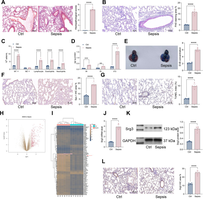
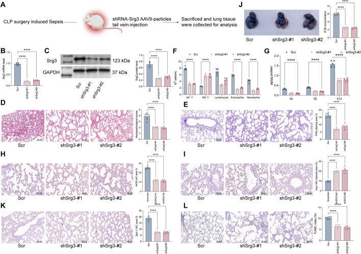
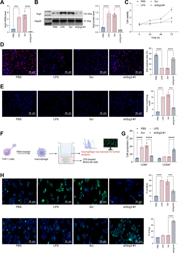
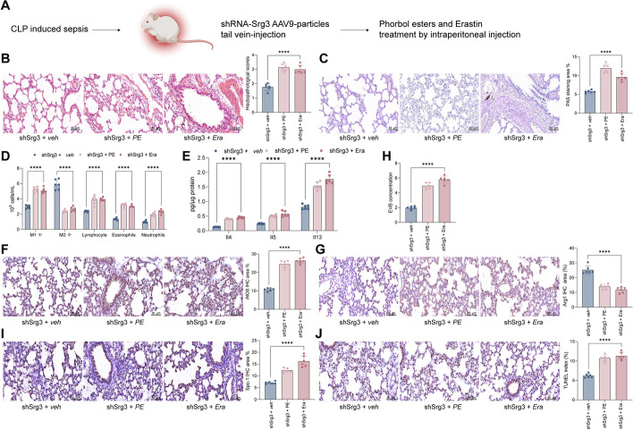
Methods: First, a sepsis-induced acute lung injury rat model was established using cecal ligation and puncture (CLP). RNA sequencing (RNA-seq) was used to screen for highly expressed genes in sepsis-induced acute lung injury (ALI), and the results showed that Srg3 was significantly upregulated. Then, SWI3-related gene 3 (Srg3) was knocked down using AAV9 vector in vivo, and changes in ALI symptoms in rats were analyzed. In vitro experiments were conducted by establishing a cell model using lipopolysaccharide (LPS)-induced BEAS-2B cells and coculturing them with phorbol 12-myristate 13-acetate (PMA)-treated THP-1 cells to analyze macrophage polarization. Next, downstream signaling pathways regulated by Srg3 and transcription factors involved in regulating Srg3 expression were analyzed using the KEGG database. Finally, gain-of-loss functional validation experiments were performed to analyze the role of downstream signaling pathways regulated by Srg3 and transcription factors involved in regulating Srg3 expression in sepsis-induced acute lung injury.

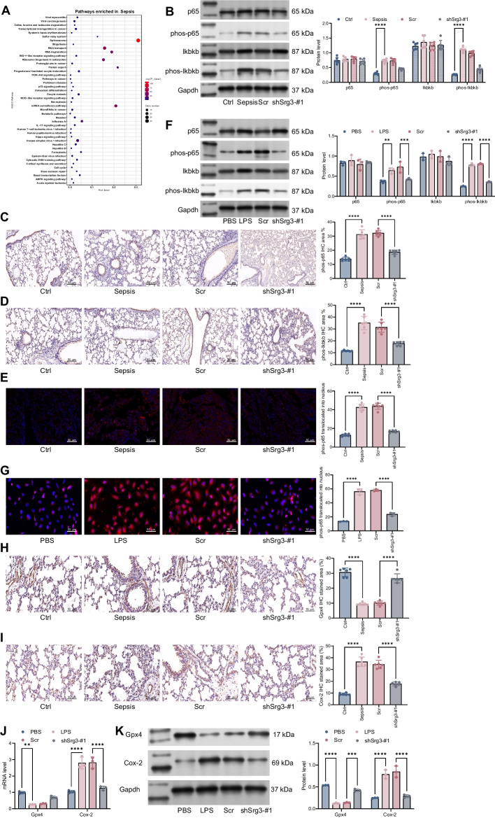
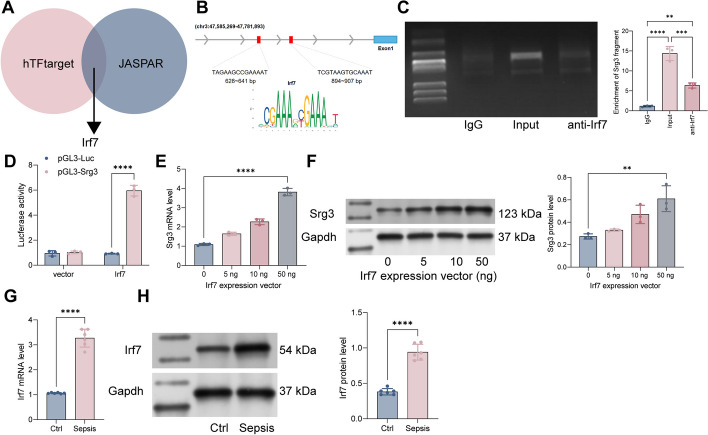
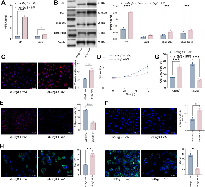
Results: Srg3 was significantly upregulated in sepsis-induced acute lung injury, and knocking down Srg3 significantly improved the symptoms of ALI in rats. Furthermore, in vitro experiments showed that knocking down Srg3 significantly weakened the inhibitory effect of LPS on the viability of BEAS-2B cells and promoted alternative activation phenotype (M2) macrophage polarization. Subsequent experiments showed that Srg3 can regulate the activation of the NF-κB signaling pathway and promote ferroptosis. Specific activation of the NF-κB signaling pathway or ferroptosis significantly weakened the effect of Srg3 knockdown. It was then found that Srg3 can be transcriptionally activated by interferon regulatory factor 7 (Irf7), and specific inhibition of Irf7 significantly improved the symptoms of ALI.

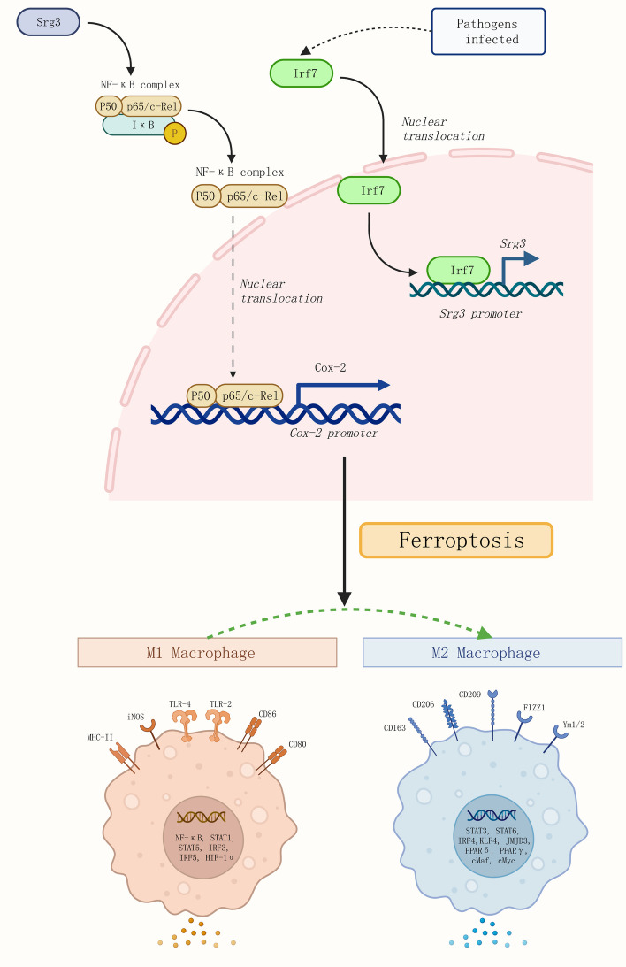
Conclusions: Irf7 transcriptionally activates the expression of Srg3, which can promote ferroptosis and activate classical activation phenotype (M1) macrophage polarization by regulating the NF-κB signaling pathway, thereby exacerbating the symptoms of septic lung injury.

Keywords: Ferroptosis; Irf7; NF-κB signaling pathway; Sepsis-induced acute lung injury; Srg3.


Conflict of interest statement

The authors declare that they have no known competing financial interests or personal relationships that could have appeared to influence the work reported in this paper.

Figures

Fig. 1
Fig. 1
Srg3 is highly expressed in sepsis-induced lung injury. A Histopathology of lung tissues from rats examined by H&E staining. B Proportions of glycogen in lung tissues of rats examined by PAS staining. C Numbers of various immune cells in bronchoalveolar lavage fluid (BALF) of rats analyzed by flow cytometry. D Levels of inflammatory factors in BALF of rats detected by ELISA. E Permeability of blood vessels in lung tissues of rats examined by EB staining. F Intensity of Spc-1 staining in lung tissues of rats examined by IHC staining. G Proportions of apoptotic cells in lung tissues of rats examined by TUNEL staining. H, I Differential gene expression analysis of lung tissues between control and sepsis-induced rats using RNA-seq. J, K mRNA and protein expression levels of Srg3 in lung tissues of rats detected by qPCR and WB, respectively. L Intensity of Srg3 staining in lung tissues of rats examined by IHC staining. Each group contained six rats, with each point representing one rat. Significant differences were detected using one-way ANOVA and Tukey’s multiple comparison test, **P < 0.01, ***P < 0.001, ***P < 0.0001
Fig. 2
Fig. 2
Knockdown of Srg3 suppresses lung injury symptoms in septic rats. A Schematic diagram of injecting the constructed AAV9–shRNA vector targeting Srg3 into rats via the tail vein. B, C qRT–PCR and WB detection of Srg3 mRNA and protein expression levels in rat lung tissue. D H&E staining to detect pathological changes in rat lung tissue. E PAS staining to observe the proportion of glycogen in rat lung tissue. F Flow cytometry analysis of the number of various immune cells in rat BALF. G ELISA detection of inflammatory factors in rat BALF. H, I IHC staining to observe the staining intensity of iNOS and Arg1 in rat lung tissue. J EB staining to observe vascular permeability in rat lung tissue. K IHC staining to observe the staining intensity of Spc-1 in rat lung tissue. L TUNEL staining to detect the proportion of apoptotic cells in lung tissue. Each group contained six rats, with each point representing one rat. Significant differences were detected using one-way ANOVA and Tukey’s multiple comparison test, **P < 0.01, ***P < 0.001, ***P < 0.0001
Fig. 3
Fig. 3
Srg3 knockdown inhibited LPS-induced BEAS-2B cell death. A, B qRT–PCR and western blot analysis were performed to detect the mRNA and protein expression levels of Srg3 in BEAS-2B cells. C, D CCK-8 and EdU staining were used to analyze the growth activity of BEAS-2B cells. E DAPI staining was used to analyze the nuclear morphology of BEAS-2B cells. F BEAS-2B and PMA-treated THP-1 cells were cocultured in a Transwell system, and THP-1 cells were collected for subsequent experiments. G Flow cytometry was used to detect the proportion of CD86 and CD206 in THP-1 cells after coculture. H Immunofluorescence staining was used to detect the fluorescence intensity of iNOS and Arg1 in THP-1 cells after coculture. Each experiment was repeated three times, and the data were presented in the form of mean plus or minus standard deviation. One-way ANOVA or two-way ANOVA was used for significance analysis between the data. After ANOVA, Tukey’s multiple comparison test was used for post hoc test. **P < 0.01, ***P < 0.001, ***P < 0.0001
Fig. 4
Fig. 4
Srg3-regulated NF-κB signaling pathway and ferroptosis. A We performed KEGG enrichment analysis to identify signaling pathways associated with Srg3-regulated genes. B Protein expression levels of p65, phos-p65, Ikbkb, and phos-Ikbkb were measured in rat lung tissue. C, D Immunohistochemistry was used to determine the staining intensity of phos-p65 and phos-Ikbkb in rat lung tissue. E Immunofluorescence was used to determine the nuclear localization of phos-p65 in rat lung tissue. F Protein expression levels of p65, phos-p65, Ikbkb, and phos-Ikbkb were measured in BEAS-2B cells by WB. G Immunofluorescence was used to determine the nuclear localization of phos-p65 in BEAS-2B cells. H, I Immunohistochemistry was used to determine the staining intensity of Gpx4 and Cox-2 in rat lung tissue. J, K qRT–PCR and WB were used to measure mRNA and protein expression levels of Gpx4 and Cox-2 in BEAS-2B cells. Each experiment was repeated three times, and the data were presented in the form of mean plus or minus standard deviation. Each group contained six rats, with each point representing one rat. One-way ANOVA or two-way ANOVA was used for significance analysis between the data. After ANOVA, Tukey’s multiple comparison test was used for post hoc test. **P < 0.01, ***P < 0.001, ***P < 0.0001
Fig. 5
Fig. 5
Phorbol esters and erastin treatment weakened shSrg3 function on septic lung injury. A Schematic diagram of injecting the constructed AAV9–shRNA vector targeting Srg3 into rats via the tail vein, then phorbol esters and erastin treatment. B H&E staining to detect pathological changes in rat lung tissue. C PAS staining to observe the proportion of glycogen in rat lung tissue. D Flow cytometry analysis of the number of various immune cells in rat BALF. E ELISA detection of inflammatory factors in rat BALF. F, G IHC staining to observe the staining intensity of iNOS and Arg1 in rat lung tissue. H EB staining to observe vascular permeability in rat lung tissue. I IHC staining to observe the staining intensity of Spc-1 in rat lung tissue. J TUNEL staining to detect the proportion of apoptotic cells in lung tissue. Each group contained six rats, with each point representing one rat. Significant differences were detected using one-way ANOVA and Tukey’s multiple comparison test, **P < 0.01, ***P < 0.001, ***P < 0.0001
Fig. 6
Fig. 6
Irf7 transcriptionally regulates Srg3 and activates its transcriptional activity in sepsis-induced acute lung injury. A Prediction of transcription factors that can bind to the Srg3 promoter using hTFtarget and JASPAR websites. B Identification of the conserved binding sequence of Irf7. C Verification of the binding relationship between Irf7 and the Srg3 promoter using ChIP–qPCR experiments. D Construction of a luciferase reporter vector containing the Srg3 promoter sequence and transfection into HEK293T cells along with an overexpression vector of Irf7 to measure luciferase activity. E, F Measurement of Srg3 mRNA and protein expression levels in BEAS-2B cells after overexpression of Irf7. G, H Measurement of Irf7 mRNA and protein expression levels in rat lung tissue using qRT–PCR and WB, respectively. Each experiment was repeated three times, and the data were presented in the form of mean plus or minus standard deviation. Each group contained six rats, with each point representing one rat. One-way ANOVA or two-way ANOVA was used for significance analysis between the data. After ANOVA, Tukey’s multiple comparison test was used for post hoc test. **P < 0.01, ***P < 0.001, ***P < 0.0001
Fig. 7
Fig. 7
The overexpression of Irf7 weakens the beneficial effect of Srg3 knockdown on improving sepsis-induced acute lung injury. A qRT–PCR analysis was performed to detect the mRNA expression levels of Srg3 and Irf7 in BEAS-2B cells. B WB analysis was performed to detect the protein level of Srg3, Irf7, phos-p65, and phos-Ikbkb. C Immunofluorescence was used to determine the nuclear localization of phos-p65 in BEAS-2B cells. D, E CCK-8 and EdU staining were used to analyze the growth activity of BEAS-2B cells. F DAPI staining was used to analyze the nuclear morphology of BEAS-2B cells. G Flow cytometry was used to detect the proportion of CD86 and CD206 in THP-1 cells after coculture. H Immunofluorescence staining was used to detect the fluorescence intensity of iNOS and Arg1 in THP-1 cells after coculture. Each experiment was repeated three times, and the data were presented in the form of mean plus or minus standard deviation. One-way ANOVA or two-way ANOVA was used for significance analysis between the data. After ANOVA, Tukey’s multiple comparison test was used for post hoc test. **P < 0.01, ***P < 0.001, ***P < 0.0001
Fig. 8
Fig. 8
Mechanism illustration. Irf7 can activate the expression of Srg3 by transcription, thereby increasing the activity of the NF-κB signaling pathway, activating ferroptosis in lung epithelial cells, promoting M1 macrophage polarization, and exacerbating the symptoms of acute lung injury in sepsis

References

    1. Fleischmann-Struzek C, Mellhammar L, Rose N, Cassini A, Rudd KE, Schlattmann P, et al. Incidence and mortality of hospital- and ICU-treated sepsis: Results from an updated and expanded systematic review and meta-analysis. Intensive Care Med. 2020;46(8):1552–1562. doi: 10.1007/s00134-020-06151-x. - DOI - PMC - PubMed
    1. Gotts JE, Matthay MA. Sepsis: Pathophysiology and clinical management. BMJ (Clinical research ed) 2016;353:i1585. - PubMed
    1. De Freitas CN, Gaudet A, Portier L, Tsicopoulos A, Mathieu D, Lassalle P. Endocan, sepsis, pneumonia, and acute respiratory distress syndrome. Critical Care (London, England) 2018;22(1):280. doi: 10.1186/s13054-018-2222-7. - DOI - PMC - PubMed
    1. Zoulikha M, Xiao Q, Boafo GF, Sallam MA, Chen Z, He W. Pulmonary delivery of siRNA against acute lung injury/acute respiratory distress syndrome. Acta Pharm Sin B. 2022;12(2):600–620. doi: 10.1016/j.apsb.2021.08.009. - DOI - PMC - PubMed
    1. Wang W. The SWI/SNF family of ATP-dependent chromatin remodelers: Similar mechanisms for diverse functions. Curr Top Microbiol Immunol. 2003;274:143–169. - PubMed