

Deep histopathology genotype-phenotype analysis of focal cortical dysplasia type II differentiates between the GATOR1-altered autophagocytic subtype IIa and MTOR-altered migration deficient subtype IIb
- PMID: 37946310
- PMCID: PMC10633947
- DOI: 10.1186/s40478-023-01675-x
Deep histopathology genotype-phenotype analysis of focal cortical dysplasia type II differentiates between the GATOR1-altered autophagocytic subtype IIa and MTOR-altered migration deficient subtype IIb
Abstract
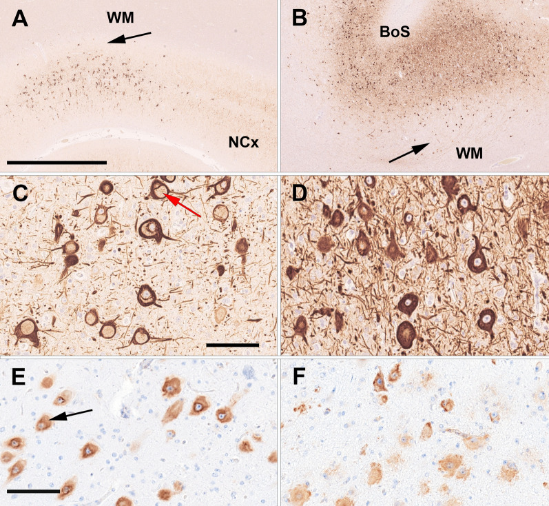
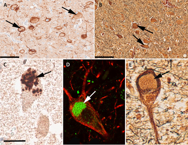
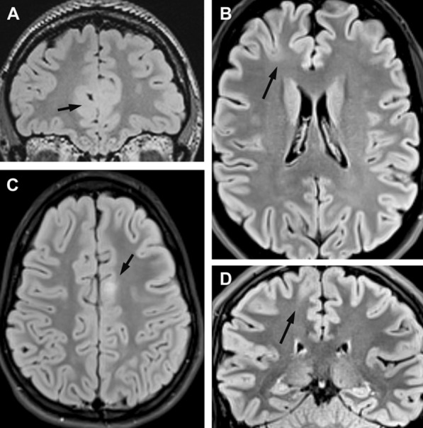
Focal cortical dysplasia type II (FCDII) is the most common cause of drug-resistant focal epilepsy in children. Herein, we performed a deep histopathology-based genotype-phenotype analysis to further elucidate the clinico-pathological and genetic presentation of FCDIIa compared to FCDIIb. Seventeen individuals with histopathologically confirmed diagnosis of FCD ILAE Type II and a pathogenic variant detected in brain derived DNA whole-exome sequencing or mTOR gene panel sequencing were included in this study. Clinical data were directly available from each contributing centre. Histopathological analyses were performed from formalin-fixed, paraffin-embedded tissue samples using haematoxylin-eosin and immunohistochemistry for NF-SMI32, NeuN, pS6, p62, and vimentin. Ten individuals carried loss-of-function variants in the GATOR1 complex encoding genes DEPDC5 (n = 7) and NPRL3 (n = 3), or gain-of-function variants in MTOR (n = 7). Whereas individuals with GATOR1 variants only presented with FCDIIa, i.e., lack of balloon cells, individuals with MTOR variants presented with both histopathology subtypes, FCDIIa and FCDIIb. Interestingly, 50% of GATOR1-positive cases showed a unique and predominantly vacuolizing phenotype with p62 immunofluorescent aggregates in autophagosomes. All cases with GATOR1 alterations had neurosurgery in the frontal lobe and the majority was confined to the cortical ribbon not affecting the white matter. This pattern was reflected by subtle or negative MRI findings in seven individuals with GATOR1 variants. Nonetheless, all individuals were seizure-free after surgery except four individuals carrying a DEPDC5 variant. We describe a yet underrecognized genotype-phenotype correlation of GATOR1 variants with FCDIIa in the frontal lobe. These lesions were histopathologically characterized by abnormally vacuolizing cells suggestive of an autophagy-altered phenotype. In contrast, individuals with FCDIIb and brain somatic MTOR variants showed larger lesions on MRI including the white matter, suggesting compromised neural cell migration.
© 2023. The Author(s).
Conflict of interest statement
The authors declare that they have no competing interest.
Figures
References
-
- Baldassari S, Ribierre T, Marsan E, Adle-Biassette H, Ferrand-Sorbets S, Bulteau C, Dorison N, Fohlen M, Polivka M, Weckhuysen S, et al. Dissecting the genetic basis of focal cortical dysplasia: a large cohort study. Acta Neuropathol. 2019;138:885–900. doi: 10.1007/s00401-019-02061-5. - DOI - PMC - PubMed
-
- Bar-Peled L, Chantranupong L, Cherniack AD, Chen WW, Ottina KA, Grabiner BC, Spear ED, Carter SL, Meyerson M, Sabatini DM. A tumor suppressor complex with GAP activity for the Rag GTPases that signal amino acid sufficiency to mTORC1. Science. 2013;340:1100–1106. doi: 10.1126/science.1232044. - DOI - PMC - PubMed
-
- Bernasconi A, Cendes F, Theodore WH, Gill RS, Koepp MJ, Hogan RE, Jackson GD, Federico P, Labate A, Vaudano AE, et al. Recommendations for the use of structural magnetic resonance imaging in the care of patients with epilepsy: a consensus report from the International League Against Epilepsy Neuroimaging Task Force. Epilepsia. 2019;60:1054–1068. doi: 10.1111/epi.15612. - DOI - PubMed
Publication types
MeSH terms
Substances
Supplementary concepts
Grants and funding
LinkOut - more resources
Full Text Sources
Medical
Miscellaneous
