. 2023 Oct;43(6):1126-1139.
doi: 10.19852/j.cnki.jtcm.20230517.003.

Yangqing Chenfei formula alleviates crystalline silica induced pulmonary inflammation and fibrosis by suppressing macrophage polarization

Affiliations

Yangqing Chenfei formula alleviates crystalline silica induced pulmonary inflammation and fibrosis by suppressing macrophage polarization

Tian Xinrong et al. J Tradit Chin Med. 2023 Oct.

Abstract

Objective: To explore the underlying mechanisms of the effects of Yangqing Chenfei formula (, YCF) on inflammation and fibrosis in silicosis via inhibition of macrophage polarization.

Methods: A silicotic rat model was established via a single intratracheal instillation of silica particles on the first day of week 0. Subsequently, YCF was administered intragastrically to silicotic rats during weeks 0-2 and 5-8 twice daily. The mouse-derived alveolar macrophage cell line was used to investigate the mechanisms of YCF in M1/M2 polarization.

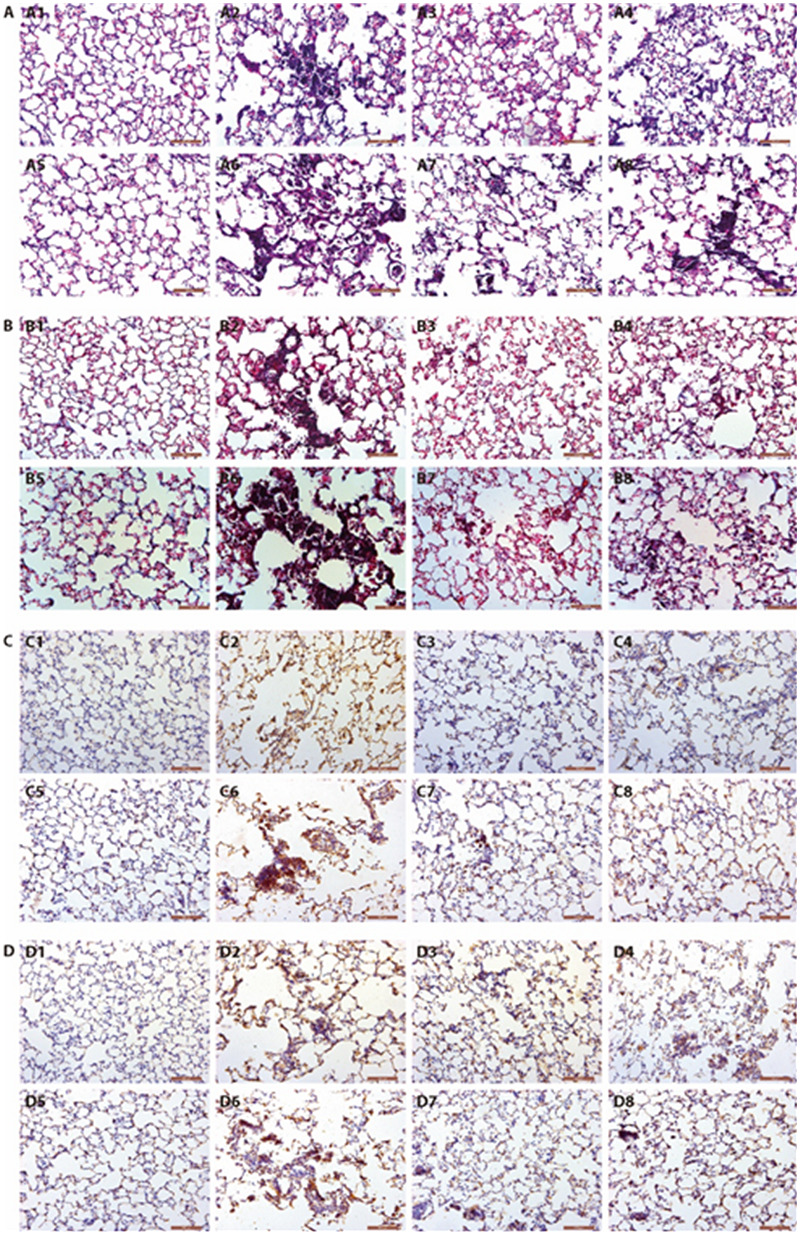
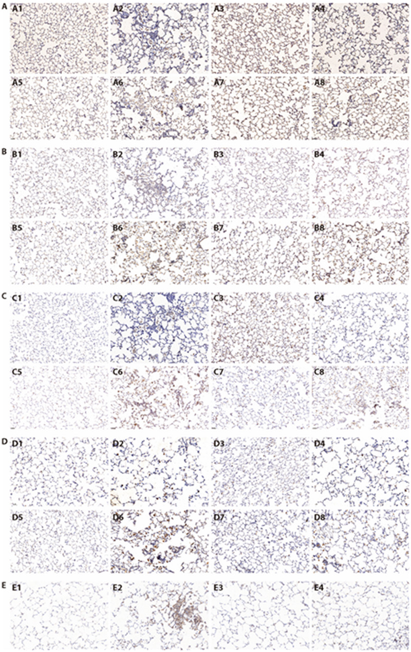
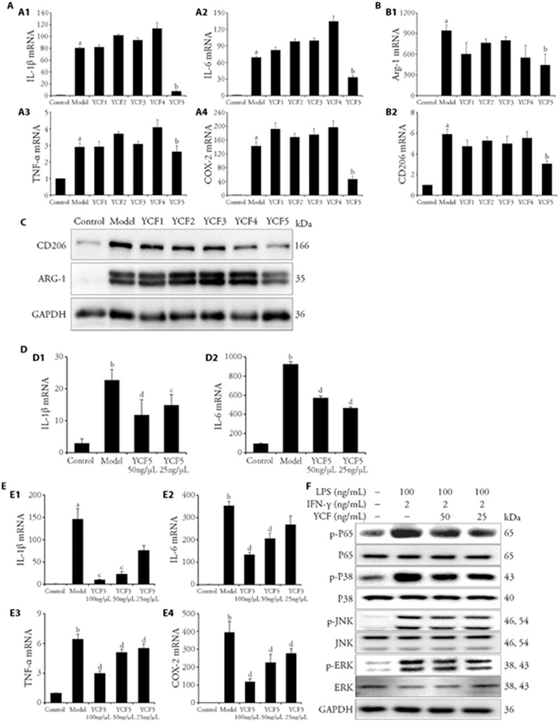
Results: YCF treatment effectively inhibited lung pathological changes, including inflammatory cell infiltration and tissue damage, and increased the forced expiratory volume in the first 0.3 s, functional residual capacity, and maximal mid-expiratory flow in weeks 2 and 8. Furthermore, the treatment improved lung functions by upregulating tidal volume, pause increase, and expiratory flow at 50% tidal volume from weeks 5 to 8. Moreover, YCF could significantly suppressed the progression of inflammation and fibrosis, by reducing the levels of inflammatory cytokines, as well as collagen- I and III. YCF treatment also decreased the numbers of macrophages and M1/M2 macrophages and the level of transforming growth factor-β (TGF-β). Additionally, YCF5, the effective substance in YCF, decreased lipopolysaccharide and interferon-γ-induced M1 macrophage polarization in a concentration-dependent manner. The mechanism of anti-M1 polarization might be related to a decrease in extracellular signal-regulated kinase, c-JUN N-terminal kinase, P38, and P65 phosphorylation. Furthermore, YCF5 inhibited interleukin-4-induced M2 macrophages by decreasing the protein and mRNA expressions of arginase-1 and CD206 as well as the levels of profibrotic factors, such as TGF-β and connective tissue growth factor. The mechanisms underlying the anti-M2 polarization of YCF5 were primarily associated with the inhibition of the nuclear translocation of phosphorylated signal transducer and activator of transcription 6 (p-STAT6).

Conclusion: YCF significantly inhibits inflammation and fibrosis in silicotic rats probably via the suppression of M1/M2 macrophage polarization mediated by the inhibition of mitogen-activated protein kinase and nuclear factor kappa B signaling pathways and Janus kinase/STAT6 pathways.

Keywords: Yin deficiency; cohort studies; fur; heart failure; risk factors.


Figures

Figure 1
Figure 1. YCF enhanced pulmonary pathological changes and alleviated collagen deposition on silicosis
A: HE staining of lung tissue on silicosis rats (× 200); A1-A4: the inflammatory cell infiltration of Normal, Model, YCF and TET in week 2; A5-A6: the inflammatory cell infiltration of Normal, Model, YCF and TET in week 8. B: MASSON staining of lung tissue on silicosis rats (× 200); B1-B4: the pulmonary pathological changes of Normal, Model, YCF and TET in week 2; B5-B6: the pulmonary pathological changes of Normal, Model, YCF and TET in week 8. C: expression level of COL-1 (× 200); C1-C4: the collagen type Ⅰ (Col Ⅰ) expression of Normal, Model, YCF and TET in week 2; C5-C6: the collagen type Ⅰ (Col Ⅰ) expression of Normal, Model, YCF and TET in week 8. D: expression level of COL-3 (× 200); D1-D4: the collagen type Ⅲ (Col Ⅲ) expression of Normal, Model, YCF and TET in week 2; D5-D6: the collagen type Ⅲ (Col Ⅲ) expression of Normal, Model, YCF and TET in week 8. Normal: healthy control rats; Model: silicosis rats; YCF: YCF treatment rats were intragastrically administered with 3.3663 g·kg-1·d-1 or 0.84 mL/100 g, once daily, from weeks 0-2 and 5-8; TET: tetrandrine treatment rats were intragastrically administered with 27 mg·kg-1·d-1 TET, from weeks 0-2 and 5-8 (n = 6). YCF: Yangqing Chenfei formula; TET: tetrandrine; HE: hematoxylin-eosin; COL-I: collage-1.
Figure 2
Figure 2. YCF inhibited the macrophages polarization on silicosis rats
A: expression level of CD68 (× 200) in silicosis rats on week 2 and 8; A1-A4: the CD68 expression of Normal, Model, YCF and TET in week 2; A5-A6: the CD68 expression of Normal, Model, YCF and TET in week 8. B: expression level of iNOS (× 200) in silicosis rats on week 2 and 8; B1-B4: the iNOS expression of Normal, Model, YCF and TET in week 2; B5-B6: the iNOS expression of Normal, Model, YCF and TET in week 8. C: expression level of CD206 (× 200) in silicosis rats on week 2 and 8; C1-C4: the CD206 expression of Normal, Model, YCF and TET in week 2; C5-C6: the CD206 expression of Normal, Model, YCF and TET in week 8. D: expression level of Arg-1 (× 200) in silicosis rats on week 2 and 8; D1-D4: the Arg-1 expression of Normal, Model, YCF and TET in week 2; D5-D6: the Arg-1 expression of Normal, Model, YCF and TET in week 8. E: expression level of TGF-β (× 200) in silicosis rats on week 8. E1-E4: the TGF-β expression of Normal, Model, YCF and TET in week 8. Normal: healthy control rats; Model: silicosis rats; YCF: YCF treatment rats were intragastrically administered with 3.3663 g·kg-1·d-1 or 0.84 mL/100 g, once daily, from weeks 5-8; TET: tetrandrine treatment rats were intragastrically administered with 27 mg·kg-1·d-1 TET, from weeks 5-8. n = 6. YCF: Yangqing Chenfei formula; TET: tetrandrine; Arg-1: Arginase-1; iNOS: inducible nitric oxide synthase; TGF-β: transforming growth factor-beta.
Figure 3
Figure 3. Effects of the substances of YCF and the effective segment of YCF (YCF5) suppressed macrophage polarization in MH-S
MH-S were treated with different segments of YCF (100 μg/mL YCF1-5) for 3-6 h, and exposed to IFN-γ (2 ng/mL) + LPS (100 ng/mL) or IL-4 (20 ng/mL) for 12 h. A: RT-qPCR was applied to determine the mRNA expressions of IL-1β, IL-6, TNF-α and COX-2 in M1 macrophages. MH-S were treated with different segments of YCF (100 μg/mL YCF1-5) for 3-6 h, and exposed to IFN-γ (2 ng/mL) + LPS (100 ng/mL) for 12 h. B: RT-qPCR was applied to determine the mRNA expressions of Arg-1 and CD206 in M2 macrophages; C: Protein expressions of CD206 and Arg-1 in M2 macrophages were detected by Western blotting; MH-S were treated with different segments of YCF (100 μg/mL YCF1-5) for 3-6 h, and exposed to IL-4 (20 ng/mL) for 12 h. GAPDH was used as the control. All data were showed as the mean ± standard deviation (n = 3). aP < 0.01, vs control macrophages; bP < 0.01, vs model macrophages. D: protein levels of IL-1β and IL-6 in the culture medium were determined by ELISA; E: mRNA expressions of IL-1β, IL-6, TNF-α, and COX-2 in M1 macrophages. F: Western blotting assay was used to determine the protein expressions of p-JNK, JNK, p-ERK, ERK, p-P38, P38, p-P65, and P65 in M1 macrophages; MH-S were treated with different concentrations of YCF5 (100, 50, 25 μg/mL) for 3-6 h, and exposed to IFN-γ (2 ng/mL) + LPS (100 ng/mL) for 12 h. GAPDH was used as the control. Control: normal macrophages; Model: IFN-γ + LPS or IL-4 induced macrophages; YCF5 100, 50, 25 μg/mL: Macrophages treated with different concentrations of YCF5 (100, 50, 25 μg/mL). YCF: Yangqing Chenfei formula; TET: tetrandrine; MH-S: murine alveolar macrophage cell line; LPS: lipopolysaccharides; IFN-γ: interferon-gamma; IL-4: interleukin-4; RT-qPCR: real time quantitative polymerase chain reaction; GAPDH: glyceraldehyde phosphate dehydrogenase; ELISA: enzyme linked immunosorbent assay; p-JNK: phosphorylated C-Jun kinase enzyme; JNK: C-Jun kinase enzyme; p-ERK: phosphorylated extracellular signal regulated kinases; ERK: extracellular signal regulated kinases; IL-1β: interleukin-1 beta; IL-6: interleukin-6; TNF-α: tumor necrosis factor-alpha; COX-2: cyclooxygenase-2. Arg-1: arginase-1; CD206: macrophage mannose receptor 1. All data were showed as the mean ± standard deviation (n = 3). aP < 0.05, bP < 0.01, vs Control macrophages; cP < 0.05, dP < 0.01, vs model macrophages.
Figure 4
Figure 4. YCF attenuated IL-4-induced macrophage polarization in MH-S
The MH-S cells were treated with different concentrations of YCF5 (50, 25 μg/mL) for 3-6 h, and exposed to IL-4 (20 ng/mL) for 12 h. A: immunofluorescence analysis of Arg-1 expression in M2 macrophages. A1: DAPI expression of Control group; A2: Arg-1 expression of Control group; A3: merged expression of Control group; A4: DAPI expression of Model group; A5: Arg-1 expression of Model group; A6: merged expression of Model group; A7: DAPI expression of 50 μg/mL YCF5 group; A8: Arg-1 expression of 50 μg/mL YCF5 group; A9: merged expression of 50 μg/mL YCF5 group. B: immunofluorescence analysis of CD206 expression in M2 macrophages. B1: DAPI expression of Control group; B2: CD206 expression of Control group; B3: merged expression of Control group; B4: DAPI expression of Model group; B5: CD206 expression of Model group; B6: merged expression of Model group; B7: DAPI expression of 50 μg/mL YCF5 group; B8: CD206 expression of 50 μg/mL YCF5 group; B9: Merged expression of 50 μg/mL YCF5 group. C: immunofluorescence analysis of p-STAT6 expression in M2 macrophages. C1: DAPI expression of Control group; C2: p-STAT6 expression of Control group; C3: merged expression of Control group; C4: DAPI expression of Model group; C5: p-STAT6 expression of Model group; C6: merged expression of Model group; C7: DAPI expression of 50 μg/mL YCF5 group; C8: p-STAT6 expression of 50 μg/mL YCF5 group; C9: merged expression of 50 μg/mL YCF5 group. D: immunofluorescence analysis of TGF-β expression in M2 macrophages. D1: DAPI expression of Control group; D2: TGF-β expression of Control group; D3: merged expression of Control group; D4: DAPI expression of Model group; D5: TGF-β expression of Model group; D6: Merged expression of Model group; D7: DAPI expression of 50 μg/mL YCF5 group; D8: TGF-β expression of 50 μg/mL YCF5 group; D9: merged expression of 50 μg/mL YCF5 group. E: immunofluorescence analysis of CTGF expression in M2 macrophages. E1: DAPI expression of Control group; E2: CTGF expression of Control group; E3: merged expression of Control group; E4: DAPI expression of Model group; E5: CTGF expression of Model group; E6: merged expression of Model group; E7: DAPI expression of 50 μg/mL YCF5 group; E8: CTGF expression of 50 μg/mL YCF5 group; E9: merged expression of 50 μg/mL YCF5 group. The images were taken at × 400 magnification. F: Western blot assay of Arg-1, CD206 and p-STAT6 protein expressions in M2 macrophages. GAPDH was used as control. Control: normal macrophages; Model: IL-4 induced macrophages; YCF5 50, 25 μg/mL: M2 macrophages treated with different concentrations of YCF5 (50, 25 μg/mL). Arg-1: Arginase-1; CD206: Macrophage mannose receptor 1; TGF-β: transforming growth factor-β; CTGF: connective tissue growth factor. YCF: Yangqing Chenfei formula; MH-S: murine alveolar macrophage cell line; IL-4: interleukin-4; RT-qPCR: real time quantitative polymerase chain reaction; DAPI: 4',6-diamidino-2-phenylindole; p-STAT6: phosphorylated signal transducers and activators of transcription 6; GAPDH: glyceraldehyde phosphate dehydrogenase.

Similar articles

References

    1. Fernández Álvarez R, Martínez González C, Quero Martínez A, Blanco Pérez JJ, Carazo Fernández L, Prieto Fernández A. . Guidelines for the diagnosis and monitoring of silicosis. Arch Bronconeumol 2015; 51: 86-93. - PubMed
    1. Rimal B, Greenberg AK, Rom WN. . Basic pathogenetic mechanisms in silicosis: current understanding. Curr Opin Pulm Med 2005; 11: 169-73. - PubMed
    1. Leung CC, Yu ITS, Chen W. . Silicosis. Lancet Lond Engl 2012; 379: 2008-18. - PubMed
    1. Reynolds K, Jerome J. . Silicosis. Workplace Health Saf 2021; 69: 51. - PubMed
    1. Pollard KM. . Silica, silicosis, and autoimmunity. Front Immunol 2016; 7: 97. - PMC - PubMed

Publication types

Substances