. 2024 Feb;26(2):101023.
doi: 10.1016/j.gim.2023.101023. Epub 2023 Nov 7.

Biallelic CRELD1 variants cause a multisystem syndrome, including neurodevelopmental phenotypes, cardiac dysrhythmias, and frequent infections

Collaborators, Affiliations

Biallelic CRELD1 variants cause a multisystem syndrome, including neurodevelopmental phenotypes, cardiac dysrhythmias, and frequent infections

Lauren Jeffries et al. Genet Med. 2024 Feb.

Abstract

Purpose: We sought to delineate a multisystem disorder caused by recessive cysteine-rich with epidermal growth factor-like domains 1 (CRELD1) gene variants.

Methods: The impact of CRELD1 variants was characterized through an international collaboration utilizing next-generation DNA sequencing, gene knockdown, and protein overexpression in Xenopus tropicalis, and in vitro analysis of patient immune cells.

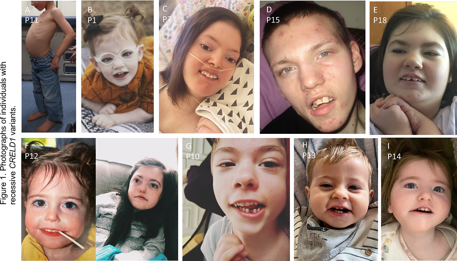
Results: Biallelic variants in CRELD1 were found in 18 participants from 14 families. Affected individuals displayed an array of phenotypes involving developmental delay, early-onset epilepsy, and hypotonia, with about half demonstrating cardiac arrhythmias and some experiencing recurrent infections. Most harbored a frameshift in trans with a missense allele, with 1 recurrent variant, p.(Cys192Tyr), identified in 10 families. X tropicalis tadpoles with creld1 knockdown displayed developmental defects along with increased susceptibility to induced seizures compared with controls. Additionally, human CRELD1 harboring missense variants from affected individuals had reduced protein function, indicated by a diminished ability to induce craniofacial defects when overexpressed in X tropicalis. Finally, baseline analyses of peripheral blood mononuclear cells showed similar proportions of immune cell subtypes in patients compared with healthy donors.

Conclusion: This patient cohort, combined with experimental data, provide evidence of a multisystem clinical syndrome mediated by recessive variants in CRELD1.

Keywords: CRELD1; Cardiac arrythmia; Developmental delay; Epilepsy; Hypotonia.


Conflict of interest statement

Conflict of Interest Two authors report part ownership of startup companies unrelated to this work: Qiyas Higher Health (Saquib A. Lakhani) and Victory Genomics (Saquib A. Lakhani and Mustafa K. Khokha). Kirsty McWalter is an employee of GeneDx. Kimberly Nugent is currently an employee of Cooper Surgical. Bryan Mak is currently an employee of Genome Medical. All other authors declare no conflicts of interest.

Figures

Figure 1.
Figure 1.. Photographs of individuals with recessive CRELD1 variants.
A. Lateral profile of P11 showing severe lordosis. For B to I, frequently seen features include open mouth, long palpebral fissures, tented vermillion border of upper lip, downturned corners of mouth, myopathic facies, narrow forehead with bitemporal narrowing, and tall or pointed chin. Figure F shows P12 at age 2 years (left) and 10 years (right).
Figure 2.
Figure 2.. Muscle studies from patients.
A. Axial T1 MRI of mid-thigh (top panel) and lower leg (bottom panel) from P11 showing proximal more than distal muscle atrophy and fatty replacement. Mid-thigh image shows rectus femoris atrophy (black arrow) and streaky fatty infiltrate in the vastus lateralis and deep vastus medialis (red arrow). There is extensive fatty replacement of the adductor magnus and adductor longus (blue arrow), whereas other muscles are relatively spared. In the lower leg there is diffuse streaky fatty replacement in the soleus (yellow arrow) with sparing of surrounding muscles. B. Muscle ultrasound images from P12, demonstrating relative sparing of the triceps (top panel) compared to the gastrocnemius (bottom panel), which shows increased echogenicity, indicative of fatty infiltration. C. Muscle analysis performed on autopsy sample from P12. Hematoxylin and eosin stain (top panel) demonstrating variability in fiber size. ATPase staining (middle panels) showing predominance of Type 1 muscle fibers, which stain light at pH 9.4 (left) and dark at pH 4.1 (right). Electron microscopy (lower panel) showing enlarged, pleiomorphic mitochondria with abnormal cristae, paracrystalline-like inclusions, and osmophilic bodies.
Figure 3.
Figure 3.. Family Pedigrees.
Overview of pedigrees presented in this cohort showing heterozygous parents and affected individuals. The ten distinct CRELD1 variants are color-coded as indicated in the legend.
Figure 4.
Figure 4.. Gene variants in CRELD1.
A. Architecture of CRELD1 gene, based on the predominantly-expressed isoform 2 (NP_056328.3). Amino acid lengths of the 10 exons are shown at top. Locations of functional domains are also shown (colored squares; see text for descriptions of domains). Location of frameshift (orange text) and missense (blue text) variants from patients are indicated. B. Multiple species alignment showing reference sequence surrounding CRELD1 amino acid residues altered in missense variants found in this cohort (blue arrows). Note complete conservation across examined species at all residues that are altered by patient missense variants.
Figure 5.
Figure 5.. Developmental defects and increased susceptibility to induced seizures in X. tropicalis tadpoles with creld1-knockdown.
A. X. tropicalis embryos were either uninjected controls (UiC) or injected with either morpholino targeting creld1 for knockdown (CRELD1-MO) or one of two sgRNAs plus CRISPR for direct creld1 gene knockout (CRELD1-CRISPR with sgRNA-1 or sgRNA-2). Tadpoles were then observed at set stages for phenotypes that would prevent scoring for seizures: at stage 22 for gastrulation defects or at stage 42 for edema/late lethality or tail defects. Results showed a high proportion of CRELD1-CRISPR embryos compared to UiC or creld1 MO with developmental defects preventing scoring for seizures. Data is shown from a representative experiment with n=indicating the number of embryos scored. B. X. tropicalis CRELD1-CRISPR embryos and UiC embryos observed for either spontaneous seizures or pilocarpine-induced seizures over time. All embryos showed minimal spontaneous seizure behaviors (<5%). By 30 minutes, approximately 70% of CRELD1-CRISPR embryos showed seizure behaviors in the presence of pilocarpine compared to approximately 40% of control embryos. Symbols indicate mean and error bars indicate standard deviation. Figure represents compilation of three biological replicates, each with >70 tadpoles per experimental condition.
Figure 6.
Figure 6.. Diminished ability of missense variants of human CRELD1 to induce craniofacial defects in X. tropicalis tadpoles.
A. As an in vivo assay of CRELD1 protein function, X. tropicalis embryos were either uninjected controls (top panel) or injected with reference sequence human mRNA for CRELD1 (bottom panel). Tadpoles injected with reference CRELD1 were noted to have defective craniofacial development with microcephaly (head area indicated by dashed white lines) and shorter interocular distance (yellow line between eyes) as shown in these representative images. B. Violin plots showing quantification of findings illustrated in A, Head Area (left plot) and Interocular Distance (right plot) for reference sequence CRELD1 and patient variants as noted. All patient variants showed a reduced ability to induce craniofacial defects in tadpoles as compared to reference sequence CRELD1, and this difference was statistically significant. Figure represents compilation of eight experimental replicates for uninjected controls and reference sequence CRELD1, and three experimental replicates for each patient variant; each experimental replicate analyzed ~20 tadpoles. For statistical significance (unpaired Welch’s t tests), *p<10−2; **p<10−3; ***p<10−4; ****p<10−5.
Figure 7.
Figure 7.. Baseline immune cell subsets.
A. Analysis of baseline peripheral blood mononuclear cells (PBMCs) gated on live singlets identified similar distribution of B cells (CD19+), T cells (CD3+), CD4+ T cells (CD4+), CD8+ T cells (CD8+), and monocytes (CD14+) in CRELD1 patients and healthy donors, who were matched by sex and age (+/− one year). CD4+ and CD8+ data is expressed as a percentage of total T cells. B. Delineation of various CD4+ and CD8+ T cell populations among CD3+ live singlets revealed similar subsets in CRELD1 patients and healthy donors. Anti-CCR7 and anti-CD45RA antibodies were used to define naïve (double positive), central memory (CM; CCR7+CD45RA−), effector memory (EM; double negative), or T effector memory cells that re-express CD45RA (TEMRA; CCR7-CD45RA+).

References

    1. Rupp PA, Fouad GT, Egelston CA, et al. Identification, genomic organization and mRNA expression of CRELD1, the founding member of a unique family of matricellular proteins. Gene. 2002;293(1–2):47–57. - PubMed
    1. Bonaguro L, Kohne M, Schmidleithner L, et al. CRELD1 modulates homeostasis of the immune system in mice and humans. Nat Immunol. 2020;21(12):1517–1527. - PubMed
    1. Mass E, Wachten D, Aschenbrenner AC, Voelzmann A, Hoch M. Murine Creld1 controls cardiac development through activation of calcineurin/NFATc1 signaling. Dev Cell. 2014;28(6):711–726. - PubMed
    1. Paradis M, Kucharowski N, Edwards Faret G, et al. The ER protein Creld regulates ER-mitochondria contact dynamics and respiratory complex 1 activity. Sci Adv. 2022;8(29):eabo0155. - PMC - PubMed
    1. Tombling BJ, Wang CK, Craik DJ. EGF-like and Other Disulfide-rich Microdomains as Therapeutic Scaffolds. Angew Chem Int Ed Engl. 2020;59(28):11218–11232. - PubMed

Publication types

Substances