

Ischemia-reperfusion myocardial infarction induces remodeling of left cardiac-projecting stellate ganglia neurons
- PMID: 37947434
- PMCID: PMC11213476
- DOI: 10.1152/ajpheart.00582.2023
Ischemia-reperfusion myocardial infarction induces remodeling of left cardiac-projecting stellate ganglia neurons
Abstract
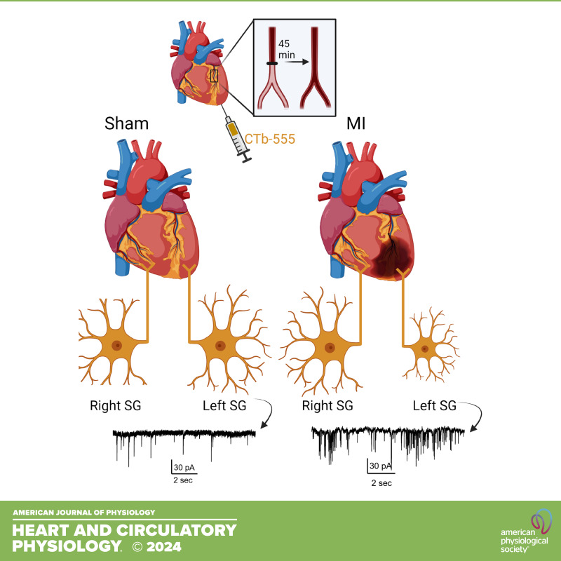
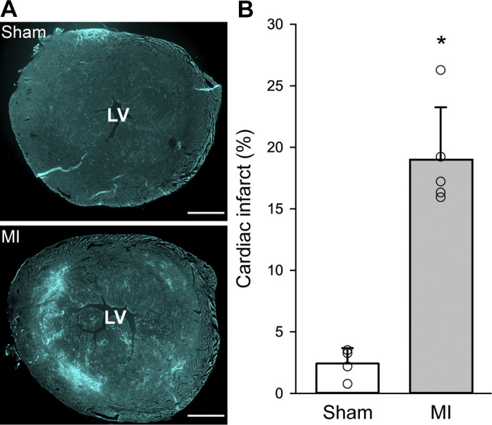
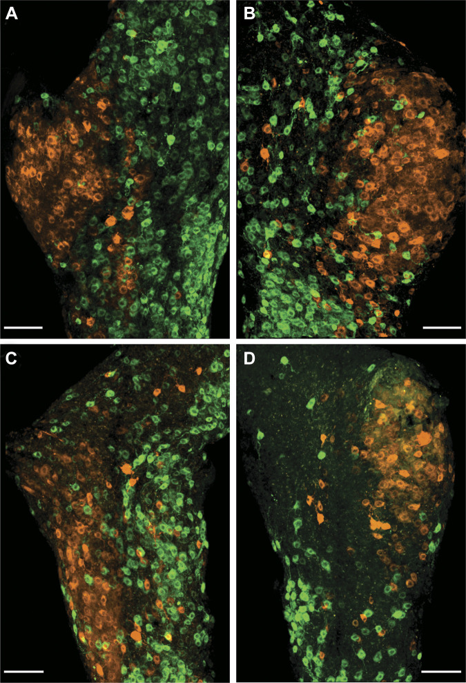
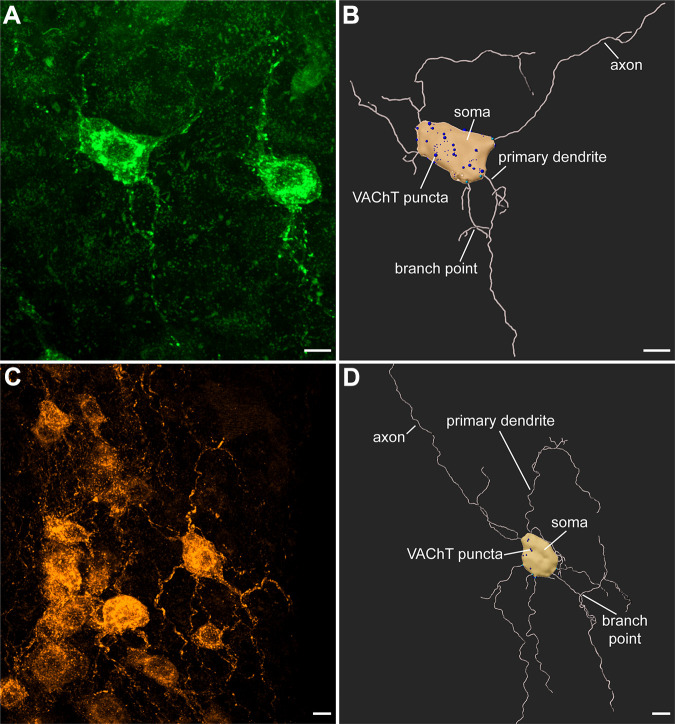
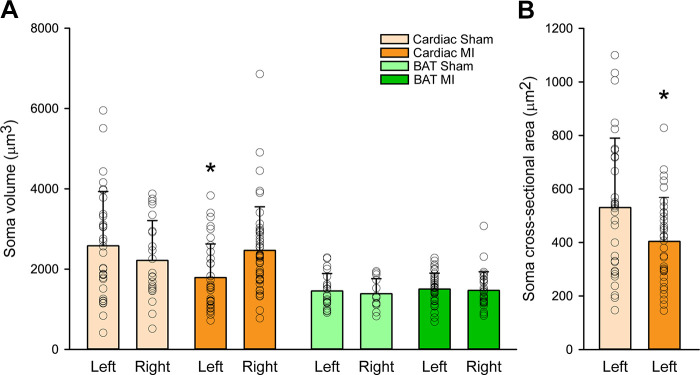
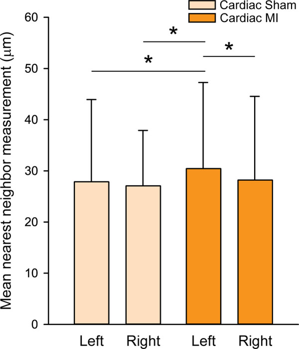
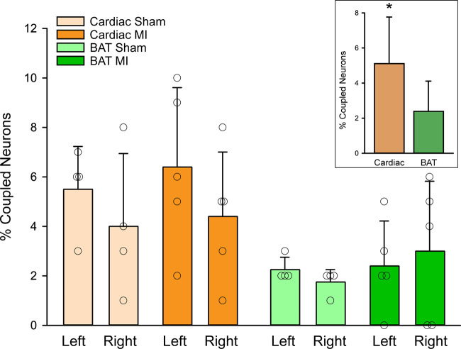
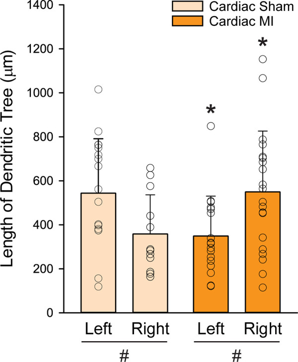
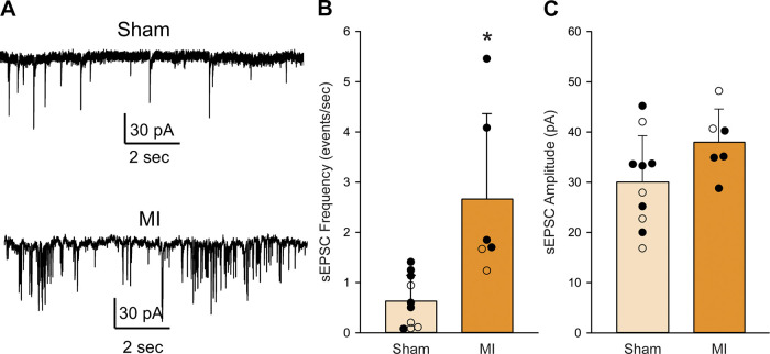
Neurons in the stellate ganglion (SG) provide sympathetic innervation to the heart, brown adipose tissue (BAT), and other organs. Sympathetic innervation to the heart becomes hyperactive following myocardial infarction (MI). The impact of MI on the morphology of cardiac sympathetic neurons is not known, but we hypothesized that MI would stimulate increased cell and dendritic tree size in cardiac neurons. In this study, we examined the effects of ischemia-reperfusion MI on sympathetic neurons using dual retrograde tracing methods to allow detailed characterization of cardiac- and BAT-projecting neurons. Different fluorescently conjugated cholera toxin subunit B (CTb) tracers were injected into the pericardium and the interscapular BAT pads, respectively. Experimental animals received a 45-min occlusion of the left anterior descending coronary artery and controls received sham surgery. One week later, hearts were collected for assessment of MI infarct and SGs were collected for morphological or electrophysiological analysis. Cardiac-projecting SG neurons from MI mice had smaller cell bodies and shorter dendritic trees compared with sham animals, specifically on the left side ipsilateral to the MI. BAT-projecting neurons were not altered by MI, demonstrating the subpopulation specificity of the response. The normal size and distribution differences between BAT- and cardiac-projecting stellate ganglion neurons were not altered by MI. Patch-clamp recordings from cardiac-projecting left SG neurons revealed increased spontaneous excitatory postsynaptic currents despite the decrease in cell and dendritic tree size. Thus, increased dendritic tree size does not contribute to the enhanced sympathetic neural activity seen after MI.NEW & NOTEWORTHY Myocardial infarction (MI) causes structural and functional changes specifically in stellate ganglion neurons that project to the heart, but not in cells that project to brown adipose fat tissue.
Keywords: morphology; neural plasticity; sympathetic; tract tracing.
Conflict of interest statement
No conflicts of interest, financial or otherwise, are declared by the authors.
Figures
Similar articles
-
Distinct morphology of cardiac- and brown adipose tissue-projecting neurons in the stellate ganglia of mice.Physiol Rep. 2022 May;10(10):e15334. doi: 10.14814/phy2.15334. Physiol Rep. 2022. PMID: 35621038 Free PMC article.
-
Targeted ablation of cardiac sympathetic neurons reduces the susceptibility to ischemia-induced sustained ventricular tachycardia in conscious rats.Am J Physiol Heart Circ Physiol. 2010 May;298(5):H1330-9. doi: 10.1152/ajpheart.00955.2009. Epub 2010 Feb 19. Am J Physiol Heart Circ Physiol. 2010. PMID: 20173045 Free PMC article.
-
Focal myocardial infarction induces global remodeling of cardiac sympathetic innervation: neural remodeling in a spatial context.Am J Physiol Heart Circ Physiol. 2013 Oct 1;305(7):H1031-40. doi: 10.1152/ajpheart.00434.2013. Epub 2013 Jul 26. Am J Physiol Heart Circ Physiol. 2013. PMID: 23893167 Free PMC article.
-
Cardiac neurones of autonomic ganglia.Microsc Res Tech. 1996 Sep 1;35(1):69-79. doi: 10.1002/(SICI)1097-0029(19960901)35:1<69::AID-JEMT6>3.0.CO;2-N. Microsc Res Tech. 1996. PMID: 8873060 Review.
-
Stellate Ganglia and Cardiac Sympathetic Overactivation in Heart Failure.Int J Mol Sci. 2022 Nov 1;23(21):13311. doi: 10.3390/ijms232113311. Int J Mol Sci. 2022. PMID: 36362099 Free PMC article. Review.
Cited by
-
Molecular and functional diversity of the autonomic nervous system.Nat Rev Neurosci. 2025 Jul 3. doi: 10.1038/s41583-025-00941-2. Online ahead of print. Nat Rev Neurosci. 2025. PMID: 40610604 Review.
-
Molecular and cellular neurocardiology in heart disease.J Physiol. 2025 Mar;603(7):1689-1728. doi: 10.1113/JP284739. Epub 2024 May 22. J Physiol. 2025. PMID: 38778747 Review.
-
Hypertension increases sympathetic neuron activity by enhancing intraganglionic cholinergic collateral connections.J Physiol. 2025 Mar;603(7):2005-2020. doi: 10.1113/JP286601. Epub 2024 Jun 21. J Physiol. 2025. PMID: 39031543
-
Hypertension-induced heart failure disrupts cardiac sympathetic innervation.Am J Physiol Heart Circ Physiol. 2024 Dec 1;327(6):H1544-H1558. doi: 10.1152/ajpheart.00380.2024. Epub 2024 Nov 1. Am J Physiol Heart Circ Physiol. 2024. PMID: 39485300
References
-
- Solomon SD, Zelenkofske S, McMurray JJ, Finn PV, Velazquez E, Ertl G, Harsanyi A, Rouleau JL, Maggioni A, Kober L, White H, Van de Werf F, Pieper K, Califf RM, Pfeffer MA; Valsartan in Acute Myocardial Infarction Trial (VALIANT) Investigators. Sudden death in patients with myocardial infarction and left ventricular dysfunction, heart failure, or both. N Engl J Med 352: 2581–2588, 2005. [Erratum in N Engl J Med 353: 744, 2005]. doi:10.1056/NEJMoa043938. - DOI - PubMed
-
- Pouleur AC, Barkoudah E, Uno H, Skali H, Finn PV, Zelenkofske SL, Belenkov YN, Mareev V, Velazquez EJ, Rouleau JL, Maggioni AP, Kober L, Califf RM, McMurray JJ, Pfeffer MA, Solomon SD; VALIANT Investigators. Pathogenesis of sudden unexpected death in a clinical trial of patients with myocardial infarction and left ventricular dysfunction, heart failure, or both. Circulation 122: 597–602, 2010. doi:10.1161/CIRCULATIONAHA.110.940619. - DOI - PubMed
-
- Vaseghi M, Gima J, Kanaan C, Ajijola OA, Marmureanu A, Mahajan A, Shivkumar K. Cardiac sympathetic denervation in patients with refractory ventricular arrhythmias or electrical storm: intermediate and long-term follow-up. Heart Rhythm 11: 360–366, 2014. doi:10.1016/j.hrthm.2013.11.028. - DOI - PMC - PubMed
Publication types
MeSH terms
Grants and funding
LinkOut - more resources
Full Text Sources
Medical