. 2023 Nov 2;12(21):2565.
doi: 10.3390/cells12212565.

Treating Parkinson's Disease with Human Bone Marrow Mesenchymal Stem Cell Secretome: A Translational Investigation Using Human Brain Organoids and Different Routes of In Vivo Administration

Affiliations

Treating Parkinson's Disease with Human Bone Marrow Mesenchymal Stem Cell Secretome: A Translational Investigation Using Human Brain Organoids and Different Routes of In Vivo Administration

Bárbara Mendes-Pinheiro et al. Cells. .

Abstract

Parkinson's disease (PD) is the most common movement disorder, characterized by the progressive loss of dopaminergic neurons from the nigrostriatal system. Currently, there is no treatment that retards disease progression or reverses damage prior to the time of clinical diagnosis. Mesenchymal stem cells (MSCs) are one of the most extensively studied cell sources for regenerative medicine applications, particularly due to the release of soluble factors and vesicles, known as secretome. The main goal of this work was to address the therapeutic potential of the secretome collected from bone-marrow-derived MSCs (BM-MSCs) using different models of the disease. Firstly, we took advantage of an optimized human midbrain-specific organoid system to model PD in vitro using a neurotoxin-induced model through 6-hydroxydopamine (6-OHDA) exposure. In vivo, we evaluated the effects of BM-MSC secretome comparing two different routes of secretome administration: intracerebral injections (a two-site single administration) against multiple systemic administration. The secretome of BM-MSCs was able to protect from dopaminergic neuronal loss, these effects being more evident in vivo. The BM-MSC secretome led to motor function recovery and dopaminergic loss protection; however, multiple systemic administrations resulted in larger therapeutic effects, making this result extremely relevant for potential future clinical applications.

Keywords: Parkinson’s disease; brain organoids; mesenchymal stem cells; neurodegeneration; pre-clinical study; stem cell secretome.


Conflict of interest statement

The authors declare no conflict of interest.

Figures

Figure 1
Figure 1
Derivation of hMOS from hvNESCs. (A) Schematic representation of the procedure of hMO culture system and the generation of the neurotoxin-induced PD model to assess the neuroprotective effects of BM-MSC secretome; and (B) growth of hMOs in culture: brightfield images of hMOS generated from hvNESCs at different days in culture (scale bar represents 100 and 200 μm), and the diameter size of hMOS per organoid line from day 2 to day 20. * Represents the specific time-point of geltrex embedding with plating and shaking in 24 well plates. Error bars represent mean ± SEM.
Figure 2
Figure 2
Characterization of hMOS at day 30 of the differentiation protocol. (a) hMO self-organization: stem cell niche in the middle (marked with SOX2 in red) and the dopaminergic neurons distributed asymmetrically (marked with TH at green) and nuclei (marked with DAPI in blue); (b) hMOs express midbrain floorplate markers like FOXA2 (red) and EN1 (gray) besides the characteristic TH (green) after 30 days in culture, and (c) express other midbrain markers besides TH such as GIRK2 (a marker of A9 neurons affected in PD) and dopamine, revealing the high specification for the midbrain, both stained in red.
Figure 3
Figure 3
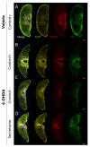
Representative images of the 6-OHDA-induced degeneration and BM-MSC secretome treatment effects. Representative maximum intensity projection of confocal images (20× magnification) of hMOS, showing TUJ1 (yellow), MAP2 (red), and TH (green) staining as well as merged images. (A) Representative organoids maintained in Control+ media without injury. (B) 6-OHDA-injured organoids for 48 h and treated with Control+ media. (C) 6-OHDA-injured organoids for 48 h and treated with Control- media. (D) 6-OHDA-injured organoids for 48 h treated with secretome.
Figure 4
Figure 4
Effects of BM-MSC secretome in the 6-OHDA-induced PD model. Extracted features from high-content image analysis to evaluate the effects of 6-OHDA exposure and the respective treatments, namely in the overall amount of (a) TUJ+ neurons, (b) MAP2+ neurons, (c) TH+ dopaminergic neurons, and on neuronal complexity, namely (d) TH fragmentation, (e) links, and (f) nodes. Control+: Differentiation medium N2B27; Control− (a basal medium without any supplementation); Secretome (BM-MSC conditioned medium). Two independent experiments (24 sections analyzed/group). Data are presented as mean ± SEM. ** p < 0.01, *** p < 0.01.
Figure 5
Figure 5
Experimental design. Schematic representation and temporal sequence of performed tasks throughout the in vivo experiment. Animals were injected with 6-OHDA into the dorsolateral striatum (2 injections); Mice were monitored daily for 14 days (post-operative care); 3 weeks after lesion, animals were characterized at motor level to validate the model; At week 5 post-lesion, animals were treated with BM-MSCs secretome using two different routes of administration: IC injections into the striatum and SNpc (2 injections into the striatum, and 1 injection into the SNpc—single administration) and repeated IV injections (3 injections in the first week, and 1 injection per week until the end of the experiment—10 injections in total). Motor behavioral assessment was performed 1, 4, and 7 weeks after treatments. Animals were sacrificed 12 weeks after lesion surgery and 9 weeks after treatments.
Figure 6
Figure 6
Motor behavioral analysis of 6-OHDA-lesioned animals after administration of BM-MSC secretome. (A) Rotameter test (apomorphine-induced rotations) as a measure of dopaminergic depletion induced by 6-OHDA lesion and compared to Sham animals; Motor performance of mice was evaluated using (B) motor swimming test, (C) pole test, and (D) beam balance walk test to assess the effects of the treatments. n = 13–15 for each group used. Statistical summary in Table S2. Data are presented as mean ± SEM. * p < 0.05, ** p < 0.01, *** p < 0.001; #: Sham group statistically different from all groups, α: Sham group statistically different form all groups except from IV Secretome; β: Sham group statistically different form all groups except from IC Secretome and IV Secretome.
Figure 7
Figure 7
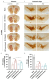
Histological analysis of the striatum and SNpc. (A) Representative images of the tyrosine hydroxylase (TH) staining on the striatum. (B) Quantification of TH+ labelling on the dorsal striatum, depicted as a percentage over the non-lesioned side. (C) Representative images of the TH staining on the ipsilateral and contralateral substantia nigra of the different experimental groups. (D) Quantification of TH+ cells on the SNpc, depicted as a percentage over the non-lesioned side. n = 7 for each group used. Statistical summary in Table S3. Data are presented as mean ± SEM. * p < 0.05, ** p < 0.01, *** p < 0.001. # p < 0.001 from all other groups. Scale bar = 2 mm.

References

    1. Poewe W., Seppi K., Tanner C.M., Halliday G.M., Brundin P., Volkmann J., Schrag A.-E., Lang A.E. Parkinson Disease. Nat. Rev. Dis. Primers. 2017;3:17013. doi: 10.1038/nrdp.2017.13. - DOI - PubMed
    1. Bloem B.R., Okun M.S., Klein C. Parkinson’s Disease. Lancet. 2021;397:2284–2303. doi: 10.1016/S0140-6736(21)00218-X. - DOI - PubMed
    1. Zahoor I., Shafi A., Haq E. Parkinson’s Disease: Pathogenesis and Clinical Aspects. Volume 311. Codon Publications; Singapore: 2018. Pharmacological Treatment of Parkinson’s Disease; pp. 129–144.
    1. Stoker T.B., Barker R.A. Recent Developments in the Treatment of Parkinson’s Disease. F1000Research. 2020;9:862. doi: 10.12688/f1000research.25634.1. - DOI - PMC - PubMed
    1. Han Y., Li X., Zhang Y., Han Y., Chang F., Ding J. Mesenchymal Stem Cells for Regenerative Medicine. Cells. 2019;8:886. doi: 10.3390/cells8080886. - DOI - PMC - PubMed

Publication types