Review
. 2023 Nov 5;12(21):2579.
doi: 10.3390/cells12212579.

Mechanisms Underlying Rare Inherited Pediatric Retinal Vascular Diseases: FEVR, Norrie Disease, Persistent Fetal Vascular Syndrome

Affiliations
Review

Mechanisms Underlying Rare Inherited Pediatric Retinal Vascular Diseases: FEVR, Norrie Disease, Persistent Fetal Vascular Syndrome

Vincent Le et al. Cells. .

Abstract

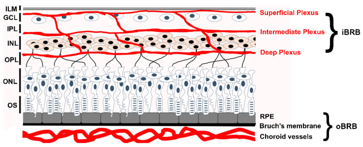
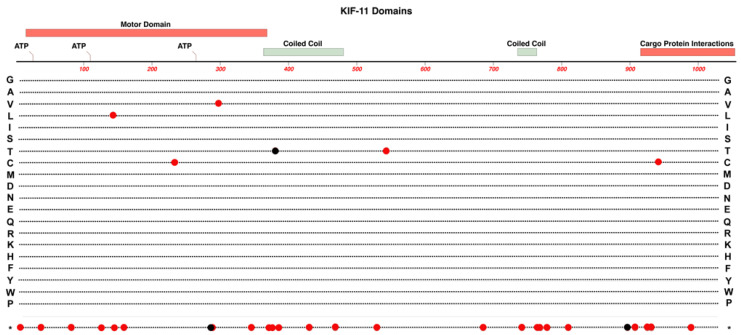
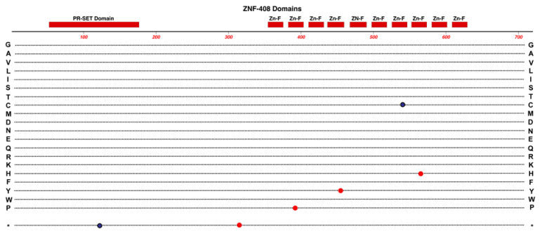
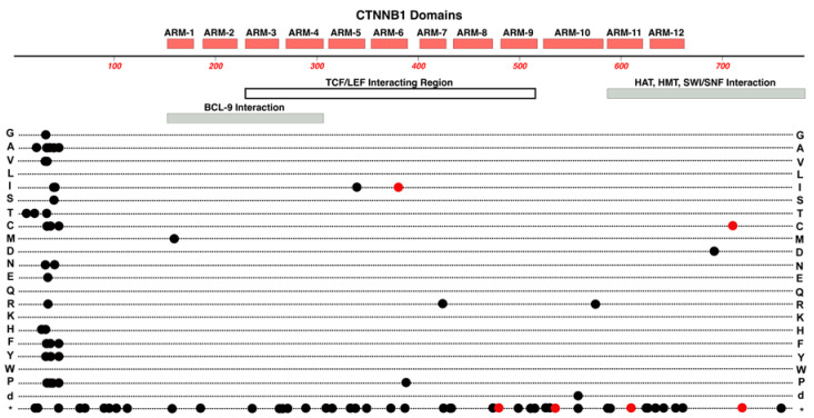
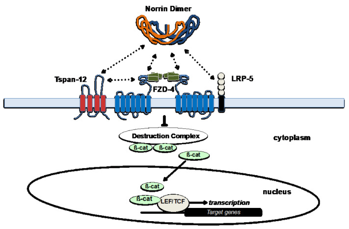
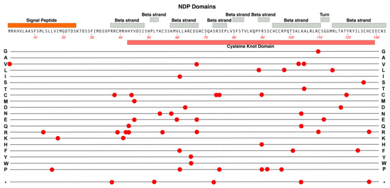
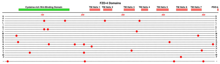
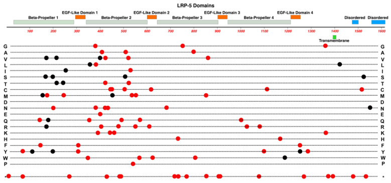
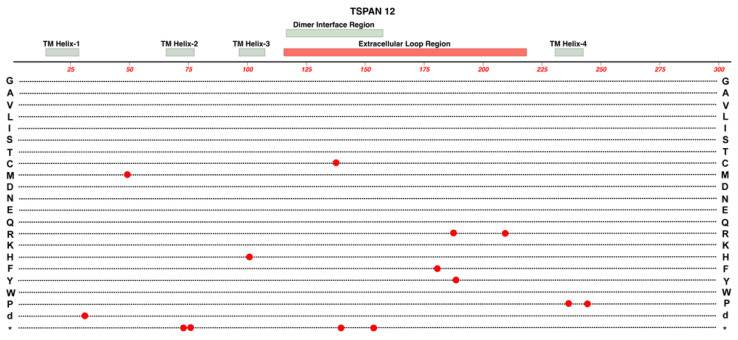
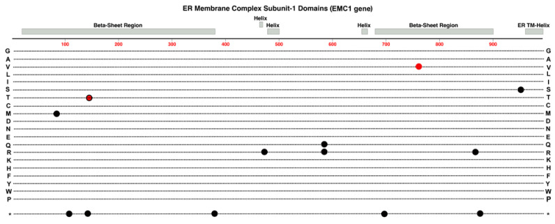
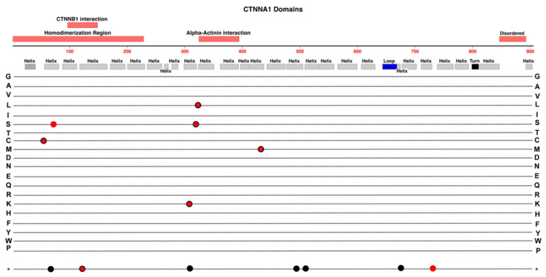
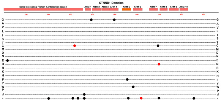
Familial Exudative Vitreoretinopathy (FEVR), Norrie disease, and persistent fetal vascular syndrome (PFVS) are extremely rare retinopathies that are clinically distinct but are unified by abnormal retinal endothelial cell function, and subsequent irregular retinal vascular development and/or aberrant inner blood-retinal-barrier (iBRB) function. The early angiogenesis of the retina and its iBRB is a delicate process that is mediated by the canonical Norrin Wnt-signaling pathway in retinal endothelial cells. Pathogenic variants in genes that play key roles within this pathway, such as NDP, FZD4, TSPAN12, and LRP5, have been associated with the incidence of these retinal diseases. Recent efforts to further elucidate the etiology of these conditions have not only highlighted their multigenic nature but have also resulted in the discovery of pathological variants in additional genes such as CTNNB1, KIF11, and ZNF408, some of which operate outside of the Norrin Wnt-signaling pathway. Recent discoveries of FEVR-linked variants in two other Catenin genes (CTNND1, CTNNA1) and the Endoplasmic Reticulum Membrane Complex Subunit-1 gene (EMC1) suggest that we will continue to find additional genes that impact the neural retinal vasculature, especially in multi-syndromic conditions. The goal of this review is to briefly highlight the current understanding of the roles of their encoded proteins in retinal endothelial cells to understand the essential functional mechanisms that can be altered to cause these very rare pediatric retinal vascular diseases.

Keywords: CTNNA1; CTNND1; ECM1; FEVR; FZD4; KIF11; LRP5; NDP; Norrie disease; TSPAN12; ZNF408; blood-brain-barrier; genetic disease mechanisms; norrin; persistent fetal vascular syndrome; retinal endothelial cell; retinal vasculature.


Conflict of interest statement

The authors declare no conflict of interest.

Figures

Figure 1
Figure 1
Location of the inner-Blood-Retinal-Barrier (iBRB) and retinal endothelial cells. The neural retina comprises several distinct layers. In the orientation of this diagram, light enters from the top and passes through the retinal layers to reach rod and cone opsins in the outer segments (OS) of photoreceptors. Photoreceptor nuclei form the Outer Nuclear Layer (ONL). Photoreceptor cells form synaptic connections with downstream inter-neurons in the Outer Plexiform Layer (OPL). Nuclei of Bipolar Cells, Horizontal Cells, and some Amacrine Cells comprise the Inner Nuclear Layer (INL). In the Inner Plexiform Layer, the Bipolar and Amacrine Cells form synaptic contacts with Ganglion Cells in the Ganglion Cell Layer (GCL). An Inner-Limiting Membrane (ILM) lays on top of the retina and the superficial plexus. Two different blood supplies support the neural retina and both have a high-barrier character, which are collectively the Blood-Retinal-Barrier (BRB). The outer-BRB (oBRB) sustains photoreceptor cells and Retinal Pigment Epithelial (RPE) cells and is formed by the fenestrated choroidal vasculature, Bruch’s membrane and the RPE. The RPE cells, not the choroidal endothelial cells, provide the high-barrier nature of the outer-BRB. The inner-BRB (iBRB) sustains neurons of the inner retina and comprises three microvascular beds, the superficial, intermediate, and deep plexus (shown by red tracts). These are collectively referred to as the neural retinal vasculature. The endothelial cells of the neural retinal vasculature are responsible for the high-barrier nature of the iBRB.
Figure 2
Figure 2
Structure and functional domain map of variants in the KIF-11 protein. Key features of the KIF-11 amino acid sequence include an ATP-binding motor-domain region, coiled-coil domains for multimer formation, and a C-terminal domain for cargo-protein interactions. Over 30 pathogenic and likely pathogenic variants are indicated by red circles with the variant amino acid indicated by the standard single-letter amino acid code. Multisyndromic with potential ocular effects (red circles) and non-ocular (black circles) disease variants are shown. The bottom row * indicates the location of nonsense (stop codon) variants. These and many other non-pathogenic variants can be explored using the UniProt database: https://www.uniprot.org/uniprotkb/P52732 (accessed on 27 October 2023).
Figure 3
Figure 3
Structure and functional domain map of variants in the ZNF-408 protein. Key features include a PR-SET domain and 10 zinc-finger domains. Several pathogenic and likely pathogenic variants are indicated with the variant of amino acid indicated by the standard single-letter amino acid code. FEVR (red circles) and Retinitis Pigmentosa-72 variants (black circles) are shown. The bottom row * indicates the location of nonsense (stop codon) variants. These variants and many non-pathogenic variants can be explored in the UniProt database: https://www.uniprot.org/uniprotkb/Q9H9D4 (accessed on 27 October 2023).
Figure 4
Figure 4
Structure and functional domain map of variants in the ß-catenin protein. Key structural features include 12 imperfect ARM repeats. The protein has numerous regulatory protein interaction partners that bind ß-catenin in separate domains but also in some overlapping regions. The N-terminus is essential for binding with alpha-Catenin as well as the beta-TrCP ubiquitin ligase that targets ß-catenin for proteosome degradation. Pathogenic and likely pathogenic variants linked to FEVR (red circles) and non-FEVR (cancer, black circles) conditions are indicated by marking the linked variant amino acid. The variant marked on row d indicates the position of a large in-frame deletion. The bottom row * indicates the location of nonsense (stop codon) variants. These and other non-pathogenic variants can be explored in the UniProt database: https://www.uniprot.org/uniprotkb/P35222 (accessed on 27 October 2023).
Figure 5
Figure 5
The Norrin Wnt-signaling pathway in retinal endothelial cells. The main functional proteins in the Norrin Wnt-signaling pathway, which is active in neural retinal endothelial cells. Norrin’s binding affinity for FZD-4 is increased when the co-receptors Tspan-12 and LRP-5 are present. Direct protein-protein interactions are indicated between various partners in the complex by dashed double-arrows. Specific extracellular domains and surfaces of proteins within this ligand/receptor complex provide the various interactions and the synergy which makes retinal endothelial cells uniquely sensitive to Norrin. Norrin binding blocks activity of the Destruction Complex, which reduces the delivery of ß-catenin to proteosome-degradation. The resulting increase in cytoplasmic ß-catenin and its translocation into the nucleus leads to interaction with LEF/TCF transcription factors to regulate the expression of target genes. General structural domains and features are illustrated for Norrin, FZD-4, LRP-5, Tspan-12, including transmembrane domains, extracellular loops, and other extracellular domains that are involved in protein interactions.
Figure 6
Figure 6
Structure and functional domain map of variants in NDP (Norrin). Key features include an N-terminal signal peptide required to target Norrin for extracellular transport. Four longer and four shorter beta-strand regions are layered in the Norrin monomer and the entire tertiary structure is stabilized by multiple disulfide bridges. The so-called cysteine knot domain involves most of the mature Norrin amino acid sequence. Over 50 pathogenic and likely pathogenic variants are indicated by red circles, marking the variant amino acid linked to Norrie disease or FEVR. The bottom row * indicates the location of nonsense (stop codon) variants. These and other non-pathogenic variants can be explored in the UniProt database: https://www.uniprot.org/uniprotkb/Q00604 (accessed on 27 October 2023).
Figure 7
Figure 7
Structure and functional domain map of variants in FZD-4 (Frizzled-4). Key features include an N-terminal Wnt-binding domain, seven transmembrane helical regions, and a PDZ-binding domain. The locations of 25 pathogenic and likely pathogenic variants are indicated (red circles), showing the pathogenic amino acid variant. The bottom row * indicates the location of nonsense (stop codon) variants. These and other non-pathogenic variants can be explored in the UniProt database: https://www.uniprot.org/uniprotkb/Q9ULV1 (accessed on 27 October 2023).
Figure 8
Figure 8
Structure and functional domain map of variants in LRP-5. Key features include the transmembrane domain, two intracellular disordered domains, and four extracellular Beta-Propeller/EGF-Like domain regions. Over 90 pathogenic and likely pathogenic amino acid variants are indicated, affecting the retina (red circles) and non-ocular syndromes (black circles, bone density). The bottom row * indicates the location of nonsense (stop codon) variants. These and many non-pathogenic variants can be explored in the UniProt database: https://www.uniprot.org/uniprotkb/O75197 (accessed on 27 October 2023).
Figure 9
Figure 9
Structure and functional domain map of variants in TSPAN-12. Key features include the four transmembrane (TM) helical domains, the large extracellular loop region, and the dimer interface region. TSPAN-12 is a member of the Tetraspanin family of membrane proteins. Both the N-terminal and C-terminal domains are intracellular. The locations of 14 pathogenic and likely pathogenic variants are indicated by red circles with the variant amino acid indicated by the standard single-letter amino acid code. Row “d” indicates the location of a multiple amino acid deletion. The bottom row * indicates the location of nonsense (stop codon) variants. Exploration of additional non-pathogenic variants can be found in the UniProt database: https://www.uniprot.org/uniprotkb/O95859 (accessed on 27 October 2023).
Figure 10
Figure 10
Structure and functional domain map of variants in Endoplasmic Reticulum Membrane Complex Protein Subunit-1. Key features include a C-terminal endoplasmic reticulum transmembrane helix and two large complex beta-sheet barrels joined by smaller helical domains. Pathogenic and likely pathogenic variants are indicated for those linked to FEVR (red circle), Retinitis Pigmentosa (red circle with black outline), and those linked to other developmental conditions (black circles). The bottom row * indicates the location of nonsense (stop codon) variants. Exploration of additional non-pathogenic variants can be found in the UniProt database: https://www.uniprot.org/uniprotkb/Q8N766 (accessed on 27 October 2023).
Figure 11
Figure 11
Structure and functional domain map of variants in CTNNA1 (Catenin alpha-1). Secondary structures are derived from Alphafold data (https://alphafold.ebi.ac.uk/entry/P35221 (accessed on 27 October 2023).) [68,69]. Pathogenic and likely pathogenic variants are indicated for those linked to FEVR (red circles), patterned macular dystrophy (red circles with black outlines), and those linked to non-retinal conditions including cancers (black circles). The bottom row * indicates the location of nonsense (stop codon) variants. Exploration of additional non-pathogenic variants can be found in the UniProt database: https://www.uniprot.org/uniprotkb/P35221 (accessed on 27 October 2023).
Figure 12
Figure 12
Structure and functional domain map of variants in CTNND1 (Catenin delta-1). Pathogenic and likely pathogenic variants are indicated for those linked to FEVR (red circles) and those linked to other developmental conditions (black circles). The bottom row * indicates the location of nonsense (stop codon) variants. Exploration of additional non-pathogenic variants can be found in the UniProt database: https://www.uniprot.org/uniprotkb/O60716 (accessed on 27 October 2023).

References

    1. Fruttiger M. Development of the Mouse Retinal Vasculature: Angiogenesis versus Vasculogenesis. Investig. Ophthalmol. Vis. Sci. 2002;43:522–527. - PubMed
    1. Zuercher J., Fritzsche M., Feil S., Mohn L., Berger W. Norrin Stimulates Cell Proliferation in the Superficial Retinal Vascular Plexus and Is Pivotal for the Recruitment of Mural Cells. Hum. Mol. Genet. 2012;21:2619–2630. doi: 10.1093/hmg/dds087. - DOI - PubMed
    1. Selvam S., Kumar T., Fruttiger M. Retinal Vasculature Development in Health and Disease. Prog. Retin. Eye Res. 2018;63:1–19. doi: 10.1016/j.preteyeres.2017.11.001. - DOI - PubMed
    1. Tokunaga C.C., Mitton K.P., Dailey W., Massoll C., Roumayah K., Guzman E., Tarabishy N., Cheng M., Drenser K.A. Effects of Anti-VEGF Treatment on the Recovery of the Developing Retina Following Oxygen-Induced Retinopathy. Investig. Ophthalmol. Vis. Sci. 2014;55:1884–1892. doi: 10.1167/iovs.13-13397. - DOI - PubMed
    1. Dailey W.A., Drenser K.A., Wong S.C., Cheng M., Vercellone J., Roumayah K.K., Feeney E.V., Deshpande M., Guzman A.E., Trese M., et al. Ocular Coherence Tomography Image Data of the Retinal Laminar Structure in a Mouse Model of Oxygen-Induced Retinopathy. Data Brief. 2017;15:491–495. doi: 10.1016/j.dib.2017.09.075. - DOI - PMC - PubMed

Publication types

Supplementary concepts